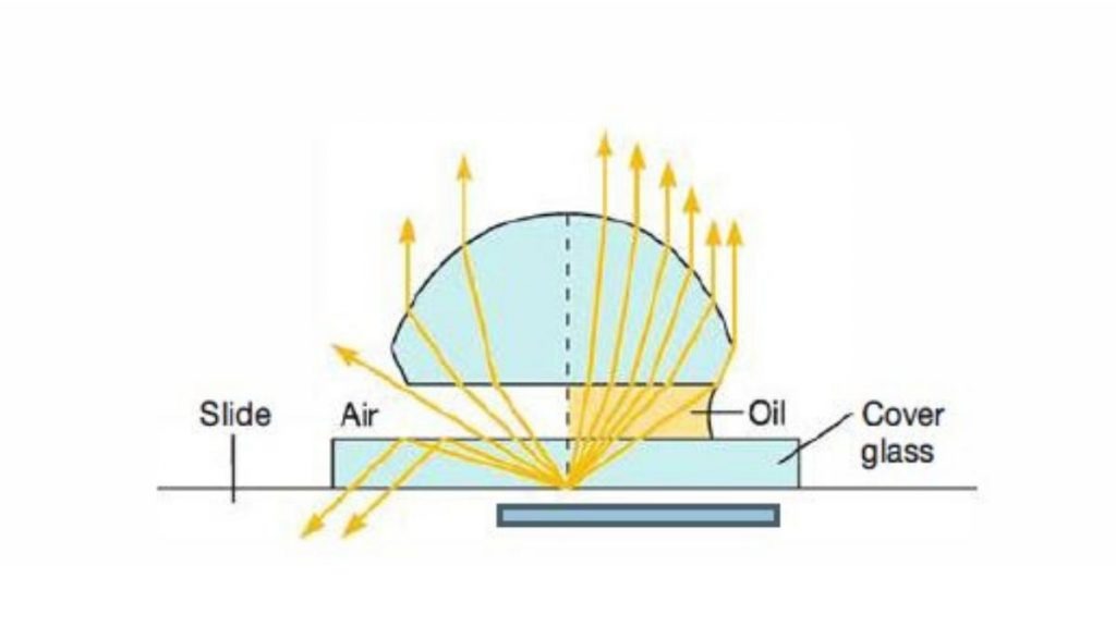
The oil immersion method enhances the resolving power of a microscope, enabling light microscopy to distinguish finer details. Immersion oil—clear and colorless, having a refractive index of about 1.515—is placed in between the lens and the specimen. The refractive index is higher; therefore, less light is refracted. If less light is refracted, then less is diverted from viewing a slide, and since more enters the objective lens, the result is a clearer image. This is especially true for the higher power magnifications; 100x is an oil immersion objective. The refractive index of immersion oil and that of the glass of the objective lens are similar, which increases the numerical aperture of the lens and thus the resolving power.
What is Oil Immersion?
Ever looked through a microscope and wondered how people take such high-resolution photographs of little things—cells, germs, for example? While the answer to this question may be plentiful and varied, one possibility is that they use something called oil immersion. Oil? Yes. Not the vegetable oil in your kitchen, but one type of oil (usually called “immersion oil”) serves to give a better view. But why? Because, at such high magnification, when the light from the objective lens has to travel through air (the space between the lens and the specimen), dispersion occurs.
Think of shining a flashlight beam in the fog. The beam doesn’t only weaken, but the image is blurry. Yet, when oil fills the space between the lens and the slide, no dispersion occurs. That’s because oil’s refractive index (just like glass) is the same as that of glass (which the microscope lenses are made of). Therefore, the light travels through without dispersion. This isn’t just any old magic trick—it’s a phenomenon.
You’d NEVER see bacteria—or portions of certain blood cells—without oil immersion, much like you would have difficulty reading through a fogged-up window. This is what brings microscopes to those standards of magnification necessary to bring everything into focus, and therefore, it’s the championed procedure in every lab and hospital from here to Timbuktu. What’s more fascinating, however, is that this isn’t just some modern-day discovery. When microscopes were first created to image on minuscule levels in the 1800s, Carl Zeiss and his peer Ernst Abbe (the first microscope lovers) discovered that viewing their creations through air allowed for uncontrollable aberrations in their images. Oil, in their devices, was the fix. More than a century later, it’s still the fix.
Whether diagnosing diseases, proving skin issues, or just figuring out what that bump might be on one’s epidermis—whether from micro milliliters of pond slime to a particle of dust—this thin layer of oil is a quasi-invisible protector of one of mankind’s greatest contraptions. And all from something people think is just vegetable oil!
What is Microscopic Resolution?
Microscopic resolution refers to a microscope’s ability to distinguish between two closely spaced points as separate entities. In other words, it is the smallest distance between two distinct points that can still be identified as separate under the microscope. This capability is crucial for observing fine details in specimens, such as cells and microorganisms, which are often not visible to the naked eye.
What influences resolution in a microscope? How does light relate to resolution? For example, the numerical aperture of a lens influences microscope resolution.
The numerical aperture (NA) of the objective lens is a unitless value relative to the amount of light the lens can gather and the lens’s capacity to resolve detail at a given distance. Therefore, the higher the value of the NA, the higher the resolution. In addition, some wavelengths are more resolute than others; for instance, blue light is more resolute than red light because blue has a shorter wavelength than red. It’s an interesting notion that a gradual increase in resolution over time would occur.
The resolution limit set in the 19th century by Ernst Abbe was the diffraction limit to resolution. It defines the theoretical maximum level of resolution attained with optical microscopes. Essentially, it means that if something is too close together than about one half of the wavelength of light used to perceive it, it cannot be perceived. Thus, with ranges of wavelengths in visible light, 200 nanometers is the practically resolved nanometer limit/separation. Yet, this has been surpassed by current microscopy—from electron microscopy to fluorescence microscopy.
Equation of Microscopic resolution
The microscopic resolution can be calculated by using Abbe Equation. Resolution is described mathematically by an equation developed in the 1870s by Ernst Abbe, a German physicist responsible for much of the optical theory underlying microscope Design.
The Abbe equation is;

Where,
d = Distance between two objects.
𝞴 = Wavelength of light.
nsin𝝧 = Numerical Aperture.
When d becomes smaller, the resolution increases, therefore the resolution (r );

Immersion oil Objectives
- Immersion oil increases the numerical aperture of the lens.
- This results in better light gathering and resolution.
- Higher resolution allows viewing finer details in specimens.
- It improves light transmission for clearer and brighter images.
- Using immersion oil reduces optical distortions like aberrations.
- It matches the refractive index for sharper images.
Why is Oil Immersion used?

Bending of light happens when light rays transition from one medium to another while being independent due to a natural difference in refractive index. This is relevant to the operation of a light microscope because light rays pass through two independent media with two different refractive indices: air and glass (the objective lens and the cover slip).
For example, as the light emitted from the specimen slide travels up into the objective lens, it simultaneously travels through air in the space between the lens and the slide. As a result, some of the light is lost and dissipated in the space between glass and air because of the difference in refractive indexes. The index of refraction of air is about 1.0, and the index of refraction of glass is about 1.5. Therefore, there is a refraction of light where glass and air meet.
This means that things are distorted when they are brought into more focus—that is, if you attempt to focus even more through the eyepiece after attempting to view the image through the enhanced focus provided by the objective lens. This distortion, this challenge of light refraction, occurs only with the 100x objective lens. It does not occur with the lower power objectives—4x, 10x, 40x—which are also not as powerful.
However, if light refraction decreases, more light enters the objective lens of the specimen slide and thus the image will form. Light refraction decreases when there is a drop of immersion oil between the objective and the slide because it equals the refractive index of glass and the air in between. Therefore, more light approaches the objective lens, and a better image is produced.
Why do we use oil immersion in microscope 100x?
What is the benefit of oil immersion? Why is it particularly helpful with a 100x objective? Using a 100x objective refers to “oil immersion,” where a drop of oil is placed on top of the slide one is viewing, where the slide happens to be, where the specimen is located. This drop of immersion oil enables the 100 objective to be “immersed” in the same medium as the specimen.
Oil and glass have similar refractive indices, so by adding a layer of oil as the medium between the slide and specimen and the objective, it minimizes the refraction at the air/glass and glass/oil boundaries. This facilitates an increase in the lens’s numerical aperture; with less impediment to light at this boundary, more points are visible that would otherwise not be visible because their dimensions would interfere without.
The 100x is the only objective lens meant for oil immersion; it’s meant for oil—or else. If one were to apply oil to any of the dry objective lenses, optical or mechanical disaster would ensue. This is because the dry lenses have no gaskets to keep oil out, and once the oil seeps into the lenses, it messes with the mechanics and makes the objective useless.
Therefore, it’s clear that following the manufacturer’s intent is crucial because applying oil is for the 100x—and only the 100x objective. Ultimately, oil immersion in conjunction with the 100x objective lens pushes a microscope’s resolution to astounding heights. There’s less light dissipated through refraction, the images become crisper, and it’s much easier to get into the details of smaller subcellular and microscopic components.
The precision with which such a technique operates with microscopy suggests that the optical configuration must be flawless and the application must be constructed with just as much accuracy.
Immersion oil Types
- Standard Immersion Oil– Characterized by a refractive index (RI) of approximately 1.515–1.518, this oil is composed of paraffin derivatives and synthetic polymers. Commonly employed in conjunction with 100x oil immersion objectives in brightfield microscopy, it optimizes light transmission by minimizing refractive index disparities between glass slides and objectives.
- Synthetic Immersion Oil– Synthesized from non-natural hydrocarbons, this variant shares a comparable RI (≈1.515) with standard oils. Enhanced chemical stability and reduced evaporation rates render it suitable for prolonged microscopy sessions in automated or temperature-controlled systems.
- Type A Immersion Oil – A mineral oil base modified with viscosity-enhancing additives defines this category. With an RI of ~1.515, it serves general-purpose applications at lower magnifications (e.g., 40x–60x), where moderate resolution and cost efficiency are prioritized.
- Type B Immersion Oil– Distinguished by an elevated RI (~1.520) and increased viscosity, this formulation enhances image resolution in high-magnification workflows (e.g., 100x). Its thickened consistency reduces optical aberrations, making it preferable for detailed histological or cytological analyses.
- Fluorescence-Specific Immersion Oil – Engineered for fluorescence microscopy, these oils exhibit reduced autofluorescence and a variable RI (1.40–1.50). Their low fluorescence background prevents signal interference, ensuring accurate detection of fluorophore emissions in confocal or epifluorescence systems.
- High Refractive Index Immersion Oil– Specialized oils with an RI of 1.56–1.58 are utilized in advanced microscopy techniques (e.g., super-resolution or 3D imaging). Their elevated RI and viscosity enhance numerical aperture, enabling superior light-gathering capacity and resolution at ultrahigh magnifications (>100x).
Procedure of Immersion oil Technique
- Slide Preparation– Position the specimen on a sterilized glass slide and secure it with a coverslip using standardized mounting techniques. Proper sealing prevents environmental contaminants and mechanical degradation of the sample during observation.
- Microscope Configuration– Initiate observation using low-magnification objectives (4x–10x) to calibrate the field of view and locate the specimen. Achieve preliminary focus via coarse and fine adjustment knobs before transitioning to high-magnification optics.
- Immersion Oil Application -After achieving optimal focus at lower magnifications, dispense a single droplet (1–2 µL) of immersion oil onto the coverslip. Excess oil should be avoided, as it may scatter light, degrade resolution, or infiltrate objective housings.
- Engagement of Oil Immersion Objective – Rotate the nosepiece to align the 100x oil immersion objective with the specimen. Ensure the lens maintains near-contact with the oil droplet without physical collision, preserving both sample integrity and optical alignment.
- Image Refinement Process– Utilize fine-focus controls to enhance image sharpness. Minor adjustments to the condenser aperture or illumination intensity may be required to optimize contrast and numerical aperture alignment.
- Post-Imaging Maintenance– Post-observation, disengage the objective and remove the slide. Delicately cleanse the objective’s front lens using lint-free, non-abrasive lens paper moistened with ethanol (70%). Residue removal from slides and coverslips is critical to prevent oil crystallization or long-term optical damage.
Advantages of Immersion oil Technique
- It helps to improves the resolution of images.
- It reduces light refraction, making the images clearer.
- It enhances the numerical aperture for better image detail.
- It allows more light to reach the objective lens.
- It is essential for observing finer details at high magnification.
- It helps capture sharper and more accurate images.
Limitations of Immersion oil Technique
- It requires careful handling to prevent contamination.It can be challenging to clean the objective lens after use.
- It is not suitable for live cell imaging.
- It may cause spherical aberrations when used with live specimens.
- It can damage microscope components if not cleaned properly.
- It requires additional preparation steps, increasing the time needed for observation.
- It is not ideal for specimens that are not mounted on slides.
- https://en.wikipedia.org/wiki/Oil_immersion
- https://www.microscopeworld.com/t-using_microscope_immersion_oil.aspx
- https://www.microscopyu.com/tutorials/immersion