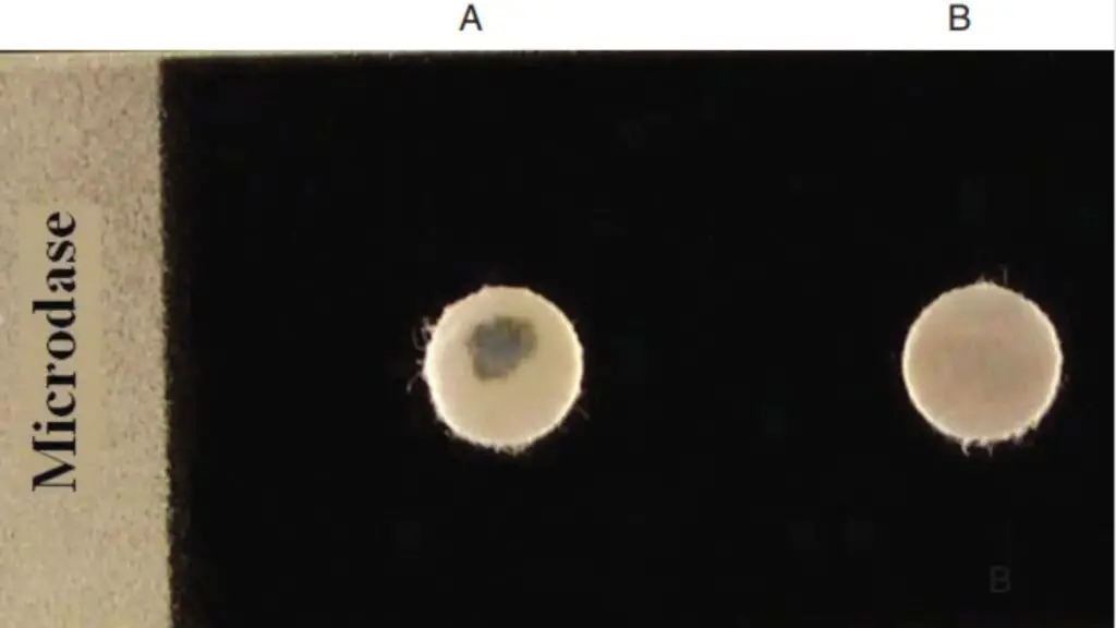
Microdase test is a rapid modified oxidase test that is used in clinical microbiology for differentiation of Micrococcus and Staphylococcus. It is based on detection of cytochrome c oxidase enzyme. It is mainly applied for Gram positive cocci that are catalase positive. This test is considered as a simple and quick screening method in routine laboratory diagnosis.
The principle of Microdase test is based on presence of cytochrome c oxidase enzyme in bacterial cells. This enzyme is present in all micrococci but generally absent in most staphylococci. The reagent used is tetramethyl-p-phenylenediamine (TMPD) which is dissolved in dimethyl sulfoxide (DMSO). DMSO helps in making the thick cell wall of Gram positive bacteria permeable to the reagent and also prevents spontaneous oxidation. When cytochrome c oxidase is present the reagent gets oxidized and produces a blue to purple-blue colour.
A filter paper disk impregnated with Microdase reagent is taken. A small amount of a young bacterial colony (15–24 hours old) is rubbed gently on the disk using a wooden applicator stick. The reaction is observed for a maximum period of two minutes. Metal loops should not be used as they may give false results. The colour development is carefully noted within the given time
Appearance of blue or purple-blue colour within two minutes indicates a positive Microdase test. This positive reaction confirms the organism as Micrococcus species. No colour change indicates a negative result and the organism is identified as Staphylococcus species. However some members of Staphylococcus sciuri group may also give positive reaction. Thus Microdase test is useful but results should be interpreted with other biochemical tests.
Objective of Microdase Test
- To differentiate between Staphylococcus and Micrococcus genera.
- To detect the presence of cytochrome c oxidase enzyme in bacteria.
- To help in identification of aerobic Gram positive and catalase positive cocci.
- To distinguish micrococci showing blue colour reaction from most staphylococci showing negative reaction except Staphylococcus sciuri group.
Principle of Microdase Test
The principle of Microdase test is based on detection of cytochrome c oxidase enzyme in bacteria. It is the process used for differentiation of Micrococcus and Staphylococcus. Cytochrome c oxidase is present in micrococci but it is generally absent in most staphylococci. This difference is used as the basis of the test.
In this test, filter paper discs are impregnated with tetramethyl-p-phenylenediamine (TMPD) which is dissolved in dimethyl sulfoxide (DMSO). The DMSO helps in permeabilizing the thick cell wall of Gram positive bacteria and allows the reagent to reach the enzyme inside the cell. It also stabilizes the reagent and prevents its auto-oxidation in air.
When the bacterial cells possess cytochrome c oxidase, the TMPD reagent is oxidized in presence of atmospheric oxygen. This oxidation results in formation of a coloured compound known as indophenol. The development of blue or purple-blue colour indicates a positive Microdase test and confirms Micrococcus species. If no colour change occurs, the reaction is negative and the organism is identified as Staphylococcus species.
Requirement of Microdase Test
- Microdase discs impregnated with tetramethyl-p-phenylenediamine (TMPD) dissolved in dimethyl sulfoxide (DMSO).
- Young bacterial culture of aerobic Gram positive and catalase positive cocci.
- Blood agar medium for growth of organism.
- Culture age of about 15–24 hours is required.
- Wooden applicator stick for transferring and rubbing the colony on disc.
- Sterile forceps for handling the Microdase disc.
- Clean glass slide or empty petri plate to place the disc.
- Control strains such as Micrococcus luteus as positive control and Staphylococcus aureus as negative control.
Procedure of Microdase Test
- A Microdase disc is taken from the container using sterile forceps and placed on a clean dry petri plate or glass slide.
- The disc should not be moistened or rehydrated before use as dilution of reagent may occur.
- A few isolated colonies are selected from a young pure culture grown on blood agar (15–18 hours old).
- The bacterial growth is picked using a clean wooden applicator stick and metal loops are avoided.
- The collected growth is rubbed gently on the surface of the Microdase disc to form a visible smear.
- The disc is kept at room temperature and observed for colour development.
- The reaction is read within 2 minutes for appearance or absence of blue or purple-blue colour.
Interpretation and Result of the Test
- Positive result (+)
- Blue or purple-blue colour develops on the disc.
- Colour change appears within 2 minutes.
- It indicates presence of cytochrome c oxidase enzyme.
- The organism is identified as Micrococcus species.
- Staphylococcus sciuri group may also show positive reaction.
- Negative result (–)
- No colour change is observed on the disc.
- The disc remains white or grey even after 2 minutes.
- It indicates absence of cytochrome c oxidase enzyme.
- The organism is identified as Staphylococcus species.

Quality Control
- Micrococcus luteus is used as positive control strain.
- Positive control must show blue or purple-blue colour within 2 minutes.
- Staphylococcus aureus is used as negative control strain.
- Negative control should show no colour change and disc remains white or grey.
- Microdase discs should not be used if they appear purple inside the container.
- Desiccant inside the vial should remain blue and not turn pink.
- Quality control should be performed for each new lot or shipment of discs.
- Control cultures must be fresh and 15–24 hours old grown on blood agar.
- Wooden applicator sticks or plastic loops should be used and metal loops are avoided.
Uses of Microdase Test
- It is used for differentiation between Staphylococcus and Micrococcus genera.
- It is used to detect presence of cytochrome c oxidase enzyme in bacteria.
- It helps in identification of aerobic Gram positive and catalase positive cocci.
- It is used to distinguish micrococci showing positive blue or purple colour from most staphylococci showing negative reaction except Staphylococcus sciuri group.
- It is used as a rapid screening test in clinical microbiology laboratories.
Advantages of Microdase Test
- It is used as a primary test to differentiate Micrococcus from Staphylococcus.
- It gives rapid result and reaction is obtained within 2 minutes.
- Dimethyl sulfoxide helps in increasing permeability of bacterial cell wall to reagent.
- The reagent remains stable and does not undergo rapid auto-oxidation.
- It is more sensitive than routine oxidase test for Gram positive bacteria.
- The test is simple to perform and ready-to-use discs are available.
Limitations of Microdase Test
- Staphylococcus sciuri group may give positive reaction and may be misidentified as micrococci.
- Some related genera such as Macrococcus and Kocuria species also show positive Microdase test.
- The test is limited only for differentiation of aerobic Gram positive catalase positive cocci.
- The reagent is unstable and may undergo auto-oxidation if exposed to air moisture or light.
- Discs showing colour change inside the vial should not be used.
- Use of metal loops may produce false positive reaction.
- Result must be read within 2 minutes as delayed reading may give false positivity.
- Old or very young cultures may give false results.
- Acidic media or high glucose containing media may interfere with oxidase activity.
- The disc should not be rehydrated before use as it reduces sensitivity.
- Acharya, T. (n.d.). Modified Oxidase Test (Microdase): Procedure, Uses. Microbe Online.
- Acharya, T. (n.d.). Oxidase Test: Principle, Procedure, Results. Microbe Online.
- Acharya, T. (n.d.). Staphylococcus vs. Micrococcus. Microbe Online.
- Genetic Science Learning Center. (n.d.). Low-Tech Microbiology Tools. University of Utah.
- Götz, F., Bannerman, T., & Schleifer, K.-H. (2006). The genera Staphylococcus and Macrococcus. In M. Dworkin, S. Falkow, E. Rosenberg, K.-H. Schleifer, & E. Stackebrandt (Eds.), The Prokaryotes (pp. 5–75). Springer.
- Hafezi, A., & Khamar, Z. (2024). The method and analysis of some biochemical tests commonly used for microbial identification: A review. Comprehensive Health and Biomedical Studies, 3(2), e160199.
- Baker, J. S. (1984). Comparison of various methods for differentiation of staphylococci and micrococci. Journal of Clinical Microbiology, 19(6), 875–879.
- Couto, I., Sanches, I. S., Sá-Leão, R., & de Lencastre, H. (2000). Molecular characterization of Staphylococcus sciuri strains isolated from humans. Journal of Clinical Microbiology, 38(3), 1136–1143.
- Faller, A., & Schleifer, K. H. (1981). Modified oxidase and benzidine tests for separation of staphylococci from micrococci. Journal of Clinical Microbiology, 13(6), 1031–1035.
- Gielas, A. (n.d.). Staphylococcus vs Micrococcus Differences. Scribd.
- ITW Reagents. (2017). Oxidase Sticks for microbiology [Product Information]. PanReac AppliChem.
- Michigan State University. (2021). Differential Media. Virtual Interactive Bacteriology Laboratory.
- Public Health England. (2014). Identification of Staphylococcus species, Micrococcus species and Rothia species (UK Standards for Microbiology Investigations ID 7, Issue 2.3).
- Tarrand, J. J., & Gröschel, D. H. M. (1982). Rapid, modified oxidase test for oxidase-variable bacterial isolates. Journal of Clinical Microbiology, 16(4), 772–774.
- The Microdase Test: Biochemical Principles, Clinical Methodologies, and Taxonomic Applications in the Differentiation of Catalase-Positive Cocci. (n.d.).
- Thermo Fisher Scientific. (2020). Microdase Disk [Instructions for Use]. Remel.
- UK Health Security Agency. (2025). Oxidase test (UK Standards for Microbiology Investigations TP 26, Issue 4.1).
- Vedantu. (n.d.). Micrococcus: Classification, Infections, Tests & Study Guide.
- Text Highlighting: Select any text in the post content to highlight it
- Text Annotation: Select text and add comments with annotations
- Comment Management: Edit or delete your own comments
- Highlight Management: Remove your own highlights
How to use: Simply select any text in the post content above, and you'll see annotation options. Login here or create an account to get started.