Digestion is a complex process that allows our bodies to break down the food we consume into smaller, more manageable components. This crucial process enables the absorption of nutrients and energy necessary for our overall health and well-being. Digestion occurs through a combination of mechanical and chemical processes, working together harmoniously to ensure the efficient breakdown of food particles.
Mechanical digestion involves the physical breakdown of food into smaller fragments, increasing the surface area available for chemical digestion to take place. It begins in the mouth, where the teeth chew and grind the food, breaking it down into smaller pieces. The tongue helps in mixing the food with saliva, forming a cohesive bolus that can be easily swallowed and transported through the esophagus.
Once in the stomach, the process of mechanical digestion continues as the muscular walls contract and churn the food, further breaking it down. This muscular action, known as peristalsis, facilitates the mixing of food with gastric juices, creating a semi-liquid mixture called chyme. The chyme is then gradually released into the small intestine, where the majority of chemical digestion takes place.
Chemical digestion involves the action of enzymes, specialized proteins that accelerate chemical reactions in the body, breaking down complex molecules into simpler substances. Enzymes are secreted by various organs throughout the digestive system, including the salivary glands, stomach, pancreas, and small intestine. Each enzyme has a specific role in digesting different types of nutrients.
In the mouth, salivary amylase begins the breakdown of carbohydrates by converting starches into simpler sugars. In the stomach, gastric juice containing hydrochloric acid and the enzyme pepsin helps break down proteins into smaller peptide chains. The pancreas plays a vital role by releasing pancreatic enzymes, such as amylase, lipase, and proteases, into the small intestine. These enzymes continue the digestion process by breaking down carbohydrates, fats, and proteins into their respective building blocks, such as glucose, fatty acids, and amino acids.
As the chyme passes through the small intestine, the walls of the organ release additional enzymes to facilitate further digestion. The small intestine also absorbs the resulting nutrients, vitamins, and minerals into the bloodstream, where they can be transported to various cells and tissues throughout the body.
In conclusion, the process of digestion involves both mechanical and chemical processes, working in tandem to break down food into its essential components. Mechanical digestion begins in the mouth and continues in the stomach, while chemical digestion occurs primarily in the small intestine with the help of enzymes. Understanding the intricate mechanisms of mechanical and chemical digestion provides insights into how our bodies extract nutrients from the food we consume, ensuring optimal nutrition and overall well-being.
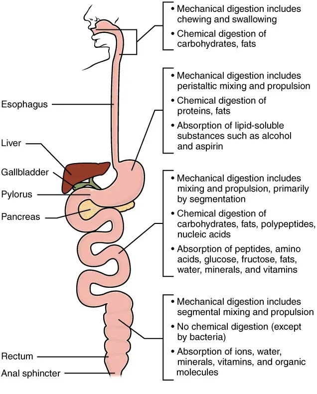
Mechanical digestion of food
- Mechanical digestion refers to the physical breakdown of food into smaller fragments, increasing the surface area available for chemical digestion to occur. It begins in the mouth and continues in the stomach, aiding in the overall process of digestion.
- In the mouth, mechanical digestion starts with the process of chewing. The teeth tear and grind the food, reducing it into smaller pieces. This action not only breaks down the food into more manageable sizes but also mixes it with saliva, forming a cohesive bolus that can be easily swallowed and transported down the esophagus.
- Once the food reaches the stomach, mechanical digestion continues. The muscular walls of the stomach contract and churn the food, thoroughly mixing it with gastric juices. This action, known as peristalsis, helps to break down the food further into smaller particles, creating a semi-liquid mixture called chyme. Peristalsis also helps to mix the food with gastric enzymes and aids in the chemical digestion process.
- The mechanical digestion process in the stomach, along with the presence of chyme, helps prepare the food for further digestion in the small intestine. The chyme is gradually released from the stomach into the small intestine, where it undergoes additional mechanical and chemical processes.
- In summary, mechanical digestion involves the physical breakdown of food through actions such as chewing, grinding, and churning. These processes increase the surface area of the food particles, making it easier for enzymes to access and break them down chemically. Mechanical digestion is an essential initial step in the overall digestive process, facilitating the efficient absorption of nutrients from the food we consume.
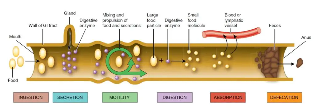
Components invole in Mechanical digestion of food
The mechanical digestion of food involves various components in the digestive system. Here are the key components involved in mechanical digestion:
- Teeth: The teeth play a vital role in the mechanical breakdown of food. They tear, grind, and crush the food into smaller pieces, increasing its surface area and making it easier to swallow and digest. Different types of teeth, such as incisors, canines, premolars, and molars, have specific functions in breaking down different types of food.
- Tongue: The tongue assists in mechanical digestion by manipulating the food within the mouth. It helps in moving the food around, mixing it with saliva, and forming it into a cohesive bolus that can be easily swallowed.
- Saliva: Saliva, produced by the salivary glands, aids in the mechanical digestion of food. It contains an enzyme called salivary amylase, which begins the breakdown of complex carbohydrates into simpler sugars. The moistening action of saliva also helps soften the food, making it easier to chew and swallow.
- Esophagus: Although the esophagus is primarily responsible for the transportation of food from the mouth to the stomach, it also contributes to mechanical digestion. The walls of the esophagus undergo rhythmic contractions called peristalsis, which propel the food bolus forward into the stomach.
- Stomach: The stomach plays a significant role in mechanical digestion. Its muscular walls contract and churn the food, mixing it with gastric juices and breaking it down into smaller particles. This mechanical action, along with the presence of gastric enzymes, helps to further break down proteins and other food components.
- Small Intestine: The small intestine continues the process of mechanical digestion. Peristaltic contractions propel the chyme, the partially digested food, along the length of the small intestine. These contractions mix and churn the chyme, facilitating further breakdown and increasing contact with the intestinal walls for nutrient absorption.
- Bile: Bile, produced by the liver and stored in the gallbladder, is released into the small intestine. It plays a mechanical role in digestion by emulsifying fats. Bile breaks down large fat globules into smaller droplets, increasing the surface area and facilitating the action of digestive enzymes.
- Segmentation: Segmentation is a mechanical process that occurs in the small intestine. It involves rhythmic contractions of the circular muscles in the intestinal wall, causing the chyme to be mixed and moved back and forth. This action enhances the contact between the chyme and the intestinal walls, promoting further breakdown and nutrient absorption.
These components work together to perform mechanical digestion, breaking down food into smaller particles, increasing the surface area, and preparing it for further chemical digestion and nutrient absorption in the digestive system.
Process of Mechanical digestion of food
Mechanical digestion of food involves several steps that occur in different parts of the digestive system. Here are the key steps of mechanical digestion:
- Chewing (Mastication): The process of mechanical digestion begins in the mouth with the action of chewing. The teeth tear and grind the food, breaking it down into smaller, more manageable pieces. The tongue also helps in moving the food around the mouth and mixing it with saliva.
- Swallowing: After chewing, the tongue pushes the food to the back of the mouth, triggering the swallowing reflex. The food bolus is then propelled down the esophagus and into the stomach through a series of muscular contractions called peristalsis.
- Mixing and churning in the stomach: Once in the stomach, the food undergoes further mechanical digestion. The muscular walls of the stomach contract and relax, mixing and churning the food with gastric juices and digestive enzymes. This process breaks down the food into smaller particles and forms a semi-liquid mixture called chyme.
- Peristalsis in the small intestine: The chyme is gradually released from the stomach into the small intestine. The small intestine is lined with smooth muscle that undergoes peristaltic contractions, propelling the chyme through the length of the small intestine. This mixing and propelling action help further break down the food and facilitate the absorption of nutrients.
- Segmentation in the small intestine: Along with peristalsis, another mechanical process called segmentation occurs in the small intestine. Segmentation involves rhythmic contractions of the circular muscles in the intestinal wall, causing the chyme to be mixed and moved back and forth. This action enhances the contact between the chyme and the intestinal walls, aiding in the absorption of nutrients.
- Mechanical breakdown by bile: Bile, produced by the liver and stored in the gallbladder, is released into the small intestine. Bile helps emulsify fats, breaking them down into smaller droplets. This mechanical breakdown increases the surface area of fats, allowing enzymes to act more effectively during chemical digestion.
Overall, mechanical digestion involves chewing, swallowing, mixing, churning, peristalsis, and segmentation. These processes work together to physically break down the food, increase its surface area, and facilitate the subsequent chemical digestion and absorption of nutrients in the digestive system.
Why Mechanical digestion of food is Important?
Mechanical digestion of food is important for several reasons:
- Increased Surface Area: Mechanical digestion breaks down food into smaller particles, increasing its surface area. This increased surface area allows for more efficient chemical digestion by exposing a larger area to digestive enzymes. It enhances the accessibility of enzymes to the food particles, enabling them to break down complex molecules into simpler forms.
- Facilitates Chemical Digestion: Mechanical digestion prepares the food for chemical digestion. By breaking down food into smaller fragments, mechanical digestion helps enzymes access the food particles more easily. This allows the enzymes to work more effectively in breaking down proteins, carbohydrates, and fats into their respective building blocks, such as amino acids, glucose, and fatty acids.
- Enhances Nutrient Absorption: Mechanical digestion aids in the efficient absorption of nutrients from the food we consume. By breaking down food into smaller particles, mechanical digestion increases the surface area available for nutrient absorption in the small intestine. This increased surface area allows for better contact between the digested food and the lining of the small intestine, facilitating the absorption of nutrients into the bloodstream.
- Eases Swallowing and Digestion: Mechanical digestion begins in the mouth with chewing, which helps break down food into smaller, more manageable pieces. Chewing also mixes the food with saliva, which contains enzymes that initiate the digestion of carbohydrates. Properly chewed and moistened food is easier to swallow, ensuring smooth passage through the digestive tract and minimizing the risk of choking.
- Enhances Efficiency and Speed of Digestion: Mechanical digestion accelerates the overall digestive process. The breakdown of food into smaller particles through chewing, mixing, churning, and peristalsis helps move the food along the digestive tract more efficiently. This allows for a faster rate of digestion, enabling the body to process larger quantities of food in a shorter amount of time.
In summary, mechanical digestion of food is essential for optimal digestion and nutrient absorption. It increases the surface area of food particles, facilitates chemical digestion, enhances nutrient absorption, eases swallowing and digestion, and improves the efficiency and speed of the digestive process. Mechanical digestion sets the stage for effective nutrient extraction from the food we consume, supporting overall health and well-being.
Chemical digestion of food
Chemical digestion refers to the complex process in which large food molecules, such as proteins, lipids, nucleic acids, and starches, are broken down into smaller subunits that can be absorbed and utilized by the cells of the body. This process involves the action of various enzymes that catalyze specific reactions, resulting in the hydrolysis of food molecules.
Enzymes are protein molecules that act as biological catalysts, speeding up chemical reactions without being consumed in the process. Different enzymes are responsible for breaking down specific types of food molecules. For example, salivary enzymes such as lingual lipase and salivary amylase are produced in the lingual glands and salivary glands, respectively. Lingual lipase breaks down triglycerides into free fatty acids, monoacylglycerides, and diglycerides, while salivary amylase begins the breakdown of polysaccharides into disaccharides and trisaccharides.
In the stomach, gastric enzymes play a role in chemical digestion. Gastric lipase, produced by chief cells, acts on triglycerides, breaking them down into fatty acids and monoacylglycerides. Pepsin, also produced by chief cells, targets proteins and breaks them down into peptides.
The small intestine is a crucial site for chemical digestion and absorption. Brush border enzymes, located on the surface of the small intestine’s epithelial cells, play a significant role in further breaking down food molecules. For example, lactase breaks down lactose into glucose and galactose, while sucrase breaks down sucrose into glucose and fructose. Other brush border enzymes, such as maltase, dextrinase, and peptidases, act on maltose, α-dextrins, and peptides, respectively, breaking them down into their respective subunits.
The pancreas also plays a vital role in chemical digestion. Pancreatic enzymes, produced by pancreatic acinar cells, are released into the small intestine. These enzymes include carboxy-peptidase, chymotrypsin, elastase, nucleases, pancreatic amylase, pancreatic lipase, and trypsin. They act on proteins, nucleic acids, and polysaccharides, breaking them down into smaller peptides, amino acids, nucleotides, and glucose.
It is important to note that some of these enzymes, such as pepsin and trypsin, are initially produced in an inactive form and require activation by other substances in the digestive system.
Overall, chemical digestion is a complex process involving a variety of enzymes that work together to break down food molecules into smaller subunits, allowing for their absorption and utilization by the body’s cells. It is an essential step in the overall digestive process, ensuring that nutrients are properly broken down and made available for energy production and other cellular functions.
| Table 1. The Digestive Enzymes | ||||
|---|---|---|---|---|
| Enzyme Category | Enzyme Name | Source | Substrate | Product |
| Salivary Enzymes | Lingual lipase | Lingual glands | Triglycerides | Free fatty acids, and mono- and diglycerides |
| Salivary Enzymes | Salivary amylase | Salivary glands | Polysaccharides | Disaccharides and trisaccharides |
| Gastric enzymes | Gastric lipase | Chief cells | Triglycerides | Fatty acids and monoacylglycerides |
| Gastric enzymes | Pepsin* | Chief cells | Proteins | Peptides |
| Brush border enzymes | α-Dextrinase | Small intestine | α-Dextrins | Glucose |
| Brush border enzymes | Enteropeptidase | Small intestine | Trypsinogen | Trypsin |
| Brush border enzymes | Lactase | Small intestine | Lactose | Glucose and galactose |
| Brush border enzymes | Maltase | Small intestine | Maltose | Glucose |
| Brush border enzymes | Nucleosidases and phosphatases | Small intestine | Nucleotides | Phosphates, nitrogenous bases, and pentoses |
| Brush border enzymes | Peptidases | Small intestine | Aminopeptidase: amino acids at the amino end of peptidesDipeptidase: dipeptides | Aminopeptidase: amino acids and peptidesDipeptidase: amino acids |
| Brush border enzymes | Sucrase | Small intestine | Sucrose | Glucose and fructose |
| Pancreatic enzymes | Carboxy-peptidase* | Pancreatic acinar cells | Amino acids at the carboxyl end of peptides | Amino acids and peptides |
| Pancreatic enzymes | Chymotrypsin* | Pancreatic acinar cells | Proteins | Peptides |
| Pancreatic enzymes | Elastase* | Pancreatic acinar cells | Proteins | Peptides |
| Pancreatic enzymes | Nucleases | Pancreatic acinar cells | Ribonuclease: ribonucleic acidsDeoxyribonuclease: deoxyribonucleic acids | Nucleotides |
| Pancreatic enzymes | Pancreatic amylase | Pancreatic acinar cells | Polysaccharides (starches) | α-Dextrins, disaccharides (maltose), trisaccharides (maltotriose) |
| Pancreatic enzymes | Pancreatic lipase | Pancreatic acinar cells | Triglycerides that have been emulsified by bile salts | Fatty acids and monoacylglycerides |
| Pancreatic enzymes | Trypsin* | Pancreatic acinar cells | Proteins | Peptides |
| *These enzymes have been activated by other substances. | ||||
Digestive chemicals
Digestive chemicals play a crucial role in the process of digestion. While digestive enzymes are primarily responsible for chemical digestion, there are other important chemicals that contribute to maintaining the optimal environment for enzymatic reactions and perform additional functions. These chemicals include water, bile, gastric acid, and bicarbonate.
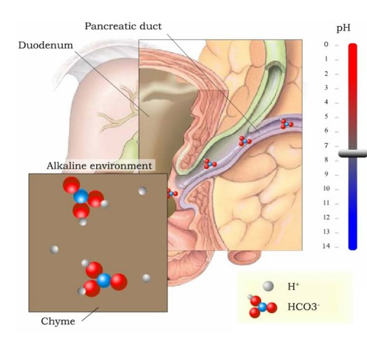
- Water, being the most abundant molecule in ingested fluids, has several vital functions in digestion. It plays a primary role in hydrolytic digestive reactions by assisting in the breakdown of food particles. Water helps liquefy and transport the digested food along the digestive tract. It also aids in the absorption of nutrients and facilitates the transport of secretions from accessory digestive organs to the gastrointestinal (GI) tract.
- Gastric acid, produced by the stomach mucosa, is a strong acid known as hydrochloric acid. Its main role is to break hydrogen bonds and modify the globular shape of proteins. This process promotes the production of pepsin, an important gastric enzyme, and facilitates the destruction of microbial proteins. As a result, the resulting polypeptides are more easily broken down by enzymes into smaller peptides.
- Bile, which is produced by the liver, is composed mostly of bile salts derived from cholesterol and water. Its primary function is the emulsification of fatty globules. Emulsification is the process of breaking down fat molecules into smaller droplets, allowing lipase enzymes to efficiently break them down further during digestion.
- Bicarbonate is a chemical that is secreted into the intestine. Its primary role is to buffer the acidic chyme (partially digested food) from the stomach, thereby protecting the intestinal mucosa. Additionally, bicarbonate promotes an alkaline pH level in the small intestine, creating a suitable environment for intestinal enzymes to function effectively.
In summary, digestive chemicals are essential for the process of digestion. Water helps in the breakdown, transport, and absorption of nutrients. Gastric acid aids in protein digestion and microbial protein destruction. Bile facilitates the breakdown of fats. Bicarbonate buffers the acidic chyme and promotes an alkaline environment for optimal enzymatic activity in the small intestine. Together, these digestive chemicals contribute to the efficient digestion and absorption of nutrients in the body.
Carbohydrate Digestion
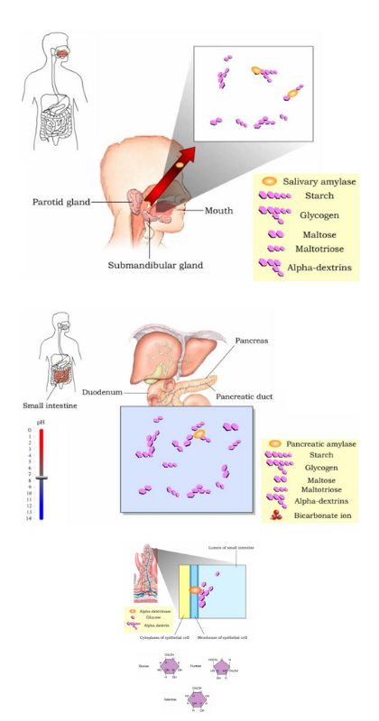
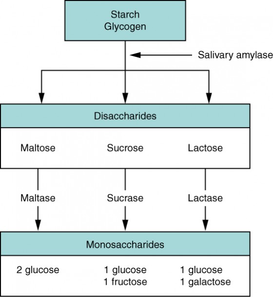
- Carbohydrate digestion is a complex process that begins in the mouth and continues in the small intestine. The average American diet consists of carbohydrates, which can be classified into simple sugars (monosaccharides and disaccharides) and complex sugars (polysaccharides).
- In the mouth, salivary amylase, produced by the salivary glands, starts the digestion of carbohydrates. It converts starch and glycogen into maltose, maltotriose, and alpha-dextrins, which are starch fragments. However, only a small portion of starch or glycogen is fully digested in the mouth.
- In the stomach, the acidic environment halts the action of salivary amylase. No significant carbohydrate digestion takes place in this organ.
- The primary site for carbohydrate digestion is the small intestine. Pancreatic amylase, secreted by the pancreas into the duodenum, continues the breakdown of starches and glycogen into maltose, maltotriose, and alpha-dextrins. The bicarbonate ions present in pancreatic juice neutralize the acidic chyme from the stomach.
- The final steps of carbohydrate digestion occur in the microvilli of the small intestine, specifically in the brush border epithelial cells. Four brush border enzymes play a role in this process. Alpha-dextrinase breaks down alpha-dextrin chains by removing glucose units. Sucrase breaks down sucrose into glucose and fructose. Maltase breaks down maltose and maltotriose into glucose. Lactase breaks down lactose into glucose and galactose. These enzymes convert disaccharides into monosaccharides.
- The end products of carbohydrate digestion are glucose, fructose, and galactose. These monosaccharides are absorbed through the intestinal epithelium into the bloodstream. From there, they can be utilized by cells for energy production and other metabolic processes.
- It’s worth noting that some polysaccharides, such as cellulose, are indigestible by the human body. Although they do not provide nutritional value, they contribute to dietary fiber, which aids in the movement of food through the digestive system.
- In summary, carbohydrate digestion involves the action of enzymes such as salivary amylase, pancreatic amylase, and brush border enzymes. Through these processes, complex carbohydrates are broken down into simpler molecules, including monosaccharides, which can be absorbed and used by the body for energy.
Protein Digestion
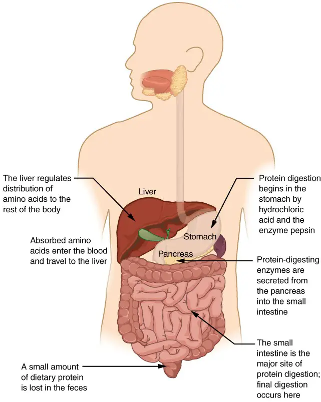
- Protein digestion is a complex process that occurs in the stomach and small intestine, where proteins are broken down into their constituent amino acids. Proteins are composed of long chains of amino acids linked together by peptide bonds. Typically, 15 to 20 percent of our total calorie intake comes from protein.
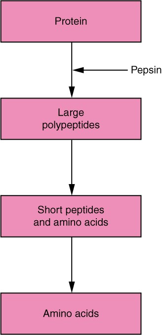
- The initial stage of protein digestion takes place in the stomach. Here, the acidic environment created by hydrochloric acid (HCl) activates pepsinogen, converting it into pepsin. Pepsin is an enzyme that catalyzes the breakdown of proteins into smaller polypeptides. The newly produced pepsin molecules further catalyze the production of more pepsin, allowing for the continued breakdown of proteins into peptides.
- Protein digestion continues in the small intestine, specifically in the duodenum. When chyme, the partially digested food, enters the duodenum, it interacts with pancreatic juice, which is a mixture of fluid and various enzymes. The pancreatic enzymes involved in protein digestion include trypsin, chymotrypsin, elastase, and carboxypeptidase.
- Trypsin, chymotrypsin, and elastase are responsible for breaking down larger peptides into smaller peptides. Each of these enzymes acts on specific bonds in the amino acid sequences of peptides. Carboxypeptidase, on the other hand, breaks the bond between the terminal amino acid and the carboxyl end of the peptide, resulting in the release of smaller peptides or even individual amino acids.
- The final stage of protein digestion occurs in the brush border of the small intestine. The brush border refers to the microvilli-covered surface of the intestinal cells. Two important enzymes found here are aminopeptidase and dipeptidase. Aminopeptidase breaks the peptide bond that attaches the terminal amino acid to the amino end of the peptide, further breaking down peptides into smaller fragments. Dipeptidase, as the name suggests, splits dipeptides into individual amino acids.
- By the end of the protein digestion process, the proteins have been broken down into their smallest units, which are amino acids. These amino acids, along with dipeptides and tripeptides, are small enough to be absorbed into the bloodstream through the intestinal lining. Once in the bloodstream, they can be transported to various tissues and organs for utilization in numerous biological processes, including the synthesis of new proteins.
- In summary, protein digestion begins in the stomach with the action of HCl and pepsin, continues in the small intestine with pancreatic enzymes, and is completed by brush border enzymes. This multi-step process breaks down proteins into smaller peptides, dipeptides, and eventually individual amino acids, which can be absorbed into the bloodstream for use in the body.
Lipid Digestion
- Lipid digestion is a process that primarily occurs in the small intestine, with minor contributions from the mouth and stomach. It involves the breakdown of dietary lipids, mainly triglycerides, into fatty acids and monoglycerides, which can be absorbed and utilized by the body. A healthy diet typically limits lipid intake to 35 percent of total calorie intake.
- The digestion of lipids begins in the mouth and stomach to some extent, where lingual lipase and gastric lipase, respectively, hydrolyze a small amount of triglycerides. However, the majority of lipid digestion takes place in the small intestine, with pancreatic lipase being the main enzyme involved.
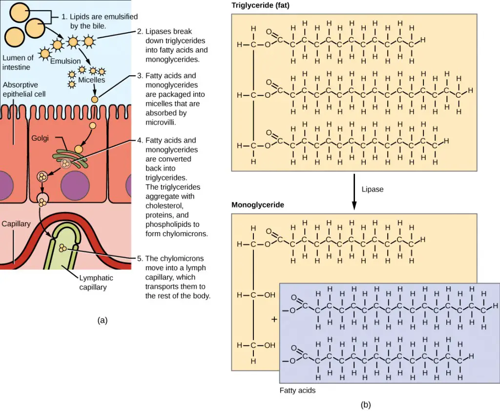
- In the duodenum, triglycerides interact with bile salts and pancreatic juice. Bile salts cling to the mono, di, and triglycerides present in fat globules, leading to the breakup of fat globules into smaller emulsion droplets. Pancreatic lipase, produced by pancreatic acinar cells, then attaches to the triglyceride molecules within these emulsion droplets.
- The role of pancreatic lipase is crucial in lipid digestion. It catalyzes the hydrolysis of triglyceride molecules, breaking them down into monoglycerides and fatty acids. This process results in the release of two free fatty acids and a monoglyceride from each triglyceride molecule. Pancreatic lipase is highly efficient and breaks down most of the triglycerides in the duodenum of the small intestine.
- The end products of lipid digestion in the small intestine are fatty acids and monoglycerides. These smaller lipid molecules are now in a form that can be easily absorbed by the intestinal cells. The breakdown of triglycerides into fatty acids and monoglycerides is essential for their absorption and subsequent utilization by the body.
- Once absorbed, fatty acids and monoglycerides can be reassembled into triglycerides within the intestinal cells and then packaged into structures called chylomicrons. These chylomicrons are transported through lymphatic vessels and eventually enter the bloodstream. From the bloodstream, lipids are delivered to various tissues and organs in the body to provide energy, serve as structural components, and participate in other vital functions.
- In summary, lipid digestion primarily occurs in the small intestine. Pancreatic lipase plays a crucial role in breaking down triglycerides into monoglycerides and fatty acids. Bile salts aid in the emulsification of fat globules, facilitating the action of lipase. The end products of lipid digestion, fatty acids, and monoglycerides, are absorbed by the intestinal cells and further utilized by the body for energy and other physiological processes.
Nucleic Acid Digestion
- Nucleic acid digestion is the process by which the nucleic acids DNA and RNA present in the foods we consume are broken down into their constituent components for absorption and utilization by the body. The main enzymes involved in nucleic acid digestion are pancreatic nucleases, including deoxyribonuclease (DNase) and ribonuclease (RNase). Further breakdown and absorption occur through brush border enzymes in the small intestine.
- Nucleic acid digestion primarily takes place in the small intestine. As the gastric chyme, which is the partially digested food, enters the duodenum of the small intestine, pancreatic juice is released. Pancreatic juice contains two important nucleases: ribonuclease, which catalyzes the breakdown of RNA into ribonucleotides, and deoxyribonuclease, which catalyzes the breakdown of DNA into deoxyribonucleotides.
- Once the nucleic acids are broken down into nucleotides, further digestion occurs at the microvilli of the epithelial cells lining the villi in the small intestine. Brush border enzymes play a crucial role in this process. Two specific enzymes are involved: phosphatases and nucleosidases. Phosphatases catalyze the cleavage of a phosphate group from the nucleotides, resulting in the formation of nucleosides, which consist of a nitrogenous base and a pentose sugar. Nucleosidases then catalyze the breaking of the covalent bond that holds the nitrogenous base to the pentose sugar, producing the final end products of nucleic acid digestion.
- The end products of nucleic acid digestion are nitrogenous bases (such as adenine, guanine, cytosine, thymine, and uracil), pentose sugars (ribose and deoxyribose), and phosphate ions. These components can be absorbed through the wall of the alimentary canal into the bloodstream, where they can be transported to various cells and tissues for utilization in processes such as DNA and RNA synthesis, energy production, and other essential functions.
- In summary, nucleic acid digestion involves the breakdown of DNA and RNA into nucleotides by pancreatic nucleases in the small intestine. Further digestion occurs through brush border enzymes, resulting in the formation of nitrogenous bases, pentose sugars, and phosphate ions. These end products are absorbed into the bloodstream for utilization in cellular processes throughout the body.
The Digestive Process
The digestive process can be broken down into the following step-by-step format:
- Mouth:
- Food is chewed into smaller pieces using specialized teeth.
- The tongue moves the food around the mouth for efficient mechanical digestion.
- Salivary glands secrete saliva, which moistens the food and begins chemical digestion of carbohydrates.
- Pharynx:
- Swallowing forces the chewed food through the pharynx.
- The epiglottis closes over the trachea to prevent choking by blocking the windpipe.
- Esophagus:
- The esophagus connects the pharynx to the stomach.
- Contractions of the esophagus push the food through a sphincter and into the stomach.
- Peristalsis, a wavelike contraction of smooth muscle, aids in moving the food along.
- Stomach:
- The stomach serves three important functions: mixing and storing food, secreting chemicals for digestion, and controlling the passage of food into the small intestine.
- Mechanical digestion continues in the stomach as it mixes and churns the food.
- Chemical digestion of proteins begins with the secretion of gastric juices containing hydrochloric acid (HCl) and pepsin.
- The stomach lining produces mucus to protect itself from the acidic environment.
- Small Intestine:
- The small intestine is the primary site for chemical digestion and nutrient absorption.
- It consists of three sections: the duodenum, jejunum, and ileum.
- Villi, finger-like projections, line the small intestine and increase the surface area for absorption.
- Most chemical digestion occurs in the duodenum, aided by secretions from the liver, pancreas, and small intestine.
- Nutrients are absorbed through the villi and transported into the bloodstream.
- Large Intestine:
- The large intestine receives the remaining undigested material from the small intestine.
- Its main function is to remove water from the undigested material through the process of absorption.
- Water is absorbed through the villi and returned to the bloodstream.
- Rectum:
- The rectum serves as a holding area for the undigested material, known as feces.
- Anus:
- Waste material leaves the body through the anus during elimination.
Accessory Organs:
- Liver:
- The liver produces bile, which helps digest fats.
- Bile is stored in the gallbladder and released into the small intestine to aid in fat digestion.
- Gallbladder:
- The gallbladder stores bile produced by the liver and releases it into the small intestine.
- Pancreas:
- The pancreas has three important functions: a) Producing enzymes for the digestion of proteins, lipids, and carbohydrates. b) Producing insulin, a hormone that regulates blood glucose levels. c) Producing sodium bicarbonate to neutralize stomach acids.
The digestive process involves both mechanical and chemical digestion, with the goal of breaking down food into smaller molecules for absorption and utilization by the body. It relies on the coordinated functioning of various organs and accessory structures to ensure efficient digestion and nutrient absorption.
Absorption
The absorption mechanism is a crucial process in the digestive system that allows the conversion of food into molecules small enough to be absorbed by the epithelial cells of the intestinal villi. Absorption occurs primarily in the small intestine, where the absorptive capacity of the alimentary canal is almost limitless.
There are five main mechanisms of absorption: active transport, passive diffusion, facilitated diffusion, co-transport (or secondary active transport), and endocytosis.
- Active transport involves the movement of substances across the cell membrane from an area of lower concentration to an area of higher concentration, requiring cellular energy in the form of ATP. Proteins within the cell membrane act as “pumps” to facilitate this process.
- On the other hand, passive diffusion refers to the movement of substances from an area of higher concentration to an area of lower concentration,
- while facilitated diffusion involves the movement of substances from an area of higher to an area of lower concentration with the help of carrier proteins in the cell membrane.
- Co-transport utilizes the movement of one molecule from higher to lower concentration to power the movement of another molecule from lower to higher concentration.
- Lastly, endocytosis is a transportation process in which the cell membrane engulfs material, requiring energy, typically in the form of ATP.
Water-soluble nutrients face a barrier due to the hydrophobic nature of the cell’s plasma membrane. To enter cells, these nutrients rely on transport molecules embedded in the membrane. Additionally, tight junctions between the epithelial cells of the intestinal mucosa prevent substances from passing between the cells. Therefore, water-soluble nutrients can only enter blood capillaries by crossing the apical surfaces of epithelial cells and entering the interstitial fluid. From there, water-soluble nutrients enter the capillary blood in the villi and travel to the liver through the hepatic portal vein.
In contrast, lipid-soluble nutrients can diffuse directly through the plasma membrane. Once inside the cell, they are packaged for transport and exit the cell through the base. They then enter the lacteals of the villi and are transported by lymphatic vessels to the systemic circulation through the thoracic duct.
The absorption of most nutrients through the mucosa of the intestinal villi requires active transport fueled by ATP. The specific mechanisms of absorption for different food categories are summarized in Table 3. Carbohydrates, such as glucose, galactose, and fructose, are absorbed through co-transport with sodium ions or facilitated diffusion. Amino acids, the breakdown products of proteins, are absorbed through co-transport with sodium ions. Lipids, including long-chain fatty acids, monoacylglycerides, short-chain fatty acids, glycerol, and nucleic acid digestion products, are absorbed through various mechanisms such as diffusion, active transport via membrane carriers, and combination with proteins to form chylomicrons.
In conclusion, the absorption mechanism in the digestive system plays a vital role in extracting nutrients from food and facilitating their entry into the bloodstream for further distribution and utilization throughout the body. Different mechanisms of absorption are employed based on the nature of the nutrients, ensuring efficient absorption and utilization of essential molecules for bodily functions.
| Table 3. Absorption in the Alimentary Canal | ||||
|---|---|---|---|---|
| Food | Breakdown products | Absorption mechanism | Entry to bloodstream | Destination |
| Carbohydrates | Glucose | Co-transport with sodium ions | Capillary blood in villi | Liver via hepatic portal vein |
| Carbohydrates | Galactose | Co-transport with sodium ions | Capillary blood in villi | Liver via hepatic portal vein |
| Carbohydrates | Fructose | Facilitated diffusion | Capillary blood in villi | Liver via hepatic portal vein |
| Protein | Amino acids | Co-transport with sodium ions | Capillary blood in villi | Liver via hepatic portal vein |
| Lipids | Long-chain fatty acids | Diffusion into intestinal cells, where they are combined with proteins to create chylomicrons | Lacteals of villi | Systemic circulation via lymph entering thoracic duct |
| Lipids | Monoacylglycerides | Diffusion into intestinal cells, where they are combined with proteins to create chylomicrons | Lacteals of villi | Systemic circulation via lymph entering thoracic duct |
| Lipids | Short-chain fatty acids | Simple diffusion | Capillary blood in villi | Liver via hepatic portal vein |
| Lipids | Glycerol | Simple diffusion | Capillary blood in villi | Liver via hepatic portal vein |
| Lipids | Nucleic acid digestion products | Active transport via membrane carriers | Capillary blood in villi | Liver via hepatic portal vein |
Carbohydrate Absorption Mechanism
Carbohydrate absorption occurs in the small intestine and involves the uptake of monosaccharides, specifically glucose, galactose, and fructose. Here is a step-by-step process of the carbohydrate absorption mechanism:
- Digestion: Carbohydrates are initially broken down into disaccharides, such as sucrose, lactose, and maltose, during the process of digestion.
- Monosaccharide Formation: Disaccharides are further broken down into their constituent monosaccharides. For example, sucrose is broken down into glucose and fructose, lactose into glucose and galactose, and maltose into two glucose molecules.
- Glucose and Galactose Absorption: a. Secondary Active Transport: Glucose and galactose are transported into the epithelial cells of the intestinal villi using common protein carriers. This transport occurs via secondary active transport, which involves co-transport with sodium ions. b. Sodium Co-Transport: The transport of glucose and galactose is coupled with the movement of sodium ions, utilizing the concentration gradient of sodium ions across the cell membrane. c. Direction of Transport: Glucose and galactose are transported from the lumen of the small intestine into the epithelial cells. This transport occurs against their concentration gradient, with the movement of sodium ions down their concentration gradient. d. Facilitated Diffusion: Once inside the epithelial cells, glucose and galactose exit these cells via facilitated diffusion, utilizing carrier proteins embedded in the cell membrane. e. Entry to Capillaries: Glucose and galactose pass through intercellular clefts and enter the capillaries located in the villi, moving into the interstitial fluid.
- Fructose Absorption: a. Facilitated Diffusion: Fructose, which is primarily found in fruits, is absorbed and transported by facilitated diffusion alone. It enters the epithelial cells of the intestinal villi using specific transport proteins. b. Carrier Proteins: Fructose combines with the transport proteins immediately after disaccharides are broken down. c. Intercellular Movement: Similar to glucose and galactose, fructose is transported out of the epithelial cells via facilitated diffusion, moving into the interstitial fluid. d. Entry to Bloodstream: From the interstitial fluid, fructose diffuses into the bloodstream without requiring ATP.
- Capillary Blood to Systemic Circulation: Once in the capillaries of the villi, the monosaccharides, including glucose, galactose, and fructose, are carried away by the capillary blood. They travel to the liver through the hepatic portal vein, where they are further processed and distributed to the systemic circulation for energy production and other metabolic functions.
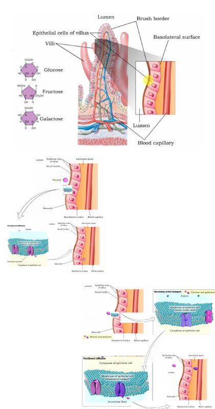
In summary, carbohydrate absorption involves the uptake of monosaccharides, with glucose and galactose being transported into the epithelial cells of the intestinal villi through secondary active transport, co-transported with sodium ions. On the other hand, fructose is absorbed solely by facilitated diffusion. Once inside the epithelial cells, glucose, galactose, and fructose exit via facilitated diffusion and enter the capillaries, eventually reaching the bloodstream and being transported to the liver for further processing.
Protein Absorption Mechanism
The protein absorption mechanism involves the uptake of amino acids, dipeptides, and tripeptides in the small intestine. Here is a step-by-step process of the protein absorption mechanism:
- Digestion: Proteins are initially broken down into smaller peptides during the process of digestion, which occurs primarily in the stomach and continues in the small intestine.
- Formation of Amino Acids, Dipeptides, and Tripeptides: The digestion of proteins results in the breakdown of peptides into their constituent amino acids, as well as the formation of dipeptides (two amino acids) and tripeptides (three amino acids).
- Active Transport of Amino Acids: a. Active Transport: Most amino acids are transported into the absorptive epithelial cells of the small intestine via active transport mechanisms. b. Carrier Proteins: Specific carrier proteins facilitate the active transport of amino acids. These carriers are linked to the active transport of sodium ions (Na+), utilizing the concentration gradient of sodium ions across the cell membrane. c. Sodium Co-Transport: The transport of amino acids occurs in conjunction with the movement of sodium ions into the epithelial cells. This process is referred to as Na+-dependent secondary active transport.
- Active Transport of Dipeptides and Tripeptides: a. Secondary Active Transport: Dipeptides and tripeptides can also be actively transported into the absorptive epithelial cells. b. Proton Co-Transport: The transport of dipeptides and tripeptides is facilitated by hydrogen ions (H+). This process is known as H+-dependent secondary active transport.
- Intracellular Hydrolysis: Once inside the absorptive epithelial cells, dipeptides and tripeptides undergo hydrolysis, where they are broken down into their constituent amino acids. This process is mediated by enzymes present within the cells.
- Diffusion into Capillary Blood: a. Amino Acids: After the intracellular hydrolysis, amino acids are transported out of the absorptive epithelial cells through facilitated diffusion. b. Intestinal Fluid: The amino acids diffuse through the intestinal fluid, which is present in the lumen of the small intestine. c. Entry into Capillaries: Amino acids enter the capillary blood located within the villi of the small intestine. From there, they are transported to the liver via the hepatic portal vein.
In summary, protein absorption primarily occurs in the duodenum and jejunum of the small intestine. Most proteins are digested and absorbed as amino acids, along with the active transport of dipeptides and tripeptides. Amino acids are transported into the absorptive epithelial cells through active transport mechanisms, either independently or co-transported with sodium ions. Dipeptides and tripeptides are transported through secondary active transport mechanisms involving hydrogen ions. Once inside the cells, dipeptides and tripeptides are hydrolyzed into amino acids. Amino acids then diffuse out of the cells, pass through the intestinal fluid, and enter the capillary blood within the villi, eventually reaching the liver for further processing and distribution throughout the body.
Lipid Absorption Mechanism
The lipid absorption mechanism involves the uptake and processing of fatty acids and monoglycerides in the small intestine. Here is a step-by-step process of the lipid absorption mechanism:
- Digestion: Lipid digestion begins in the small intestine, where bile salts emulsify large lipid droplets into smaller droplets, increasing the surface area for enzymatic action. Pancreatic lipase then breaks down triglycerides into fatty acids and monoglycerides.
- Micelle Formation: To facilitate the absorption of hydrophobic long-chain fatty acids and monoacylglycerides, bile salts and lecithin form micelles. Micelles are tiny spheres with polar ends facing the watery environment and hydrophobic tails turned inward. They enclose the long-chain fatty acids, monoacylglycerides, cholesterol, and fat-soluble vitamins, creating a receptive environment for absorption.
- Micelle Absorption: The micelles move close to the luminal cell surface of the absorptive cells (enterocytes) in the small intestine. The lipid substances exit the micelle and are absorbed by the enterocytes via simple diffusion. Short-chain fatty acids, being relatively water-soluble, can enter enterocytes directly through simple diffusion.
- Reformation of Triglycerides: Within the enterocytes, the absorbed fatty acids and monoacylglycerides are reassembled into triglycerides. They combine with phospholipids and cholesterol to form triglyceride-rich lipoproteins.
- Formation of Chylomicrons: The triglyceride-rich lipoproteins are coated with proteins, forming chylomicrons. Chylomicrons are water-soluble lipoproteins that consist of triglycerides, phospholipids, cholesterol, and proteins.
- Release and Transport: After being processed by the Golgi apparatus, chylomicrons are released from the enterocytes through exocytosis. Since chylomicrons are too large to pass through the basement membranes of blood capillaries, they enter the large pores of lacteals, specialized lymphatic vessels in the villi of the small intestine.
- Lymphatic Transport: Chylomicrons are transported within the lymphatic vessels and eventually enter the bloodstream through the thoracic duct, which empties into the subclavian vein. This allows chylomicrons to bypass the liver initially.
- Lipoprotein Lipase Action: Once in the bloodstream, the enzyme lipoprotein lipase acts on the triglycerides of the chylomicrons, breaking them down into free fatty acids and glycerol.
- Utilization or Storage: The breakdown products of chylomicrons, including free fatty acids and glycerol, can pass through capillary walls and be used by cells for energy or stored in adipose tissue as fat.
- Chylomicron Remnants: The remaining chylomicron remnants, after the action of lipoprotein lipase, are combined with proteins in the liver cells to form lipoproteins. These lipoproteins transport cholesterol in the blood.
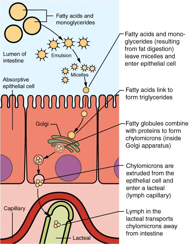
In summary, the lipid absorption mechanism involves the formation of micelles to solubilize long-chain fatty acids and monoacylglycerides, allowing for their absorption by enterocytes through simple diffusion. Within the enterocytes, the absorbed lipids are reassembled into triglycerides and packaged into chylomicrons, which are transported through the lymphatic system and eventually enter the bloodstream. Lipoprotein lipase breaks down the triglycerides of chylomicrons, and the resulting free fatty acids and glycerol can be utilized by cells or stored in adipose tissue. The liver combines the remaining chylomicron remnants with proteins, forming lipoproteins that transport cholesterol in the blood.
Nucleic Acid Absorption Mechanism
The nucleic acid absorption mechanism involves the uptake and transport of the products of nucleic acid digestion in the small intestine. Here is a step-by-step process of the nucleic acid absorption mechanism:
- Digestion: Nucleic acids, such as DNA and RNA, are digested in the small intestine, breaking them down into their constituent components. The digestion process releases pentose sugars (ribose and deoxyribose), nitrogenous bases (adenine, cytosine, thymine, guanine, and uracil), and phosphate ions.
- Villus Absorption: The products of nucleic acid digestion are absorbed in the duodenum and jejunum of the small intestine, specifically at the intestinal villi. They are transported across the villus epithelium via carrier proteins.
- Active Transport: Carriers facilitate the active transport of the products of nucleic acid digestion into the epithelial cells from the lumen of the small intestine. Active transport mechanisms require energy to move molecules against their concentration gradients.
- Secondary Active Transport: Some of the products of nucleic acid digestion are transported into the epithelial cells via secondary active transport. This process couples the transport of the products with the movement of other molecules or ions, utilizing the concentration gradient of the co-transported substance.
- Diffusion: Once inside the intestinal epithelial cells, the products of nucleic acid digestion diffuse across the basolateral membrane, which separates the cells from the interstitial fluid.
- Entry into the Bloodstream: The products of nucleic acid digestion enter the interstitial fluid and eventually the bloodstream. They are transported through the blood circulation to the liver and other tissues.
- Further Degradation: In the liver and other tissues, the products of nucleic acid digestion undergo further degradation and processing. They are utilized for various cellular processes or incorporated into new nucleic acids as needed.
In summary, the nucleic acid absorption mechanism involves the active transport and secondary active transport of the products of nucleic acid digestion across the villus epithelium in the small intestine. The absorbed products, including pentose sugars, nitrogenous bases, and phosphate ions, enter the bloodstream and are transported to the liver and other tissues for further degradation and utilization.
Mineral Absorption Mechanism
The mineral absorption mechanism involves the uptake and transport of electrolytes and specific minerals in the small intestine. Here is a step-by-step process of mineral absorption:
- Source of Minerals: Minerals are obtained from both gastrointestinal secretions and ingested foods. Electrolytes dissociate into ions in water and are absorbed as such.
- Active Transport: Most minerals are absorbed throughout the entire small intestine via active transport mechanisms. Active transport requires energy and occurs against the concentration gradient.
- Co-Transport and Anti-Port Mechanisms: During absorption, co-transport mechanisms lead to the accumulation of sodium ions inside the intestinal cells. On the other hand, anti-port mechanisms reduce the concentration of potassium ions inside the cells.
- Sodium-Potassium Pump: To restore the sodium-potassium gradient across the cell membrane, a sodium-potassium pump is involved. This pump, which requires ATP (adenosine triphosphate), actively transports sodium ions out of the cells and potassium ions into the cells.
- Iron Absorption: Ionic iron, needed for the production of hemoglobin, is absorbed into mucosal cells via active transport. Once inside the cells, ionic iron binds to the protein ferritin, forming iron-ferritin complexes. These complexes store iron until it is needed. When the body has sufficient iron, most of the stored iron is lost when worn-out epithelial cells are shed. However, during periods of increased iron demand (e.g., due to bleeding), there is increased uptake of iron from the intestine and accelerated release of iron into the bloodstream.
- Calcium Absorption: The absorption of dietary calcium is regulated by blood levels of ionic calcium. When blood levels of ionic calcium decrease, the parathyroid glands secrete parathyroid hormone (PTH). PTH stimulates the release of calcium ions from bone and increases the reabsorption of calcium by the kidneys. PTH also enhances the activation of vitamin D in the kidneys. Activated vitamin D facilitates the absorption of calcium ions in the intestine.
In general, all minerals that enter the intestine are absorbed, except for iron and calcium, which are absorbed in the duodenum based on the body’s current requirements. The absorption of these minerals is tightly regulated to meet the body’s needs. Iron is absorbed via active transport and stored in mucosal cells, while calcium absorption is influenced by blood levels of ionic calcium and regulated by parathyroid hormone and activated vitamin D.
In summary, mineral absorption involves active transport mechanisms in the small intestine. Minerals are absorbed as ions, and the absorption process is regulated to maintain appropriate mineral levels in the body. Iron and calcium absorption have specific regulatory mechanisms to ensure the body’s iron and calcium needs are met.
Vitamin Absorption
Vitamin absorption involves the uptake and transport of vitamins in the small intestine. Here is a step-by-step process of vitamin absorption:
- Source of Vitamins: Vitamins can be obtained from natural food sources as well as dietary supplements. They are present in both fat-soluble and water-soluble forms.
- Fat-Soluble Vitamins: Fat-soluble vitamins, including vitamins A, D, E, and K, are absorbed along with dietary lipids. They are transported in micelles, which are tiny spheres formed by the combination of bile salts and lipids. The fat-soluble vitamins dissolve in the lipid component of micelles and are absorbed into the enterocytes of the small intestine through simple diffusion. It is recommended to consume some fatty foods when taking fat-soluble vitamin supplements to enhance their absorption.
- Water-Soluble Vitamins: Most water-soluble vitamins, such as most B vitamins and vitamin C, are absorbed through simple diffusion. In this process, the water-soluble vitamins dissolve in the watery contents of the small intestine and passively diffuse across the enterocyte membranes into the bloodstream.
- Vitamin B12 Absorption: Vitamin B12 is an exception among water-soluble vitamins due to its large molecular size. The absorption of vitamin B12 requires a specific mechanism. In the stomach, intrinsic factor is secreted, which binds to vitamin B12 and forms a complex. This complex is resistant to digestion and travels to the terminal ileum of the small intestine. In the terminal ileum, the complex binds to mucosal receptors on the enterocyte membranes. The enterocytes then internalize the complex through endocytosis, allowing the absorption of vitamin B12.
- Transport to the Bloodstream: Once absorbed into the enterocytes, both fat-soluble and water-soluble vitamins are transported from the enterocytes into the bloodstream. From the bloodstream, they are distributed to various tissues and organs where they are utilized for various physiological processes.
In summary, vitamin absorption in the small intestine involves different mechanisms depending on the type of vitamin. Fat-soluble vitamins are absorbed along with dietary lipids through simple diffusion, while most water-soluble vitamins are also absorbed through simple diffusion. However, vitamin B12 absorption requires the presence of intrinsic factor and occurs via endocytosis in the terminal ileum. Adequate absorption of vitamins is essential for maintaining overall health and meeting the body’s vitamin requirements.
Water Absorption
Water absorption in the small intestine plays a crucial role in maintaining the body’s hydration balance. Here is a step-by-step process of water absorption in the small intestine:
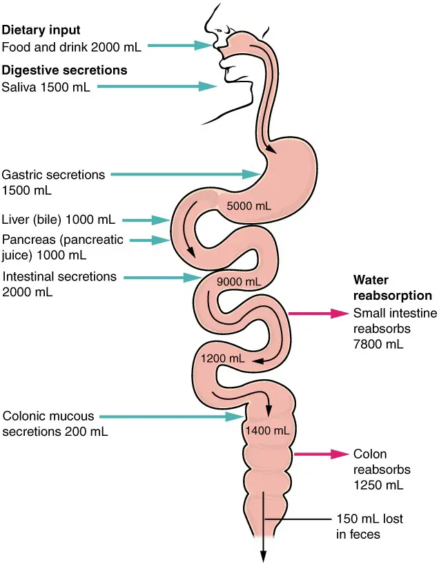
- Fluid Intake: Each day, approximately nine liters of fluid enter the small intestine. This fluid consists of approximately 2.3 liters ingested through foods and beverages, while the rest is derived from gastrointestinal secretions.
- Water Concentration Gradient: The process of water absorption is driven by the concentration gradient of water. The concentration of water is higher in the chyme, which is the partially digested food mixture in the small intestine, compared to the concentration in the epithelial cells lining the small intestine.
- Movement of Water: Due to the higher concentration of water in the chyme, water moves down its concentration gradient from the chyme into the epithelial cells of the small intestine. This movement occurs through the process of osmosis, which is the passive diffusion of water across a semi-permeable membrane.
- Absorption in Small Intestine: Approximately 90 percent of the water present in the small intestine is absorbed across the epithelial cells. As water moves into the cells, it is then transported across the epithelium and enters the bloodstream.
- Remaining Water Absorption: After the absorption in the small intestine, a significant amount of water still remains in the gastrointestinal tract. This remaining water, along with undigested materials, moves into the colon or large intestine.
- Water Absorption in the Colon: In the colon, further water absorption takes place. The colon is highly efficient at reabsorbing water, as it is specialized for concentrating waste material and reclaiming water to prevent excessive fluid loss.
Overall, water absorption in the small intestine occurs through osmosis, driven by the concentration gradient of water. The movement of water from the chyme into the epithelial cells helps maintain the body’s fluid balance. The remaining water is then absorbed in the colon, ensuring efficient water reabsorption and preventing excessive fluid loss.
Difference Between Mechanical and Chemical Digestion
Difference Between Mechanical and Chemical Digestion:
Mechanical digestion and chemical digestion are two distinct processes involved in the breakdown of food in the digestive system. Here are the key differences between them:
Occurrence:
- Mechanical Digestion: It occurs from the mouth to the stomach, involving the physical breakdown of food particles.
- Chemical Digestion: It occurs from the mouth to the intestine, involving the chemical breakdown of complex molecules.
Major Part:
- Mechanical Digestion: A major part of mechanical digestion occurs in the mouth, where food is chewed and mixed with saliva.
- Chemical Digestion: A major part of chemical digestion occurs in the stomach, where enzymes and gastric juices break down proteins.
Driven by:
- Mechanical Digestion: Mechanical digestion is driven by the action of teeth, which bite, tear, and grind food into smaller pieces.
- Chemical Digestion: Chemical digestion is driven by enzymes, which break down complex molecules into simpler substances.
Mechanism:
- Mechanical Digestion: The primary mechanism in mechanical digestion is the physical breakdown of large food particles into smaller food particles through chewing and grinding.
- Chemical Digestion: The primary mechanism in chemical digestion is the chemical breakdown of compounds with high molecular weights into smaller molecules through the action of digestive enzymes.
Role:
- Mechanical Digestion: Mechanical digestion increases the surface area of food particles, facilitating the enzymatic reactions in chemical digestion and aiding in the overall digestion process.
- Chemical Digestion: Chemical digestion breaks down complex molecules into small, absorbable compounds, enhancing the absorption of nutrients by the body.
In summary, mechanical digestion involves the physical breakdown of food particles, mainly by the teeth, while chemical digestion involves the chemical breakdown of complex molecules by enzymes. Both processes work together to prepare food for absorption and utilization by the body.
| Aspect | Mechanical Digestion | Chemical Digestion |
|---|---|---|
| Occurrence | From the mouth to the stomach | From the mouth to the intestine |
| Major Part | Mostly in the mouth | Mostly in the stomach |
| Driven by | Teeth | Enzymes |
| Mechanism | Physical breakdown of food particles | Chemical breakdown of complex molecules |
| Role | Increases surface area for enzymatic reactions | Enhances absorption of nutrients by breaking them down into small molecules |
References
- Patricia JJ, Dhamoon AS. Physiology, Digestion. [Updated 2022 Sep 12]. In: StatPearls [Internet]. Treasure Island (FL): StatPearls Publishing; 2023 Jan-. Available from: https://www.ncbi.nlm.nih.gov/books/NBK544242/
- https://training.seer.cancer.gov/anatomy/digestive/
- alth/chemical-digestion#takeaway
- https://courses.lumenlearning.com/suny-ap2/chapter/chemical-digestion-and-absorption-a-closer-look/
- https://www.sisd.net/cms/lib/TX01001452/Centricity/Domain/6705/Digestive%20Reading%20Station.pdf
- https://ib.bioninja.com.au/standard-level/topic-6-human-physiology/61-digestion-and-absorption/mechanical-digestion.html
- https://pediaa.com/difference-between-mechanical-and-chemical-digestion/
- https://pennstatehershey.adam.com/pages/guide/reftext/html/dige_sys_fin.html
- http://www.e-missions.net/cybersurgeons/?/dig_teacher/
- https://opentextbc.ca/biology/chapter/15-3-digestive-system-processes/
- https://natomasunified.org/content/uploads/sites/2/2017/02/Digestive-System-1.pptx
- https://content.patnawomenscollege.in/zoology/02%20PHYSIOLOGY%20OF%20DIGESTION%20-%20CHEMICAL%20DIGESTION.pdf
- https://www.uc.edu/content/dam/uc/ce/docs/OLLI/Page%20Content/OLLI%20-%20The%20Digestive%20System.pdf
- https://www.dentonisd.org/cms/lib/TX21000245/Centricity/Domain/1708/7.6B%20Evaluate%20-%20Physical%20Chemical%20Digestion%20Quick%20Check.pdf
- https://biology-igcse.weebly.com/mechanical-and-chemical-digestion.html
- https://humanbiology.pressbooks.tru.ca/chapter/17-3-digestion-and-absorption/
- Text Highlighting: Select any text in the post content to highlight it
- Text Annotation: Select text and add comments with annotations
- Comment Management: Edit or delete your own comments
- Highlight Management: Remove your own highlights
How to use: Simply select any text in the post content above, and you'll see annotation options. Login here or create an account to get started.