What is Lung?
- The lungs are integral organs in the respiratory systems of many terrestrial animals, particularly tetrapod vertebrates, which include mammals, reptiles, and birds. Some aquatic species, like lungfish, also possess lungs, while certain gastropods (land snails and slugs) and arachnids (like spiders and scorpions) have evolved analogous respiratory structures. The primary function of the lungs is to facilitate gas exchange: they extract oxygen from the air and transfer it into the bloodstream while simultaneously expelling carbon dioxide, a process known as respiration.
- In humans, the lungs are paired structures located on either side of the heart within the thoracic cavity. Their positioning and structure are homologous to the swim bladders found in ray-finned fish, emphasizing their evolutionary significance. The lungs are primarily responsible for the ventilation process, which refers to the movement of air in and out of the respiratory system. This process is driven by muscular actions specific to different animal groups. For instance, mammals, reptiles, and birds utilize specialized respiratory muscles, while primitive tetrapods rely on pharyngeal muscles for buccal pumping.
- The mechanics of breathing in humans involve the diaphragm and intercostal muscles as the primary respiratory muscles. During respiratory distress, other muscles, such as those in the core and limbs, may assist in ventilation. Additionally, the lungs enable vocalization, including human speech, by facilitating airflow.
- Each lung consists of distinct anatomical features. The right lung is typically larger and heavier than the left lung, due to the leftward rotation of the heart. Together, the human lungs weigh about 1.3 kilograms (2.9 pounds). Structurally, the lungs are part of the lower respiratory tract, which commences at the trachea and branches into the bronchi and bronchioles. This tract serves to channel fresh air into the lungs through the conducting zone, terminating at the terminal bronchioles.
- The conducting zone leads into the respiratory zone, where gas exchange predominantly occurs. The respiratory bronchioles transition into alveolar ducts, which culminate in alveolar sacs containing alveoli—tiny air sacs critical for gas exchange. Collectively, human lungs encompass approximately 2,400 kilometers (1,500 miles) of airways and house between 300 to 500 million alveoli. Each lung is enveloped by pleurae, serous membranes that reduce friction during lung expansion and contraction. The visceral pleura directly covers the lung surface, while the parietal pleura lines the rib cage. The two pleurae form the pleural cavity, containing a lubricating film of pleural fluid that facilitates lung movement during respiration.
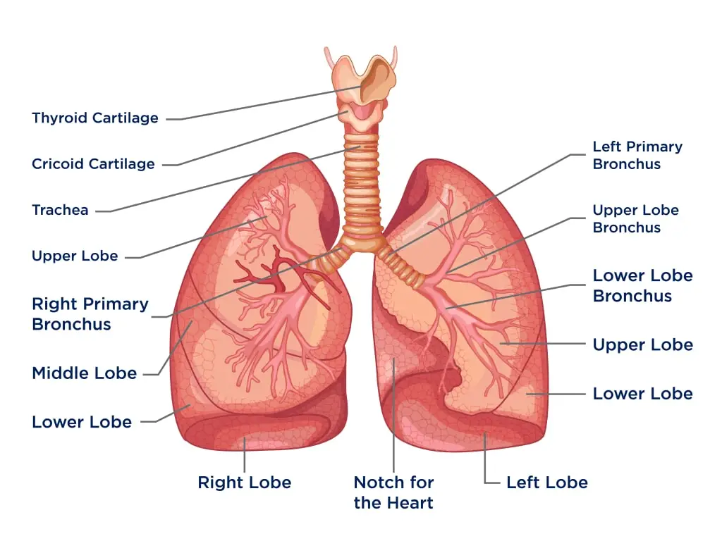
- Lung structure is further organized into lobes. The right lung is divided into three lobes, whereas the left lung has two. Each lobe can be subdivided into bronchopulmonary segments and pulmonary lobules, illustrating the complexity of lung architecture.
- The lungs receive dual blood supplies: the pulmonary circulation, which carries deoxygenated blood from the right side of the heart through pulmonary arteries to the alveolar-capillary barrier for gas exchange, and the bronchial circulation, part of the systemic circulation that supplies oxygenated blood to lung tissues. This dual blood supply is essential for maintaining lung health and function.
- Various diseases can affect lung function, including pneumonia, pulmonary fibrosis, and lung cancer. Chronic obstructive pulmonary disease (COPD) encompasses conditions like chronic bronchitis and emphysema, which are often linked to smoking or exposure to environmental pollutants. Occupational lung diseases may arise from inhaling harmful substances such as coal dust or asbestos. While asthma and acute bronchitis primarily affect airways, they can significantly impact overall lung function. The medical terminology associated with lungs often incorporates prefixes like “pulmo-” from Latin or “pneumo-” from Greek, reflecting their anatomical and functional significance.
- During embryonic development, the lungs originate as an outpouching of the foregut, a precursor to the upper digestive system. Initially, the lungs do not engage in gas exchange due to the fetus being enclosed in amniotic fluid, and blood circulation bypasses the lungs through the ductus arteriosus. At birth, as air enters the lungs and the duct closes, the lungs commence their role in respiration. Full lung development occurs in early childhood, highlighting the lungs’ growth and adaptation during the early stages of life.
Location of Lungs
The lungs are essential respiratory organs located symmetrically on either side of the thoracic cavity. Their specific positioning and anatomical features enable efficient gas exchange within the body.
- Positioning in the Body:
- The lungs are situated in the thoracic cavity, one on each side of the mediastinum, which separates them.
- Each lung rests above the diaphragm, a muscular structure that plays a crucial role in respiration.
- Protection and Covering:
- The lungs are encased within a pleural sac, which helps reduce friction during breathing movements.
- They are further safeguarded by the rib cage, providing structural support and protection from external impacts.
- Identification:
- The lungs are commonly referred to as the left lung and right lung, indicating their respective locations in the human body.
- Anatomical Features:
- Apex: This is the conical, pointed end of the lung that directs upwards, extending above the level of the first rib.
- Base: The broader end of the lung, which is directed downwards and rests upon the diaphragm.
- Three Borders:
- Anterior Margin: A thin layer that faces forward, contributing to the lung’s overall shape.
- Posterior Margin: A rounded border that faces inward, facilitating the lung’s connection to surrounding structures.
- Inferior Margin: This semilunar-shaped margin separates the costal surface from the medial surface of the lung.
- Two Surfaces:
- Costal Surface: This is the convex outer surface of the lung, directed outward and closely related to the rib cage.
- Medial Surface: A flat surface that faces inward, towards the mediastinum, playing a crucial role in the lung’s connectivity to various structures.
Structure of Lung
The lungs are vital organs characterized by their roughly cone-shaped structure. They exhibit a unique anatomy that facilitates their primary function of gas exchange. This overview presents a detailed examination of the structure of the lungs, highlighting their components and features.

- General Structure
- Shape and Orientation: The lungs are cone-shaped, featuring an apex, base, three surfaces, and three borders.
- Size Discrepancy: The left lung is smaller than the right lung, primarily due to the positioning of the heart, which occupies space on the left side of the thoracic cavity.
- Key Components of the Lungs
- Apex: This is the blunt superior end of the lung, which projects upward above the level of the first rib and extends into the neck’s floor.
- Base: The inferior surface of the lung that rests on the diaphragm, serving as the foundation for lung structure.
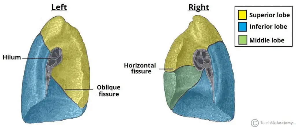
- Lobes: The lungs are divided into lobes, with the right lung containing three lobes (superior, middle, and inferior), while the left lung has two lobes (superior and inferior). The lobes are separated by fissures.
- Surfaces: The lungs have three surfaces that correspond to the areas of the thorax they face:
- Costal Surface: Smooth and convex, facing the inner chest wall, and is related to the costal pleura that separates it from the ribs and intercostal muscles.
- Mediastinal Surface: Faces the lateral aspect of the middle mediastinum and contains the lung hilum, the area where various structures enter and exit the lung.
- Diaphragmatic Surface: Forms the base of the lung, concave in shape, resting on the diaphragm; this concavity is deeper in the right lung due to the higher position of the right dome, which overlies the liver.
- Borders: Each lung has three borders:
- Anterior Border: Formed by the convergence of the mediastinal and costal surfaces. In the left lung, this border features a cardiac notch created by the apex of the heart.
- Inferior Border: Separates the base of the lung from the costal and mediastinal surfaces.
- Posterior Border: Smooth and rounded, created by the meeting of the costal and mediastinal surfaces posteriorly.
- Root and Hilum
- Lung Root: The root consists of a collection of structures that suspend the lung from the mediastinum. It includes the bronchus, pulmonary artery, two pulmonary veins, bronchial vessels, a pulmonary plexus of nerves, and lymphatic vessels.
- Hilum: A wedge-shaped area located on the mediastinal surface where all these structures enter or leave the lung.
- Bronchial Tree
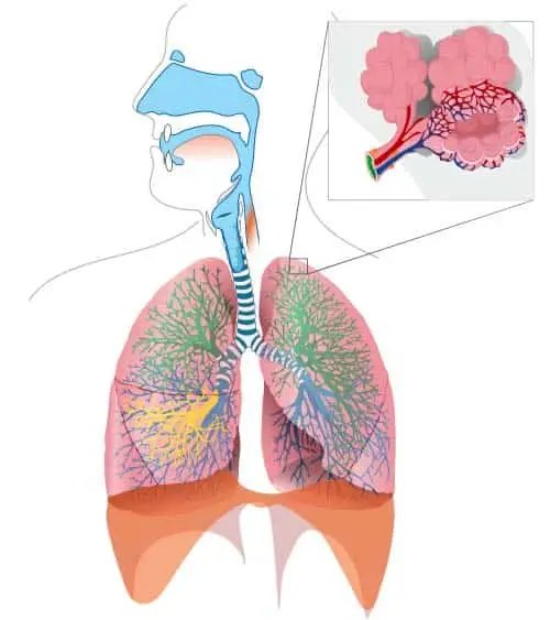
- Structure: The bronchial tree is a branching network of air passages that supplies air to the alveoli within the lungs, starting with the trachea.
- Division of Bronchi: The trachea bifurcates into the left and right bronchi. Notably, the right bronchus has a greater incidence of foreign body inhalation due to its wider shape and more vertical orientation.
- Lobar and Segmental Bronchi: Each bronchus enters the lung root at the hilum and divides into lobar bronchi—each supplying a lobe of the lung. Lobar bronchi further divide into tertiary segmental bronchi, which serve specific bronchopulmonary segments, the functional units of the lungs.
- Conducting Bronchioles: The segmental bronchi branch into conducting bronchioles, which lead to terminal bronchioles. Each terminal bronchiole gives rise to respiratory bronchioles, characterized by thin-walled outpocketings extending from their lumens, leading to the alveoli, the primary sites for gas exchange.


Functions of Lungs
The lungs serve critical functions that are essential for maintaining homeostasis and supporting life. Their primary roles encompass gas exchange, immune system support, and homeostatic regulation, all of which are vital for overall health.
- Gas Exchange:
- The lungs are primarily responsible for supplying oxygen to the body.
- During inhalation, air enters through the mouth or nose and travels down the trachea into the lungs.
- Inside the lungs, air moves through the bronchi and into smaller passages called bronchioles, ultimately filling the alveoli.
- Alveoli: These tiny air sacs are the primary sites for gas exchange. Each alveolus is surrounded by a network of capillaries, allowing oxygen to diffuse from the alveoli into the blood.
- Oxygen-rich blood is then transported to the heart, while carbon dioxide, a waste product, is transferred from the blood back into the alveoli to be expelled during exhalation.
- Immune System Support:
- The lungs play a crucial role in the body’s immune defense.
- Alveolar Macrophages: These innate immune cells reside in the distal lung tissue, specifically on the luminal surface of the alveolar space.
- They actively engage in identifying and neutralizing pathogens, such as bacteria and viruses, that enter the respiratory system, thus contributing significantly to pulmonary immunity.
- Homeostatic Functions:
- The lungs are involved in maintaining the body’s homeostasis, which refers to the stability of internal conditions.
- They help regulate blood pH by adjusting the levels of carbon dioxide through the process of respiration.
- By modifying breathing rates and depths, the lungs can influence the concentration of carbon dioxide in the blood, thus impacting overall acid-base balance.
Mechanism of Breathing
The mechanism of breathing involves a series of coordinated physiological processes that facilitate the intake of oxygen and the expulsion of carbon dioxide from the body. This vital function, known as ventilation, encompasses two primary phases: inhalation and exhalation.
- Inhalation:
- The process begins with the act of inhaling, during which oxygen is drawn into the respiratory system.
- Oxygen enters through the mouth or nose and travels down the trachea, subsequently branching into the bronchi and further into smaller bronchioles.
- Finally, the inhaled air reaches the alveoli, the tiny air sacs where gas exchange occurs.
- In the alveoli, oxygen diffuses across the alveolar-capillary barrier into the surrounding blood vessels.
- Pulmonary Veins: These vessels carry the newly oxygenated blood from the lungs back to the heart. Oxygen molecules bind to hemoglobin within red blood cells during this process.
- Exhalation:
- The process of exhalation involves expelling carbon dioxide from the body, which is a waste product of cellular metabolism.
- During exhalation, the diaphragm, a dome-shaped muscle located at the base of the thoracic cavity, relaxes and moves upward.
- Simultaneously, the intercostal muscles, which are located between the ribs, also relax and move downwards.
- The relaxation of these muscles results in a decrease in the volume of the thoracic cavity, creating increased pressure within the lungs.
- Consequently, carbon dioxide is pushed from the blood into the alveoli and then travels through the bronchi and trachea to be exhaled out of the body.
- Pressure Changes:
- The mechanics of breathing are largely influenced by pressure changes within the thoracic cavity.
- During inhalation, the downward movement of the diaphragm and upward movement of the intercostal muscles create a negative pressure in the chest cavity, drawing oxygen in.
- Conversely, during exhalation, the upward movement of the diaphragm and downward movement of the intercostal muscles increase pressure in the thoracic cavity, forcing carbon dioxide out.
Blood Circulation of the Lungs
Blood circulation in the lungs is a vital component of the respiratory system, facilitating gas exchange and providing essential nutrients to lung tissues. The circulation is divided into two main pathways: pulmonary circulation and bronchial circulation.
- Pulmonary Circulation:
- This system is primarily responsible for transporting deoxygenated blood from the heart to the lungs and returning oxygenated blood back to the heart.
- Components:
- Pulmonary Arteries: A pair of pulmonary arteries carries deoxygenated blood from the right ventricle of the heart to the lungs. As these arteries enter the lungs, they branch into smaller arterioles, eventually leading to the capillary networks surrounding the alveoli.
- Alveoli: Within the alveoli, gas exchange occurs. Oxygen from the inhaled air diffuses into the blood, while carbon dioxide in the blood diffuses into the alveoli to be exhaled.
- Pulmonary Veins: After gas exchange, the now oxygenated blood is collected by four pulmonary veins (two from each lung), which transport it back to the left atrium of the heart. This oxygenated blood is then pumped into the systemic circulation to supply oxygen to the body’s tissues.
- Bronchial Circulation:
- In contrast to pulmonary circulation, bronchial circulation is responsible for providing oxygenated blood to the lung tissues themselves.
- Components:
- Bronchial Arteries: These arteries arise from the aorta and supply oxygenated, nutritive blood to the lungs, particularly the bronchial tissues, pleura, and bronchi.
- Bronchial Veins: Following the delivery of oxygen, the deoxygenated blood is drained by the bronchial veins. This blood is then returned to the systemic circulation, either through the azygos vein or directly into the pulmonary veins.
Diseases and Disorders of the Lungs
The lungs are susceptible to a variety of diseases and disorders that can significantly impair their function and overall health. Understanding these conditions is essential for both prevention and effective management. The following are some of the most prevalent lung diseases:
- Chronic Obstructive Pulmonary Disease (COPD):
- COPD represents a progressive lung disease characterized by a gradual decline in lung function.
- It affects approximately 65 million individuals worldwide and results in about 3 million deaths annually.
- The primary risk factor for COPD is smoking, with smokers being 13 times more likely to develop this condition. Additionally, air pollution can also contribute to the disease’s onset.
- Genetic predispositions can play a role, especially in individuals with compromised immune systems.
- Complications associated with COPD may include pneumonia, lung cancer, heart failure, and various other respiratory issues.
- Asthma:
- Asthma is a common respiratory condition resulting from chronic inflammation in the lower respiratory tract.
- Despite being prevalent, asthma is often underrated and can be challenging to diagnose effectively.
- Patients with asthma commonly experience recurrent symptoms such as wheezing, coughing, chest tightness, and difficulty in breathing.
- While specific immunotherapy for asthma is not yet feasible, various therapies aim to optimize lung function.
- An effective management strategy includes the elimination of allergens that trigger asthmatic episodes. Additionally, obesity has been linked to asthma, suggesting that weight reduction can enhance lung health.
- Pulmonary Fibrosis:
- Pulmonary fibrosis is characterized by the injury to alveoli, leading to chronic inflammation and the deposition of extracellular matrix components, particularly collagen. This results in scarring of the lung tissue.
- Individuals with a genetic predisposition are more susceptible to developing pulmonary fibrosis. Other potential causes include exposure to microbial agents, certain chemotherapeutic drugs, and occupational inhalants.
- Treatment options for pulmonary fibrosis can be complex, often involving corticosteroids, immunosuppressive agents, and antifibrotic medications.
- If the condition is attributed to microbial agents, treatment with appropriate antimicrobial drugs, such as antibiotics, antivirals, or antifungals, may be necessary.
- Lung Cancer:
- Lung cancer is the leading cause of cancer-related deaths globally.
- Smoking remains the most significant risk factor for developing lung cancer, but other factors such as genetic predispositions, radiation exposure, radon gas, and various carcinogens also contribute.
- The disease arises from repeated damage to the lung cell line, causing normal cells to behave abnormally and ultimately leading to cancer.
- Common symptoms of lung cancer include shortness of breath, coughing up blood, pleural effusion, and chest pain.
- Early detection of lung cancer greatly improves treatment outcomes. Surgical interventions may include wedge resection, segmentectomy, lobectomy, and pneumonectomy, depending on the stage and extent of the disease.
Difference Between Right and Left Lungs
The lungs, essential organs in the respiratory system, are not identical in structure or function. The right and left lungs exhibit several notable differences, which are crucial for their roles in gas exchange and overall respiratory efficiency.
- Number of Lobes:
- The right lung consists of three lobes: the upper, middle, and lower lobes.
- In contrast, the left lung has only two lobes: the upper and lower lobes. This difference accommodates the space required for the heart, which is situated slightly to the left of the body’s midline.
- Number of Fissures:
- The right lung contains two fissures: the horizontal fissure and the oblique fissure. These fissures separate the three lobes from one another.
- The left lung has only one fissure, the oblique fissure, which separates its two lobes.
- Number of Bronchi:
- The right lung has two main bronchi, the right primary bronchus, which is wider and shorter, facilitating the entry of air.
- Conversely, the left lung features one primary bronchus, which is narrower and longer compared to the right.
- Weight:
- The right lung is heavier, weighing approximately 700 grams.
- The left lung is lighter, weighing about 600 grams. This weight difference is attributed to the variation in size and number of lobes.
- Structure:
- The right lung is characterized by a short and wide structure, allowing it to accommodate its three lobes and associated vasculature.
- The left lung, however, is long and narrow, shaped to provide space for the heart and associated structures.
- Cardiac Notches:
- The right lung does not possess any cardiac notches, as its structure is designed to fit snugly in the thoracic cavity.
- The left lung features a cardiac notch, a concave area that accommodates the left ventricle of the heart, further emphasizing the anatomical relationship between the lungs and the heart.
Anatomy of Lungs
The lungs are essential respiratory organs responsible for gas exchange and are anatomically complex structures. They are cone-shaped and situated in the thoracic cavity, separated by the heart and various mediastinal structures. Each lung plays a vital role in the respiratory system, with distinct features and functions.
- General Structure:
- The right lung is slightly larger and heavier than the left lung, allowing space for the heart. Together, both lungs weigh approximately 1.3 kg, with the right lung being marginally heavier.
- The trachea, or windpipe, divides into the right and left bronchi, which enter the lungs alongside blood vessels, lymphatic vessels, and nerves through an area known as the hilum.
- Bronchial Tree:
- The bronchial tree is a branching network responsible for air transport to and from the alveoli. It initiates from the trachea, which bifurcates into two primary bronchi (right and left) that enter each lung.
- These bronchi subdivide into smaller bronchi and bronchioles, ultimately leading to the alveoli, where gas exchange occurs.
- The bronchial tree is lined with ciliated epithelial cells and mucus-producing glands that trap and eliminate foreign particles and pathogens, thus ensuring efficient respiration.
- Lobes and Fissures:
- The right lung contains three lobes: superior, middle, and inferior, separated by an oblique fissure and a horizontal fissure.
- The left lung has two lobes: superior and inferior, separated only by the oblique fissure. This structural difference accommodates the cardiac notch, which houses the heart.
- Pleura:
- The lungs are enveloped by a protective double-layered membrane called the pleura.
- The parietal pleura lines the inner surface of the thoracic cavity.
- The visceral pleura adheres to the lung surface.
- The pleural cavity is the thin, fluid-filled space between these two layers, containing serous pleural fluid that lubricates the pleural surfaces, thereby reducing friction during lung expansion and contraction. This cavity is crucial for maintaining lung position and shape, as well as for facilitating breathing mechanics.
- The lungs are enveloped by a protective double-layered membrane called the pleura.
- Alveoli:
- Alveoli are tiny, hollow, cup-shaped structures within the lungs that serve as the primary site for gas exchange.
- There are millions of alveoli in each lung, collectively forming about 90% of the lung’s total space.
- They are composed of two types of alveolar cells:
- Type I pneumocytes (squamous epithelium) facilitate gas exchange.
- Type II pneumocytes (cuboidal epithelium) produce surfactant, which reduces surface tension within the alveoli and prevents their collapse.
- Alveolar macrophages are also present, serving a vital role in immune defense by engulfing pathogens and debris.
- Root and Hilum:
- The root of the lungs consists of bronchi, pulmonary arteries, pulmonary veins, and lymphatic vessels that enter or exit through the hilum. This area acts as a gateway for vital structures involved in lung function and blood circulation.
- https://teachmeanatomy.info/thorax/organs/lungs/
- https://en.wikipedia.org/wiki/Lung
- https://vocal.media/education/what-is-lungs
- https://byjus.com/biology/human-lungs-diagram/
- https://www.geeksforgeeks.org/anatomy-of-the-human-lung/
- Text Highlighting: Select any text in the post content to highlight it
- Text Annotation: Select text and add comments with annotations
- Comment Management: Edit or delete your own comments
- Highlight Management: Remove your own highlights
How to use: Simply select any text in the post content above, and you'll see annotation options. Login here or create an account to get started.