| Domain | Eukaryota |
|---|---|
| Kingdom | Animalia |
| Phylum | Chordata |
| Subphylum | Tunicata |
| Class | Ascidiacea |
| Order | Stolidobranchia |
| Family | Pyuridae |
| Genus | Herdmania |
| Authority | Lahille, 1888 |
What is Herdmania?
Herdmania is a solitary ascidian which is included under the Subphylum Tunicata of the Phylum Chordata. It is a sessile marine animal and the adult remains attached to hard substratum like rocks or shells in shallow coastal water.
The body is usually oblong or sac-like and it is enclosed by a thick test (tunic) which is leathery in texture and composed mainly of tunicin. It is reddish or pinkish in appearance and the bright red patches are formed by the terminal knobs of blood vessels situated in the test.
It is the process of filter feeding by which the animal draws water along with food particles through the branchial siphon into the branchial sac. The heart shows a characteristic reversal of heartbeat, and this is one of the important feature of tunicates. The animal is hermaphrodite and mainly cross fertilization takes place.
The life cycle shows the presence of a free-swimming tadpole larva. This larva has the typical chordate characters like notochord in the tail region, dorsal tubular nerve cord and pharyngeal gill-slits. It is the larva that helps in dispersal of the species. The larva soon settles on a substratum and then the process of retrogressive metamorphosis starts.
In this step the larva loses the tail, notochord, the larval sense organs (eye spot, statocyst) and the advanced nervous system. This is referred to as retrogressive because the highly organized larva is transformed into a simple sedentary adult which is specialized only for filter feeding.
General Characteristics of Herdmania
- It is a solitary ascidian which is included under the Subphylum Tunicata of Phylum Chordata.
- The adult is sessile and it remains attached to hard substratum like rocks or shells in shallow coastal water.
- The body is oblong or sac-like and the size usually ranges between 7–13 cm.
- The body colour is reddish or pinkish and the bright red patches are formed by terminal knobs of blood vessels present in the test.
- The body is covered by a thick leathery test (tunic) and it is mainly composed of tunicin.
- The test contains calcareous spicules which provides firmness to the body.
- Two siphons are present, the branchial siphon for intake of water and the atrial siphon for expelling water.
- The mantle lies below the test and contraction of mantle muscles helps in squirting out water.
- Feeding is ciliary type and the animal filters minute planktonic particles which are trapped by mucus secreted from endostyle.
- The pharynx is greatly enlarged to form the branchial sac which contains numerous gill-slits.
- Respiration mainly occurs through the vascular pharyngeal wall and in some extent by the vascular test.
- The circulatory system is open type and the heart is a tubular structure that shows periodic reversal of heartbeat.
- The blood contains different types of amoeboid and coloured corpuscles.
- Excretion is carried out by the neural gland and by nephrocytes present in the blood.
- The animal is hermaphrodite and the gonads are found on the right side of the body.
- Fertilization is external and usually cross-fertilization takes place due to protogynous condition.
- Development is indirect and it includes a free-swimming tadpole larva.
- The larva shows the typical chordate characters like notochord, dorsal tubular nerve cord and gill-slits.
- After attachment to substratum the larva undergoes retrogressive metamorphosis in which the advanced larval structures are lost.
- The adult becomes a simple sedentary filter-feeder specialized for its sessile mode of life.
Habit and Habitat of Herdamania
- It is a solitary ascidian and it is exclusively marine in nature.
- The adult is sessile and it remains permanently attached to hard substratum like rocks, shells or corals.
- It is generally found in shallow tropical and temperate sea water.
- The animal may occur from about 9 m depth and some species can be found in deeper regions also.
- It attaches by its flat basal region and each individual remains enclosed within its own tunic.
- Sometimes it is seen living in gregarious condition but every individual is separate.
- It is a ciliary feeder and it filters minute planktonic organisms from the incoming water current.
- The water enters through the branchial siphon and leaves through the atrial siphon.
- When disturbed the animal contracts its body and forcefully ejects water, and this is referred to as squirting behaviour.
- It acts as a hermaphrodite and external cross fertilization usually occurs in natural condition.
- In the sedentary habitat it helps in biofouling on submerged objects.
- The thick tunic may provide shelter to small marine organisms like hydroids, anemones and molluscs.
General Anatomy of Herdmania with Diagram
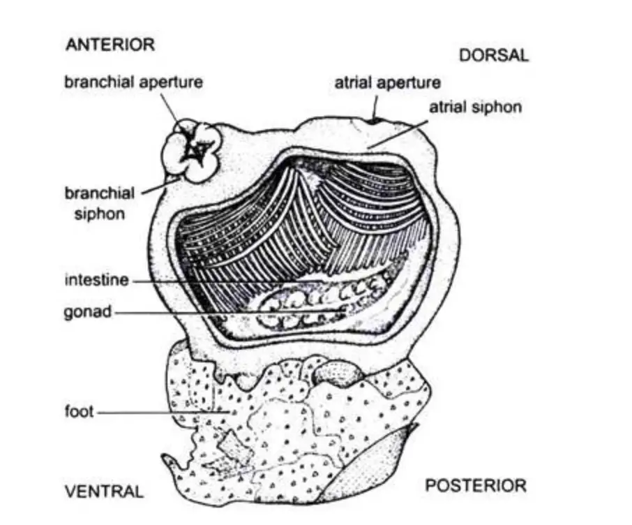
External Morphology
- The body is oblong and sac-like. It is usually 7–13 cm long and it is fixed to the substratum.
- The colour is reddish or pinkish and some bright red patches is seen on the test due to terminal knobs of blood vessels.
- It has two apertures at the free end. These is the branchial siphon and the atrial siphon.
- The branchial siphon is the incurrent opening. It is surrounded by four lobes and many branched tentacles act as a strainer.
- The atrial siphon is the excurrent opening. It is slightly longer and it is also surrounded by four lobes.
- The test is thick and leathery. It is composed of tunicin and many types of cells, fibrils and calcareous spicules. It is protective and also helps in respiration.
Body Wall and Cavities
- The mantle lies beneath the test. It is thick in the antero-dorsal part.
- The mantle has outer epidermis, middle muscular layer and inner epidermis lining the atrium.
- The muscles is non-striated longitudinal and circular fibres helping in contraction and expelling water.
- True coelom is almost absent. Only pericardial cavity is present as a remnant.
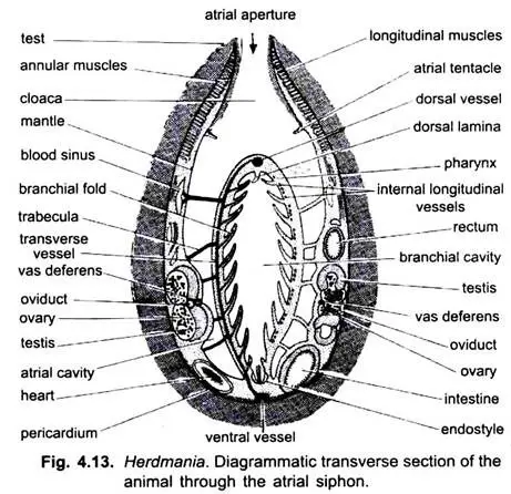
- The atrium is a large ectoderm lined space around the pharynx and it opens into the cloaca.
Digestive and Respiratory System
- The alimentary canal is complete and coiled.
- The pharynx or branchial sac is the largest part and it fills the major body cavity. Its wall is vascular and has numerous stigmata.
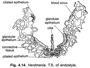
- The endostyle lies mid-ventrally. It secretes mucous net for trapping food particles.
- A dorsal lamina with many languets guide the mucous food string backwards.
- The oesophagus is short and leads into the stomach. From stomach a U-shaped intestine is present that ends in the rectum opening into cloaca.
- The liver is large and bilobed. It secretes digestive enzymes.
- Gas exchange occur mainly through vascular pharyngeal wall and the stigmata.
Circulatory System
- Circulatory system is open type. Vessels are replaced by sinuses.
- The heart is cylindrical and thin walled and it lies in the pericardium.
- A special feature is reversal of heartbeat after few minutes. It pumps blood in opposite direction.
- Major vessels is ventral aorta, dorsal aorta, branchio-visceral and cardio-visceral vessels.
- Blood is slightly reddish and transparent with many coloured corpuscles.
Excretory System
- The main excretory organ is the supra-neural gland.
- It is oval and brown and lies above the cerebral ganglion.
- It has branching tubules and a ciliated funnel.
- Nephrocytes collect waste from blood and it is carried to this gland.
- It is also believed to have endocrine function and is compared to vertebrate pituitary.
Nervous System and Sense Organs
- The larval nervous system degenerates. Adult has a single cerebral ganglion.
- The ganglion lies just below the neural gland. Nerves arise from it to siphons and other parts.
- Definite sense organs is absent.
- Sensory cells are present in test, on ampullae and in siphons.
- A dorsal tubercle is present and it acts as olfactory or gustatory organ.
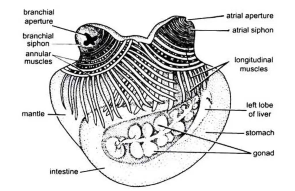
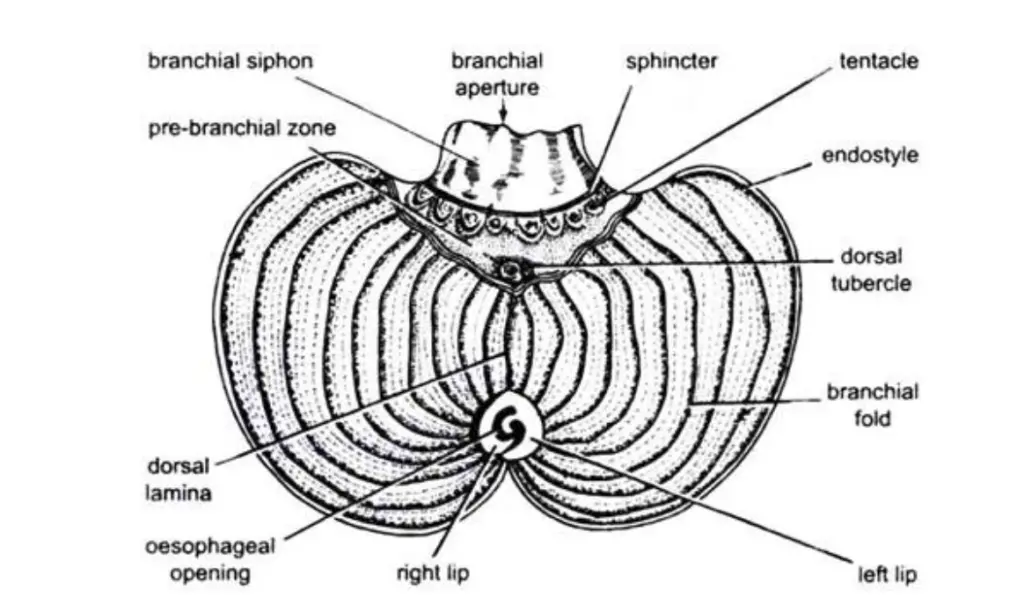
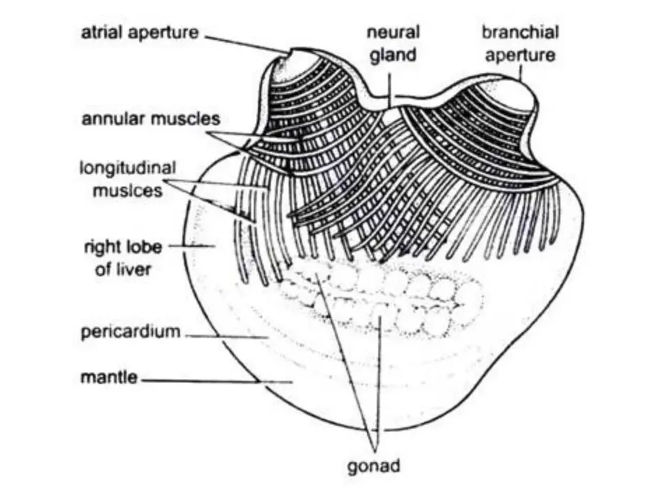
External Morphology of Herdmania (With Diagram)
Body Form, Size and Colour
- The body is oblong and sac-like. It is often compared to a flattened purse or potato-shaped form.
- Adult size usually ranges between 6–13 cm in length. In many specimens it is around 9.5 cm long, 7 cm broad and 4 cm thick.
- The body is divided into the body proper at the free end and a foot-like attached end in sandy habitats.
- Fresh specimens show reddish or pinkish colour. Some bright red patches is present which is due to vascular ampullae of the blood vessels present inside the test.
Siphons and Apertures
- Two siphons are present at the free end. These regulates the inflow and outflow of water.
- The branchial siphon is the incurrent opening. It may be about 1 cm long and it is directed laterally.
- The atrial siphon is the excurrent opening and it is generally longer. It may reach about 1.5 cm and it is directed upwards.
- Each aperture is surrounded by four lobes or lips.
- Long branched branchial tentacles is present at the base of the branchial siphon acting as a strainer.
- Small serrated atrial tentacles is present at the base of the atrial siphon.
The Test (Tunic)
- The whole animal is enclosed in a thick and leathery test.
- The test is composed mainly of tunicin which is related to cellulose.
- It is soft and transparent in young animals but it becomes thick, opaque and wrinkled in adults.
- In the matrix many types of cells, interlacing fibrils and branching blood vessels is present.
- Minute calcareous spicules occur in the test. These include microscleres and megascleres and they act as supporting structures.
- The test protects the animal and also helps in respiration and attachment.
Attachment
- Adult Herdmania is sessile and it remains attached to rocks, shells or corals.
- The attachment is by the basal or ventral region of the body.
- The foot, when present, varies in shape depending on substratum nature.
Body Wall of Herdamania
Outer Epidermis
- The outer epidermis is the outermost layer of the body wall.
- It is ectodermal in origin and made of a single layer of flattened or hexagonal cells.
- This layer secretes the test and it also covers the siphons and the apertures.
- At some regions the layer is interrupted where blood vessels and spicules pass outwards into the test.
Middle Layer (Mesenchyme / Muscular Layer)
- This layer develops from mesoderm and it forms the thickest part of the body wall.
- It is thick in the antero-dorsal region but in the postero-ventral part it becomes thin and almost transparent.
- Connective tissue elements, blood sinuses, nerve fibres and many types of cells is present in this layer.
- The musculature is non-striated and it occur as longitudinal and annular fibres. The longitudinal fibres are more numerous.
- This layer helps in contraction of the body and siphons. Sudden contraction causes water to be expelled when the animal is disturbed.
Inner Epidermis (Atrium Lining)
- The inner epidermis is the innermost layer of the mantle.
- It is composed of a single layer of flat cells.
- It lines the atrial cavity or peribranchial cavity which surrounds the pharynx.
- It maintains separation between the body wall and the pharyngeal sac.
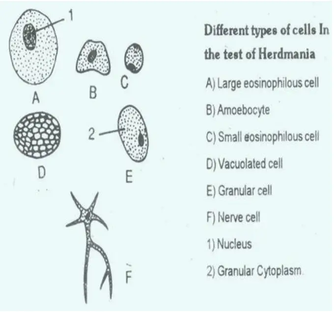
The body wall of Herdmania, with its intricate composition and structure, plays a crucial role in protecting the organism, facilitating respiration, and providing structural support.
Body Divisions of Herdamania
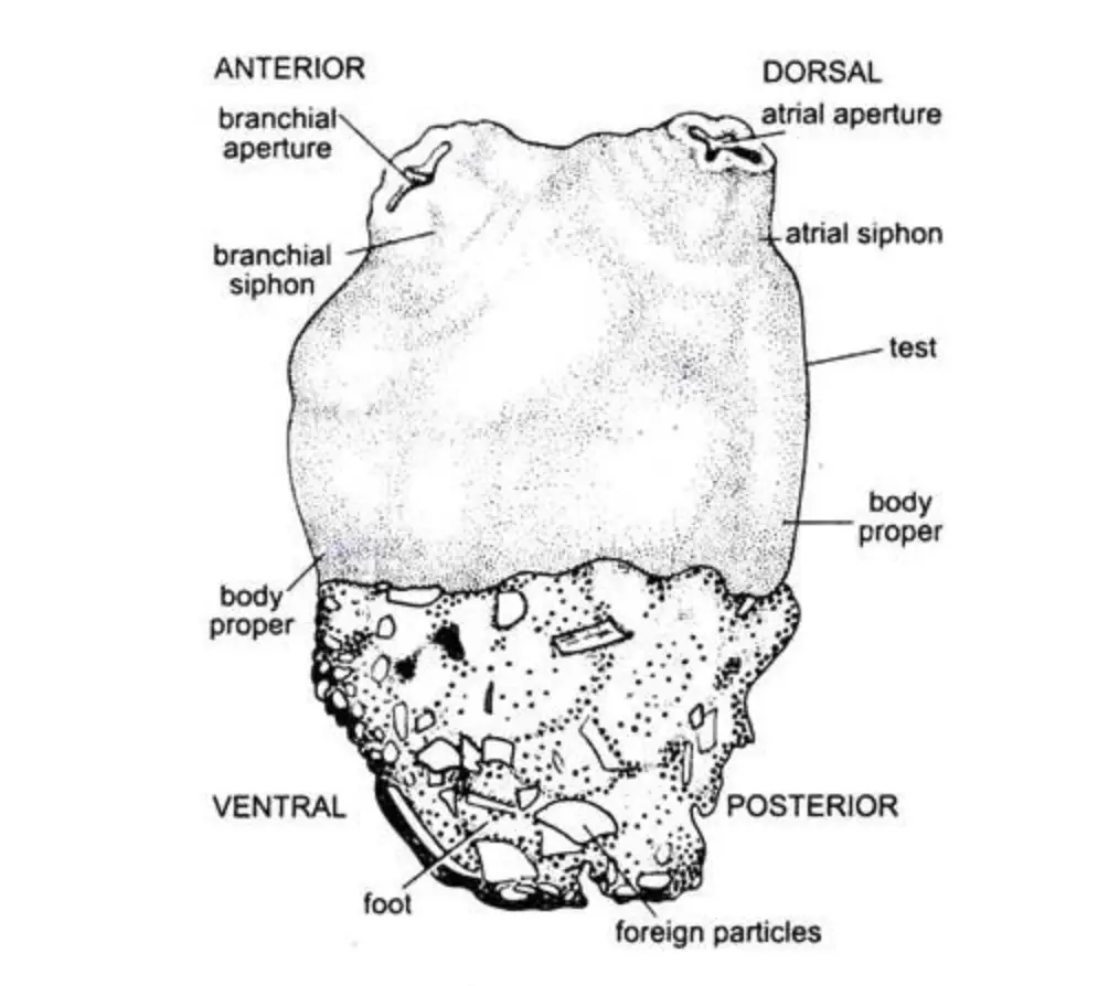
Body Proper
- The body proper is the free distal portion of the animal. It is oblong or sac-like and it is completely covered by a thick leathery test.
- The anterior end is marked by the branchial siphon. This siphon bears the oral aperture and it is generally bent outwards and directed laterally.
- The posterior end lies opposite to the branchial aperture and this end is the region which becomes attached to the substratum.
- The dorsal side is recognised by the atrial siphon. This siphon is usually longer and it is directed upwards.
- The ventral side is the opposite surface and it often remains partly attached to the substratum.
- Adult body generally measures around 9.5 cm in length, 7 cm broad and 4 cm thick but some specimens may reach about 13 cm.
Foot
- The foot is an attachment structure and it is not always present.
- It is mainly seen in species living on sandy beds. When attached to hard substratum it is generally absent.
- When present it may be 3–4 cm long.
- In fine sand the foot is oval, smooth and hard. In coarse sand or shell fragments it is irregular and soft.
- The foot is made entirely of test material.
Body Division in Larva
- The larva shows two main divisions.
- The anterior part is a short oval body or trunk.
- The posterior region is a long tail.


The body divisions of Herdmania are adapted to its marine habitat, providing both structural support and protection. The body proper, with its siphons and apertures, facilitates feeding and respiration, while the foot aids in attachment to various types of substrata.
Test or Tunic of Herdamania
- The test is the thick external covering that encloses the whole body of the animal.
- It is leathery in texture and usually translucent, and the thickness may be about 4–8 mm.
- It is mainly composed of tunicin which is a carbohydrate substance similar to cellulose.
- The test is ectodermal in origin as it is secreted by the epidermal cells of the mantle.
- Different types of cells occur within the test along with interlacing fibrils and blood vessels.
- Calcareous spicules are present in the matrix and these spicules help in providing firmness and support.
- The bright red patches on the test are formed by terminal knobs of blood vessels.
- The test protects the body from predators and mechanical injury.
- It also helps in attachment of the animal to the substratum because the basal region becomes very thick.
- The test is vascular in nature and it may act as an accessory respiratory surface.
- Some sensory receptor cells are present in the test and they respond to external stimuli.
- Small marine organisms like hydroids and gastropods may live over the test as commensals.
Structure, Appearance, and Composition
- The test is secreted by the outer epidermis of the mantle and thus it is ectodermal in origin.
- In the young animal the test is soft, leathery and transparent, but in the adult it becomes thick and opaque.
- The surface of the adult test is usually wrinkled and it shows criss-cross markings.
- The test is made of a matrix which contains proteins, salts and water along with tunicin.
- Tunicin is a cellulose-like carbohydrate, and its presence in an animal covering is a unique feature.
- The test can be described in three layers.
- The outer layer is thin in young forms but becomes thicker in adults and it contains fine fibrils.
- The middle layer consists of connective tissue and it contains spicules, blood vessels and tunic cells.
- The inner layer lies very close to the epidermis and it is bound by delicate fibrils.
- The spicules embedded in the test provide strength and support to the covering.
- The blood vessels present in the matrix make the test an accessory respiratory surface.
- The test protects the body and also helps in anchoring the animal to the substratum.
Internal Components of the Test
- Different types of cells occur inside the test and most of these cells are mesodermal in origin.
- Large and small eosinophilous cells, amoeboid cells, granular cells and vacuolated round cells are commonly present in the matrix.
- Some nerve cells with many processes may also be found embedded in the test.
- The granular cells surrounded by nerve fibres act as important receptor cells.
- Many interlacing fibrils are present and these fibrils form a fine network throughout the test.
- These fibrils look like smooth muscle fibres and they help in maintaining the structure of the matrix.
- A network of blood vessels runs through the test and these vessels branch repeatedly.
- The vessels end in swollen structures known as vascular ampullae or terminal knobs.
- These ampullae are filled with blood and they produce the bright red patches on the body surface.
- The test contains many calcareous spicules which are made of calcium carbonate.
- The smaller spicules are the microscleres which are about 50 µm in size and they usually have a spherical head with rings of spines.
- The larger spicules are the megascleres and these may be spindle-shaped or pipette-shaped and they also possess rings of spines.
- Spicule formation takes place rapidly inside special envelopes present in the tunic blood vessels.
- After formation the spicules leave the vessels and get distributed into the test matrix.
Functions of the Test
- The test acts as a protective covering and it guards the soft body from mechanical injury.
- The presence of calcareous spicules inside the test gives additional support and defence.
- It helps in firm attachment of the animal to the substratum because the basal region of the test becomes very thick.
- The test is highly vascular and it works as an accessory respiratory surface for exchange of gases.
- Some receptor cells present in the test make it an important sensory structure.
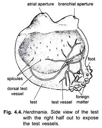
Spicules of Herdamania
- The spicules of the animal are calcareous in nature and they are mainly composed of calcium carbonate.
- These spicules are minute structures embedded inside the matrix of the test.
- The spicules give firmness to the test and act as a supporting framework.
- They also help in providing protection to the body from external injury.
- Two main types of spicules are present in the test.
- The smaller spicules are known as microscleres and their size is about 50 µm.
- Microscleres usually have a spherical head and the body bears several rings of small spines.
- The larger spicules are called megascleres and they may be spindle-shaped or pipette-shaped.
- Megascleres may reach a length of 1.5 mm to 3.5 mm and they also possess rings of spines.
- Spicule formation occurs inside special envelopes present within the tunic blood vessels.
- The spicules are produced very quickly and they may form completely within 4–5 days.
- After formation the spicules leave the vessels and are distributed throughout the test matrix.
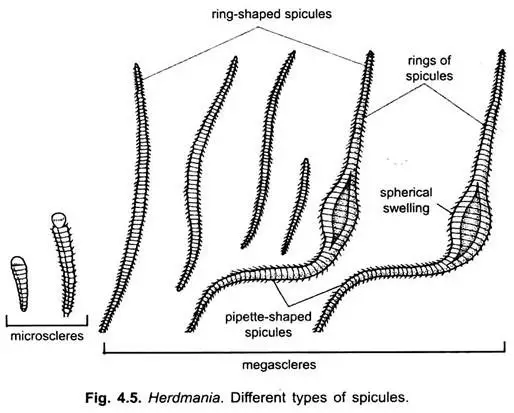
Types of Spicules
1. Microscleres
- It is found only in the test (tunic) of Herdmania.
- Each microsclere has a rounded knob-like head and an elongated tapering body.
- The head is mostly smooth but sometimes a few spines may present.
- The body has rings of spines (5–20) and these spines point towards the head.
- The size is about 50 microns on average, but it may reach nearly 80 microns depending on growth stage.
2. Megascleres
There are two kinds of megascleres–
- Spindle-shaped spicules
- Pipette-shaped spicules
a. Spindle-Shaped Spicules
- It is distributed throughout the body and usually present in connective tissue sheaths.
- These spicules are arranged in linear rows which look like strings.
- They have many rings of spines (20–60) and these spines point in the same direction.
- The size is about 1.5 mm normally but may grow up to 2.5 mm.
- These are dense around the stomach, gonads, bases of siphons and mantle, but scarce at the base of longitudinal muscles.
b. Pipette-Shaped Spicules
- It is found in different organs especially in the mantle around gonads and liver lobes.
- Each spicule has a large spherical swelling in the middle which gives a pipette-like appearance.
- Many spicules are bent into U or V shapes.
- The size is larger than spindle-shaped types and may reach about 3.5 mm.
- They have rings of spines and the whole structure is covered by connective tissue sheaths.
Formation Process of Spicules
- It is the process in which tunic spicules are formed quickly within about 4–5 days inside the tunic of Herdmania.
- The development takes place inside separate envelopes that are present in the tunic blood vessels.
- Specialized cells called sclerocytes are involved in this process. These pseudopodial sclerocytes remain connected with the neighboring body spicules and they lie over the fringing spines.
- It is thought that the spine formation occurs inside these sclerocytes.
- The mineral phase of CaCO₃ is deposited upon or within an organic matrix. This organic covering remains attached closely with the developing spicule inside the envelope.
- After the formation is completed, the tunic spicule moves out from the envelope and blood vessel.
- It then enters into the test matrix and finally protrudes outside through the tunic surface.
Functions of Spicules
- It is the main supporting element of the body as the spicules give skeletal support and help in maintaining the shape of different organs.
- Many spicules project into the test, and these projections anchor the test firmly with the mantle so the mantle does not detach during contraction.
- Spicules present around the blood vessels help in stiffening the vessel walls which prevents their collapse and thus keeps proper blood flow.
Mantle or Body Wall of Herdamania
- It is the true cellular layer present just below the test (tunic) which forms the main body covering of Herdmania.
- The mantle remains attached only at the branchial and atrial apertures where it forms the two siphons used for the entry and exit of water.
- It encloses and protects the visceral organs and also helps in maintaining the body shape.
- The mantle is composed of three layers–
1. Outer Epidermis: It is ectodermal and made of flat or hexagonal cells. This layer also forms the stomodaeum and proctodaeum at the apertures. It helps in secretion of the test, and is interrupted where blood vessels and spicules pass outwards.
2. Middle Mesoderm: It is the thickest layer and contains connective tissue, blood sinuses, nerve fibers and different types of cells. It has unstriped muscle fibers arranged in longitudinal and annular sets.
3. Inner Epidermis: It is a thin layer of flat polygonal cells lining the atrium (peribranchial cavity). - The annular muscles are arranged around the branchial and atrial siphons and help in opening and closing the siphons.
- The longitudinal muscles arise from both apertures and spread towards the middle region and these muscles assist in contraction and movement.
- A separate set of branchioatrial muscles is present between the two siphons and increase the flexibility of the body.
- The mantle becomes thick, muscular and strong in the antero-dorsal region, but it is very thin and transparent in the postero-ventral region.
- It plays an important role in contraction of the body and in forceful ejection of water from the siphons, which is why the animal is called sea squirt.
- The mantle maintains the internal atrial space around the pharynx and acts as a protective barrier against external conditions.
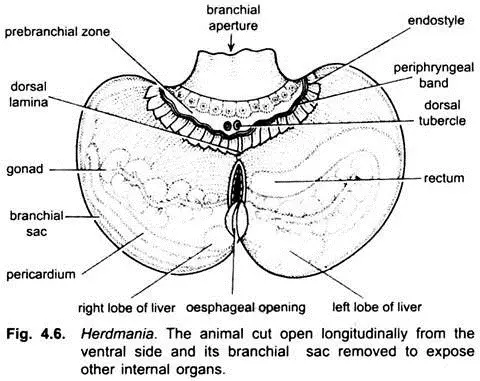
Nervous System of Herdmania (With Diagram)

- The nervous system of Herdmania is simple in structure and it is mainly formed by a brain or nerve ganglion.
- The brain is about 4 cm long and it lies mid-dorsally in the mantle between the branchial and atrial siphons.
- In the members of Pyuridae the brain is placed below the neural gland, although in most ascidians it is present above the neural gland.
- The brain contains bipolar and multipolar nerve cells.
- It gives three nerves towards the branchial siphon and two nerves towards the atrial siphon.
- The brain is considered as the degenerated remnant of the anterior part of the well-developed larval nervous system.
- The neural ganglion, the neural gland and the dorsal tubercle together form the neural complex.
- Herdmania does not have true specialized sense organs. Different receptor cells act as sensory units.
- Red pigmented cells on siphon margins and vascular ampullae act as photoreceptors.
- Sensory cells in the test, siphon margin and tentacles act as tangoreceptors responding to touch.
- Cells on the siphon margins behave as rheoreceptors for water current.
- Cells lining the siphons act as thermoreceptors detecting temperature.
- Tentacles and dorsal tubercle act as chaemoreceptors responding to chemicals.
- The dorsal tubercle is present in the pre-branchial zone near the junction of the dorsal lamina with the peripharyngeal bands.
- It has a broad base with two spirally coiled lobes and each lobe has three coils lined by ciliated cells.
- It is richly supplied with blood sinuses and nerves and functions like an olfactory and gustatory organ.

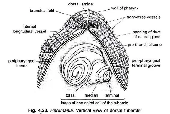
Digestive System of Herdmania (With Diagram)
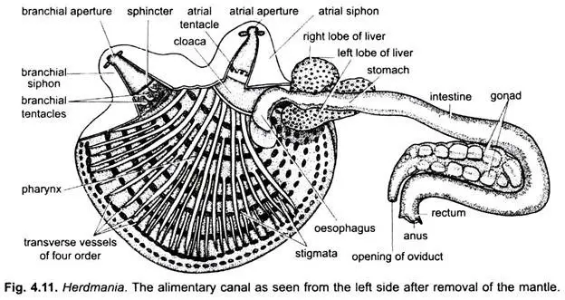
- The digestive system is complete from mouth to anus and it is adapted for a ciliary or filter-feeding habit.
- It consists of the alimentary canal and two digestive glands.
Alimentary Canal
- The canal is coiled and begins at the mouth which opens on the top of the branchial siphon.
- The mouth is surrounded by four lips of the test and leads into the branchial siphon.
- The branchial siphon forms a short narrow buccal cavity (stomodaeum) which is lined by ectoderm.
- A circlet of branching tentacles is present at its base which acts as a sieve and prevents large particles from entering the pharynx.
- The pharynx or branchial sac is the largest part and it has a pre-branchial zone with smooth walls and a posterior branchial sac with many stigmata.
- Longitudinal folds, the dorsal lamina, and the endostyle are present in the pharynx.
- The endostyle is a ventral longitudinal groove filled with ciliated and glandular tracts producing mucus.
- The dorsal lamina bears many small languets that guide the food-laden mucus cord posteriorly.
- The oesophagus is short, bent and provided with ciliated grooves that help in pushing the food into the stomach.
- The stomach is wider than the oesophagus and has thin smooth walls and a branching pyloric gland.
- The intestine is U-shaped with a descending limb and an ascending limb where absorption takes place.
- The rectum is short and ciliated and opens into the cloaca by the anus which has four lips.
Digestive Glands
- The liver is a dark brown bilobed organ lying on the stomach. It is made of many tubules embedded in connective tissue.
- It secretes amylase, protease, weak lipase and bile pigments.
- It also stores reserve food in the form of starch-like granules.
- The pyloric gland is formed of branching tubules in the walls of the stomach and intestine.
- It opens into the intestine by a single duct and probably acts both as an accessory digestive gland and an excretory organ.
Food Collection and Digestion
- Water enters through the mouth and passes into the pharynx by the action of lateral cilia.
- The tentacles filter larger particles and the endostyle secretes mucus that traps microscopic plankton.
- The mucus net with food is rolled into a cord and carried by the languets to the oesophagus.
- Digestion occurs in the stomach with enzymes from the liver and absorption occurs in the intestine.
- Undigested matter forms a mucus-covered faecal cord in the rectum and is expelled through the atrial aperture with the outgoing water current.
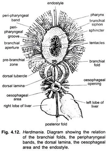
Food, Feeding and Digestion
Food
- The animal feeds on minute organisms like protozoans, zooplankton and algae.
- It also takes decaying organic matter and small animal pieces suspended in seawater.
Feeding
- Feeding is carried out mainly by ciliary action because Herdmania remains fixed.
- The cilia on the stigmata create a constant water current which enters through the mouth and buccal cavity.
- This water passes into the pharynx and then through the stigmata into the atrial cavity before leaving by the atrial aperture.
- The current brings fine food particles which settle on the pharyngeal walls.
Endostyle and Mucus Secretion
- The gland cells of the endostyle secrete mucus.
- The cilia of the endostyle lash the mucus sideways and not forwards.
- Food particles get caught in this mucus on the pharyngeal wall.
- The food-mucus mixture is carried upwards to the dorsal lamina.
- The cilia of the dorsal lamina roll this into a food string that passes into the oesophagus and then into the stomach.
Digestion
- The liver pours a yellowish-brown digestive juice into the stomach.
- This juice contains amylase for carbohydrate digestion, protease for proteins and a weak lipase for fats.
- The pyloric gland acts like an accessory digestive organ with a pancreatic nature.
- Digestion takes place mainly in the stomach and absorption occurs in the intestine.
Excretion of Waste
- The rectum is ciliated and coils the undigested matter.
- The faecal mass is expelled forcefully through the atrial siphon and may be thrown out up to about 10 cm.
- http://www.soghracollege.com/img/Lectures%2023-05-20/Zoology%20(Urochordata-2)%20B.ScPart%202.pdf
- https://www.eshiksha.mp.gov.in/mpdhe/pluginfile.php/53465/mod_resource/content/1/etxt%20deepali.pdf
- https://www.notesonzoology.com/phylum-chordata/herdmania/nervous-system-of-herdmania-with-diagram-chordata-zoology/7286
- https://www.notesonzoology.com/phylum-chordata/herdmania/digestive-system-of-herdmania-with-diagram-chordata-zoology/7298
- Text Highlighting: Select any text in the post content to highlight it
- Text Annotation: Select text and add comments with annotations
- Comment Management: Edit or delete your own comments
- Highlight Management: Remove your own highlights
How to use: Simply select any text in the post content above, and you'll see annotation options. Login here or create an account to get started.