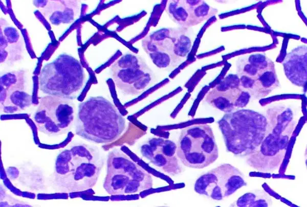
Gram-positive bacteria are a group of bacteria classified based on how they react to a lab test called the Gram stain. When stained, these bacteria hold onto a purple dye because of their thick, mesh-like cell wall made of peptidoglycan. This sturdy layer acts like a protective shield, unlike Gram-negative bacteria, which have thinner walls and an extra outer membrane. Common examples include Staphylococcus, often found on skin, and Streptococcus, which can cause infections like strep throat. While some Gram-positive bacteria are harmless or even helpful, others trigger illnesses, making them a focus in medicine and hygiene practices. Their structure also influences how antibiotics work against them.
Agnatha refers to a primitive class of jawless fish, representing some of the earliest vertebrates. Unlike most modern fish, creatures like lampreys and hagfish lack jaws, paired fins, and scales. Instead, they have smooth, slimy bodies and circular mouths lined with rows of teeth, which they use to latch onto prey or scavenge. Lampreys, for instance, attach to other fish to feed on their blood, while hagfish are known for producing slime as a defense. These ancient species offer clues about vertebrate evolution, showing how traits like backbones and basic nervous systems developed. Though often called “living fossils,” they play unique roles in aquatic ecosystems today.
What is Gram-Positive Bacteria?
- A thick peptidoglycan coating in their cell wall that holds the crystal violet stain used in the Gramme staining process defines the group of Gram-positive bacteria.
- Developed by Hans Christian Gramme in the 1880s, the Gramme staining technique sets these bacteria apart from Gram-negative bacteria depending on cell wall composition
- Teichoic acids and lipoteichoic acids found in their cell walls serve purposes in cell wall maintenance, ion control, and adherence to surfaces.
- This thick peptidoglycan structure helps the bacteria to be sensitive to certain antibiotics, including beta-lactams, which target formation of cell walls.
- Gram-positive Gram-positive several clinically important pathogens, including species from the genera Staphylococcus, Streptococcus, and Clostridium, are cause of a variety of diseases
- Furthermore crucial for their identification and categorisation in both clinical diagnostics and microbiological research are the strong structural traits of Gram-positive bacteria.
- Knowing the structural and molecular characteristics of Gram-positive bacteria helps guide the creation of focused antibiotics and supports infectious disease epidemiology research.

Definition of Gram Positive bacteria
Gram-positive bacteria are a group of bacteria that retain the crystal violet stain in the Gram stain test, appearing purple under a microscope. They have a thick peptidoglycan layer in their cell wall, which contributes to their ability to retain the stain. Gram-positive bacteria lack an outer membrane and are generally more susceptible to certain antibiotics compared to Gram-negative bacteria.
Characteristics of Gram-positive bacteria
Gram-positive bacteria possess distinct characteristics that set them apart from other types of bacteria. These features include:
- Unlike Gram-negative bacteria who have both an inner and an outside membrane, gram-positive bacteria lack an outer membrane.
- Their single cytoplasmic membrane has a thin lipid layer that supports a somewhat thicker peptidoglycan cell wall.
- Mostly made of peptidoglycan, the cell wall gives the organism structural rigidity and is absolutely important for osmotic stability.
- High concentrations of teichoic acid, particularly lipoteichoic acid that ties the cell wall to the cytoplasmic membrane, are found embedded within the peptidoglycan layer; these acids are vital for ion control and bacterial adhesion.
- Key target for beta-lactam antibiotics, crosslinking enabled by DD-transpeptidase enzymes helps to preserve the structural integrity of the peptidoglycan layer.
- Although they show a periplasmic area, it is much thinner than that of Gram-negative bacteria, therefore reducing the usefulness sometimes connected with a larger periplasm.
- Unlike the four-basal-body arrangement usually observed in Gram-negative species, certain Gram-positive bacteria are motile and have flagella with two basal bodies that offer a unique way of mobility.
- Many times, a strong polysaccharide capsule envelops Gram-positive bacteria, which helps them to be virulent by helping the host’s immune reaction to be escaped.
Shape of Gram positive bacteria
- Under microscopic study, gram-positive bacteria show different forms that help to classify and identify them.
- Their main classification is into two main forms: cocci and bacilli.
- Usually ranging in diameter from 0.5 to 1.0 micrometre, cocci are oval or circular.
- These cells might show up alone, in couples, in chains, or in clusters.
- Special arrangements of cocci include tetrads, in which cells align in square clusters of four, and sarcina, distinguished by clusters of four or cubes of eight cells.
- Usually looking like sticks, Bacilli are rod-shaped bacteria.
- Usually spanning 1 to 10 micrometres in length and 0.3 to 1.0 micrometre in width, their size
- Variations in the ends of Bacilli—round-tapered, square, or inflated terminus—help to explain their morphological variety.
- Both clinical diagnosis and microbiological research depend on the thorough morphologies seen in these bacteria, which also provide understanding of their categorisation and possible harmful behaviour.
Cell wall Structure of Gram-positive bacteria
The cell wall structure of Gram-positive bacteria is characterized by several components that provide strength, rigidity, and other functional properties. These components include peptidoglycan, teichoic acid, and a thin lipid layer.

- Peptidoglycan:
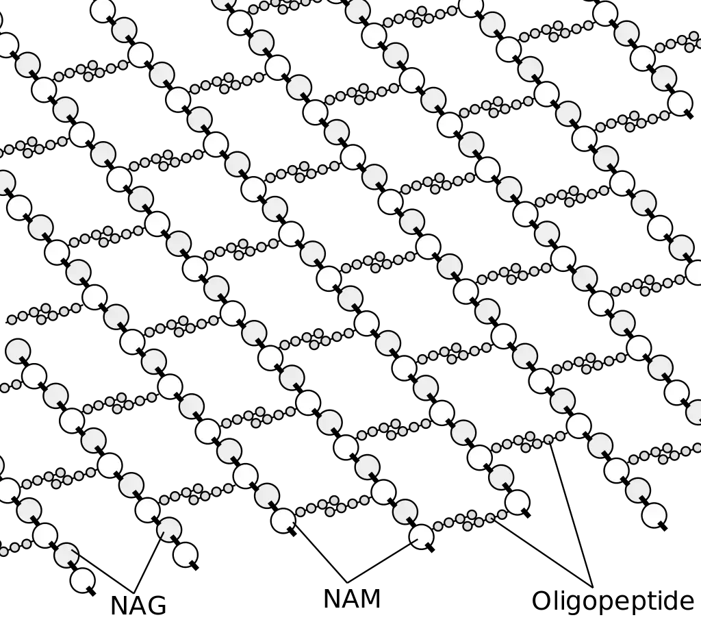
- Peptidoglycan, also known as murein, makes up the majority (about 90%) of the Gram-positive bacterial cell wall.
- It plays a crucial role in providing shape, strength, and rigidity to the cell wall.
- Peptidoglycan is a high-quality polymer composed of two sugar derivatives, N-acetylglucosamine and N-acetylmuramic acid, as well as a chain of L-amino acids and three unique D-amino acids (D-glutamic acid, D-alanine, and meso-diaminopimelic acid).
- The D-amino acids and L-amino acids form connections with N-acetylmuramic acid, and L-lysine can replace meso-diaminopimelic acid in some cases.
- This interconnection of peptidoglycan subunits contributes to the strength, integrity, and elasticity of the cell wall.
- Peptidoglycan is also permeable, allowing molecules to move in and out of the bacterial cell.
- Teichoic Acid:
- Teichoic acid is a component present in the cell walls of Gram-positive bacteria, but it is absent in Gram-negative bacteria.
- It is a polymer composed of glycerol copolymers and makes up a significant portion (up to 50%) of the total dry weight of the bacterial cell wall.
- Teichoic acid can be directly connected to the peptidoglycan through covalent bonds or to the cell membrane as lipoteichoic acid.
- The direct connection to peptidoglycan occurs through the 6-hydroxyl N-acetylmuramic acid.
- Teichoic acid carries a negative charge and extends to the surface of the peptidoglycan, contributing to the overall negative charge of the bacterial cell wall.
- It plays a role in maintaining the structure of the cell wall and has other functions depending on the bacterial species.
- Lipid:
- Gram-positive bacteria have a thin layer of lipids located below the peptidoglycan layer, comprising about 2-5% of the cell wall.
- This lipid layer functions to anchor the bacterial cell wall.

Together, these components form a complex cell wall structure in Gram-positive bacteria. The peptidoglycan provides strength and rigidity, while teichoic acid contributes to the negative charge and structural integrity of the cell wall. The lipid layer anchors the cell wall and provides additional functionality. Understanding the cell wall structure of Gram-positive bacteria is essential for identifying and targeting specific components for therapeutic interventions.
Classification of Gram Positive Bacteria
Gram-positive bacteria are classified based on various criteria, including morphology, biochemical properties, genetic relatedness, and cell wall composition. This classification aids in understanding their physiology, pathogenicity, and antibiotic susceptibility.
- Morphological Classification:
- Cocci (Spherical):
- Staphylococcus: Gram-positive cocci in clusters; catalase-positive; facultative anaerobes.
- Streptococcus: Gram-positive cocci in chains or pairs; catalase-negative; classified based on hemolytic properties (α, β, γ).
- Enterococcus: Gram-positive cocci in pairs or short chains; catalase-negative; γ-hemolytic; notable for antibiotic resistance.
- Bacilli (Rod-shaped):
- Bacillus: Aerobic or facultatively anaerobic; spore-forming; includes B. anthracis (anthrax).
- Clostridium: Anaerobic; spore-forming; includes C. botulinum, C. tetani, C. difficile.
- Listeria: Non-spore-forming; facultative anaerobe; includes L. monocytogenes (listeriosis).
- Corynebacterium: Non-spore-forming; aerobic; includes C. diphtheriae (diphtheria).
- Cocci (Spherical):
- Biochemical Properties:
- Catalase Test:
- Positive: Staphylococcus.
- Negative: Streptococcus, Enterococcus.
- Hemolysis on Blood Agar:
- α-hemolysis (partial): Streptococcus pneumoniae.
- β-hemolysis (complete): Streptococcus pyogenes.
- γ-hemolysis (none): Enterococcus faecalis.
- Catalase Test:
- Genetic Relatedness:
- Phylum Firmicutes: Includes low G+C content bacteria such as Bacillus, Clostridium, Staphylococcus, Streptococcus.
- Phylum Actinobacteria: Includes high G+C content bacteria such as Corynebacterium, Mycobacterium, Nocardia.
- Cell Wall Composition:
- Peptidoglycan Layer: Thick in Gram-positive bacteria, retaining crystal violet stain.
- Teichoic and Lipoteichoic Acids: Present in the cell wall, contributing to rigidity and antigenic specificity.
Pathogenicity of Gram Positive Bacteria
Gram-positive bacteria are responsible for a wide array of diseases in humans, ranging from mild skin infections to life-threatening systemic conditions. Their pathogenicity is attributed to various structural components and virulence factors that facilitate colonization, immune evasion, and tissue damage.
- Structural Components Contributing to Pathogenicity:
- The thick peptidoglycan layer in Gram-positive bacteria provides structural integrity and resistance to environmental stresses.
- Teichoic and lipoteichoic acids in the cell wall contribute to cell wall maintenance, ion transport, and adherence to host tissues.
- Virulence Factors:
- Exotoxins: Many Gram-positive bacteria produce exotoxins that disrupt host cell function. For instance, Staphylococcus aureus produces toxins leading to conditions like toxic shock syndrome.
- Enzymes: Enzymes such as hyaluronidase and streptokinase facilitate tissue invasion and dissemination.
- Surface Proteins: Cell wall-associated proteins play key roles in colonization and pathogenesis by mediating interactions with the host environment.
- Biofilm Formation:
- Some Gram-positive bacteria can form biofilms, complex communities that adhere to surfaces and are resistant to antibiotics, contributing to persistent infections.
- Immune Evasion Mechanisms:
- Capsule formation, as seen in Bacillus anthracis, helps in evading phagocytosis.
- Production of proteins that inactivate antibodies, aiding in immune evasion.
- Examples of Diseases Caused by Gram-Positive Bacteria:
- Staphylococcus aureus: Causes skin infections, pneumonia, and sepsis.
- Streptococcus pyogenes: Responsible for pharyngitis, scarlet fever, and rheumatic fever.
- Clostridium difficile: Leads to antibiotic-associated diarrhea and colitis.
- Bacillus anthracis: Causes anthrax, a serious infectious disease.
Virulence Factors of Gram-Positive Bacteria
- Gram-positive bacteria produce exotoxins, which are proteins that directly damage host tissues and interfere with normal cellular functions. These toxins can trigger severe inflammatory responses and cell death.
- They secrete a range of hydrolytic enzymes, such as hyaluronidase, proteases, and coagulases, that degrade extracellular matrix components, facilitating tissue invasion and dissemination within the host.
- Surface adhesion proteins on Gram-positive bacteria mediate attachment to host cells and tissues, a critical first step in colonization. For example, certain adhesins enable bacteria like Streptococcus pyogenes to evade immune recognition by interfering with opsonization.
- The formation of a polysaccharide capsule is a key virulence factor, serving as a protective barrier against phagocytosis. This capsule helps bacteria persist in the bloodstream and tissues by limiting the effectiveness of the host’s immune response.
- Biofilm formation is another significant virulence mechanism. Biofilms create structured microbial communities that enhance bacterial survival, promote resistance to antimicrobial agents, and hinder clearance by the immune system.
- Components of the cell wall, including teichoic and lipoteichoic acids, not only support cell structure but also contribute to immune activation by triggering pro-inflammatory responses, which can exacerbate tissue damage and disease severity.
- In some Gram-positive bacteria, the expression of superantigens leads to non-specific T-cell activation and massive cytokine release, resulting in conditions such as toxic shock syndrome, which are characterized by severe systemic inflammatory responses.
- The thick peptidoglycan layer characteristic of Gram-positive bacteria plays a dual role in structural integrity and immune modulation, by acting as a pathogen-associated molecular pattern (PAMP) that is recognized by host immune receptors, leading to inflammation.
Ecological and Industrial Roles of Gram-Positive Bacteria
- Gram-positive bacteria are ubiquitous in nature and contribute significantly to ecological balance by degrading organic matter, thereby facilitating nutrient recycling and carbon cycling
- In soil ecosystems, these bacteria participate in the breakdown of complex polymers such as cellulose and chitin, enhancing soil fertility and supporting plant growth
- Some species form symbiotic relationships with plants, aiding in nutrient uptake and providing a natural defense against certain soil-borne pathogens
- Certain Gram-positive bacteria, especially those from the actinobacteria group, are instrumental in the natural production of antibiotics, which helps suppress competing microorganisms in their environment
- In industrial settings, Gram-positive bacteria are harnessed for the production of fermented foods—lactic acid bacteria, for instance, are critical in yogurt, cheese, and sourdough production
- They produce a range of industrial enzymes, such as proteases and amylases, which are employed in food processing, detergent formulation, textile manufacturing, and biofuel production
- Bioactive metabolites produced by these bacteria, including antibiotics and immunomodulatory compounds, have been exploited for pharmaceutical applications
- Some strains are used in bioremediation processes to degrade pollutants and detoxify contaminated environments, leveraging their ability to metabolize complex organic compounds
- The robust nature and diverse metabolic capabilities of Gram-positive bacteria make them valuable for the production of biopolymers and single-cell protein, contributing to sustainable industrial biotechnology practices
Gram positive bacteria examples and diseases
| Bacterium | Associated Diseases |
|---|---|
| Staphylococcus aureus | Skin infections (boils, impetigo), pneumonia, endocarditis, toxic shock syndrome |
| Streptococcus pyogenes | Pharyngitis (strep throat), scarlet fever, rheumatic fever, necrotizing fasciitis |
| Streptococcus pneumoniae | Pneumonia, meningitis, otitis media, sinusitis |
| Enterococcus faecalis | Urinary tract infections, endocarditis, intra-abdominal infections |
| Bacillus anthracis | Anthrax (cutaneous, inhalational, gastrointestinal) |
| Clostridium tetani | Tetanus |
| Clostridium botulinum | Botulism |
| Clostridium perfringens | Gas gangrene, food poisoning |
| Listeria monocytogenes | Listeriosis (especially dangerous during pregnancy) |
| Corynebacterium diphtheriae | Diphtheria |
Antibiotics for Gram positive bacteria
Gram-positive bacteria are susceptible to various antibiotics, which are essential for treating bacterial infections caused by these pathogens. Here are some commonly used antibiotics and their mechanisms of action against Gram-positive bacteria:
- ß-lactam antibiotics (Amoxicillin, Methicillin, Oxacillin, Ampicillin, Penicillin G):
- Mechanism of action: Disruption of bacterial cell wall synthesis by inhibiting the formation of peptidoglycan, leading to cell lysis.
- Effective against: Staphylococcus aureus, Streptococcus pneumoniae, Streptococcus pyogenes, Corynebacterium diphtheriae, Bacillus anthracis, Clostridium botulinum.
- Vancomycin, Erythromycin, Azithromycin:
- Mechanism of action: Inhibition of cell wall synthesis by preventing the crosslinking of peptidoglycan peptidases.
- Effective against: Staphylococcus spp, Streptococcus spp, Bacillus spp, Clostridium difficile.
- Bacitracin:
- Mechanism of action: Inhibition of cell wall synthesis by preventing the movement of cytoplasmic membrane and peptidoglycan precursors.
- Effective against: Corynebacterium spp, Bacillus anthracis.
- Macrolides (Azithromycin, Clarithromycin):
- Mechanism of action: Inhibition of bacterial protein synthesis by preventing polypeptide elongation at the 50S ribosomes.
- Effective against: Streptococcus pyogenes.
- Cephalosporin:
- Mechanism of action: Inhibition of cell wall synthesis by disrupting peptidoglycan synthesis.
- Effective against: Streptococcus pneumoniae, Bacillus anthracis.
- Aminoglycosides (Gentamicin):
- Mechanism of action: Inhibition of bacterial protein synthesis by producing aberrant peptide chains at the 30S ribosomes.
- Effective against: Staphylococcus aureus, Streptococcus pneumoniae, Streptococcus pyogenes, Enterococcus spp.
- Oxazolidinone:
- Mechanism of action: Inhibition of protein synthesis at the 50S ribosomes.
- Effective against: Enterococcus spp.
- Rifampicin:
- Mechanism of action: Inhibition of nucleic acid synthesis by preventing the transcription of binding DNA-dependent RNA polymerase.
- Effective against: Bacillus anthracis.
- Sulfonamides (Sulfamethoxazole):
- Mechanism of action: Inhibition of folic acid synthesis by blocking the enzyme dihydropteroate synthase.
- Effective against: Listeria monocytogenes.
- Trimethoprim:
- Mechanism of action: Inhibition of folic acid synthesis by blocking the enzyme dihydrofolate reductase.
- Effective against: Listeria monocytogenes.
Low G+ C (Firmicutes): General characteristics with suitable examples
Low G+C Gram-positive bacteria, also known as Firmicutes, are a phylum of bacteria characterized by a low guanine-cytosine (G+C) content in their genomic DNA. They exhibit diverse characteristics and include several notable examples. Here are the general characteristics of Firmicutes along with suitable examples:
- Cell Wall Structure:
- Firmicutes have a thick peptidoglycan cell wall that provides structural integrity.
- The peptidoglycan layer is composed of alternating N-acetylglucosamine and N-acetylmuramic acid residues.
- Shape and Morphology:
- Firmicutes exhibit a variety of shapes, including cocci (spherical), bacilli (rod-shaped), and filamentous forms.
- Some examples of Firmicutes with specific shapes include Streptococcus (cocci in chains or pairs) and Bacillus (rod-shaped).
- Metabolism:
- Firmicutes display diverse metabolic capabilities, including fermentative, anaerobic, and some aerobic metabolism.
- They can utilize a wide range of carbon sources and produce various end products such as acids, alcohols, and gases.
- Examples of Firmicutes:
- Staphylococcus aureus: This bacterium is a significant human pathogen responsible for various infections, including skin and soft tissue infections, pneumonia, and septicemia.
- Streptococcus pyogenes: It causes diseases such as strep throat, impetigo, cellulitis, and invasive infections like necrotizing fasciitis and toxic shock syndrome.
- Bacillus subtilis: A model organism extensively studied for its physiology and genetics. It is commonly found in soil and has diverse metabolic capabilities.
- Clostridium difficile: This bacterium is associated with antibiotic-associated diarrhea and pseudomembranous colitis.
- Listeria monocytogenes: It is responsible for the foodborne illness listeriosis, which can lead to septicemia, meningitis, and other severe infections.
- Enterococcus faecalis: Found in the gastrointestinal tract, it can cause opportunistic infections, including urinary tract infections and endocarditis.
| Example Genus | Microscopic Morphology | Unique Characteristics |
|---|---|---|
| Bacillus | Large, gram-positive bacillus | Aerobes or facultative anaerobes; form endospores; B. anthracis causes anthrax in cattle and humans, B. cereus may cause food poisoning |
| Clostridium | Gram-positive bacillus | Strict anaerobes; form endospores; all known species are pathogenic, causing tetanus, gas gangrene, botulism, and colitis |
| Enterococcus | Gram-positive coccus; forms microscopic pairs in culture (resembling Streptococcus pneumoniae) | Anaerobic aerotolerant bacteria, abundant in the human gut, may cause urinary tract and other infections in the nosocomial environment |
| Lactobacillus | Gram-positive bacillus | Facultative anaerobes; ferment sugars into lactic acid; part of the vaginal microbiota; used as probiotics |
| Leuconostoc | Gram-positive coccus; may form microscopic chains in culture | Fermenter, used in food industry to produce sauerkraut and kefir |
| Mycoplasma | The smallest bacteria; appear pleomorphic under electron microscope | Have no cell wall; classified as low G+C Gram-positive bacteria because of their genome; M. pneumoniae causes “walking” pneumonia |
| Staphylococcus | Gram-positive coccus; forms microscopic clusters in culture that resemble bunches of grapes | Tolerate high salt concentration; facultative anaerobes; produce catalase; S. aureus can also produce coagulase and toxins responsible for local (skin) and generalized infections |
| Streptococcus | Gram-positive coccus; forms chains or pairs in culture | Diverse genus; classified into groups based on sharing certain antigens; some species cause hemolysis and may produce toxins responsible for human local (throat) and generalized disease |
| Ureaplasma | Similar to Mycoplasma | Part of the human vaginal and lower urinary tract microbiota; may cause inflammation, sometimes leading to internal scarring and infertility |
High G+C (Actinobacteria): General characteristics with suitable examples
High G+C Gram-positive bacteria, also known as Actinobacteria, are a phylum of bacteria characterized by a high guanine-cytosine (G+C) content in their genomic DNA. They are a diverse group of bacteria with unique characteristics. Here are the general characteristics of Actinobacteria along with suitable examples:
- Cell Wall Structure:
- Actinobacteria have a complex cell wall structure.
- The cell wall contains peptidoglycan, but it also includes other components such as mycolic acids, arabinogalactan, and various sugars.
- Morphology:
- Actinobacteria exhibit a variety of morphologies, including cocci, bacilli, and filamentous forms.
- Some actinobacteria form branching filaments called mycelia.
- Metabolism:
- Actinobacteria have diverse metabolic capabilities.
- They are known for their ability to produce a wide range of secondary metabolites, including antibiotics, enzymes, and pigments.
- Actinobacteria are involved in the decomposition of organic matter and nutrient cycling in various ecosystems.
- Examples of Actinobacteria:
- Streptomyces spp.: This genus is well-known for its ability to produce a vast array of antibiotics, such as streptomycin, tetracycline, and erythromycin. Streptomyces species are abundant in soil and play a crucial role in the decomposition of organic matter.
- Mycobacterium tuberculosis: The causative agent of tuberculosis, one of the deadliest infectious diseases worldwide. It has a unique cell wall structure containing mycolic acids, which contribute to its virulence and resistance to antibiotics.
- Nocardia spp.: These bacteria can cause opportunistic infections in humans, particularly in individuals with weakened immune systems. Nocardia species are commonly found in soil and can cause pulmonary and cutaneous infections.
- Corynebacterium diphtheriae: The bacterium responsible for diphtheria, a respiratory disease characterized by the formation of a pseudomembrane in the throat. It produces a potent exotoxin that can cause severe complications if left untreated.
- Actinomyces spp.: These bacteria form characteristic filamentous structures and can cause chronic infections in humans, such as actinomycosis. They are part of the normal flora in the oral cavity and gastrointestinal tract.
| Table 1. Actinobacteria: High G+C Gram-Positive | ||
|---|---|---|
| Example Genus | Microscopic Morphology | Unique Characteristics |
| Actinomyces | Gram-positive bacillus; in colonies, shows fungus-like threads (hyphae) | Facultative anaerobes; in soil, decompose organic matter; in the human mouth, may cause gum disease |
| Arthrobacter | Gram-positive bacillus (at the exponential stage of growth) or coccus (in stationary phase) | Obligate aerobes; divide by “snapping,” forming V-like pairs of daughter cells; degrade phenol, can be used in bioremediation |
| Bifidobacterium | Gram-positive, filamentous actinobacterium | Anaerobes commonly found in human gut microbiota |
| Corynebacterium | Gram-positive bacillus | Aerobes or facultative anaerobes; form palisades; grow slowly; require enriched media in culture; C. diphtheriae causes diphtheria |
| Frankia | Gram-positive, fungus-like (filamentous) bacillus | Nitrogen-fixing bacteria; live in symbiosis with legumes |
| Gardnerella | Gram-variable coccobacillus | Colonize the human vagina, may alter the microbial ecology, thus leading to vaginosis |
| Micrococcus | Gram-positive coccus, form microscopic clusters | Ubiquitous in the environment and on the human skin; oxidase-positive (as opposed to morphologically similar S. aureus); some are opportunistic pathogens |
| Mycobacterium | Gram-positive, acid-fast bacillus | Slow growing, aerobic, resistant to drying and phagocytosis; covered with a waxy coat made of mycolic acid; M. tuberculosis causes tuberculosis; M. leprae causes leprosy |
| Nocardia | Weakly gram-positive bacillus; forms acid-fast branches | May colonize the human gingiva; may cause severe pneumonia and inflammation of the skin |
| Propionibacterium | Gram-positive bacillus | Aerotolerant anaerobe; slow-growing; P. acnes reproduces in the human sebaceous glands and may cause or contribute to acne |
| Rhodococcus | Gram-positive bacillus | Strict aerobe; used in industry for biodegradation of pollutants; R. fascians is a plant pathogen, and R. equi causes pneumonia in foals |
| Streptomyces | Gram-positive, fungus-like (filamentous) bacillus | Very diverse genus (>500 species); aerobic, spore-forming bacteria; scavengers, decomposers found in soil (give the soil its “earthy” odor); used in pharmaceutical industry as antibiotic producers (more than two-thirds of clinically useful antibiotics) |
Importance of Gram Positive bacteria
- Decomposition: Many Gram-positive bacteria decompose organic matter, recycling nutrients in ecosystems.
- Antibiotic Production: Species like Streptomyces produce antibiotics such as streptomycin and tetracycline.
- Industrial Enzyme Production: Bacillus species are utilized to produce enzymes for detergents and food processing.
- Probiotic Benefits: Lactobacillus and Bifidobacterium species support gut health and enhance immunity.
- Fermentation Processes: These bacteria are essential in fermenting foods like yogurt, cheese, and sauerkraut.
- Bioremediation: Certain Gram-positive bacteria degrade pollutants, aiding in environmental cleanup efforts.
- Agricultural Applications: Some species promote plant growth and protect against pathogens.
- Scientific Research: Bacillus subtilis serves as a model organism in molecular biology studies.
- Vitamin Synthesis: They contribute to the synthesis of essential vitamins like B12 and K in the human gut.
- Pathogenic Studies: Understanding pathogenic Gram-positive bacteria aids in developing treatments for diseases they cause.
- https://www.merckmanuals.com/home/infections/bacterial-infections-gram-positive-bacteria/overview-of-gram-positive-bacteria
- https://www.antibioticresearch.org.uk/about-antibiotic-resistance/bacterial-infections/types-of-bacteria/
- https://www.antibioticresearch.org.uk/about-antibiotic-resistance/bacterial-infections/types-of-bacteria/
- https://www.exampleslab.com/20-examples-of-gram-positive-and-gram-negative-bacteria/
- https://microbeonline.com/gram-positive-bacilli-rods-and-diseases/
- https://www.healthline.com/health/gram-positive
- https://www.jotscroll.com/gram-positive-bacteria-examples-and-structure
- https://www.labtestsguide.com/gram-positive-bacteria
- https://www.medicalnewstoday.com/articles/gram-positive-bacteria
- https://bio.libretexts.org/Bookshelves/Microbiology/Microbiology_%28OpenStax%29/04%3A_Prokaryotic_Diversity/4.04%3A_Gram-positive_Bacteria
- https://www.examples.com/biology/gram-positive-bacteria.html
- https://biologynotesonline.com/gram-positive-bacteria/
- Text Highlighting: Select any text in the post content to highlight it
- Text Annotation: Select text and add comments with annotations
- Comment Management: Edit or delete your own comments
- Highlight Management: Remove your own highlights
How to use: Simply select any text in the post content above, and you'll see annotation options. Login here or create an account to get started.