What is Extra-embryonic Membranes?
- Extra-embryonic membranes, also known as foetal membranes or extra-embryonic sacs, are specialized structures that develop during embryonic development in vertebrates. They are formed from embryonic tissues and serve various functions to support and protect the developing embryo.
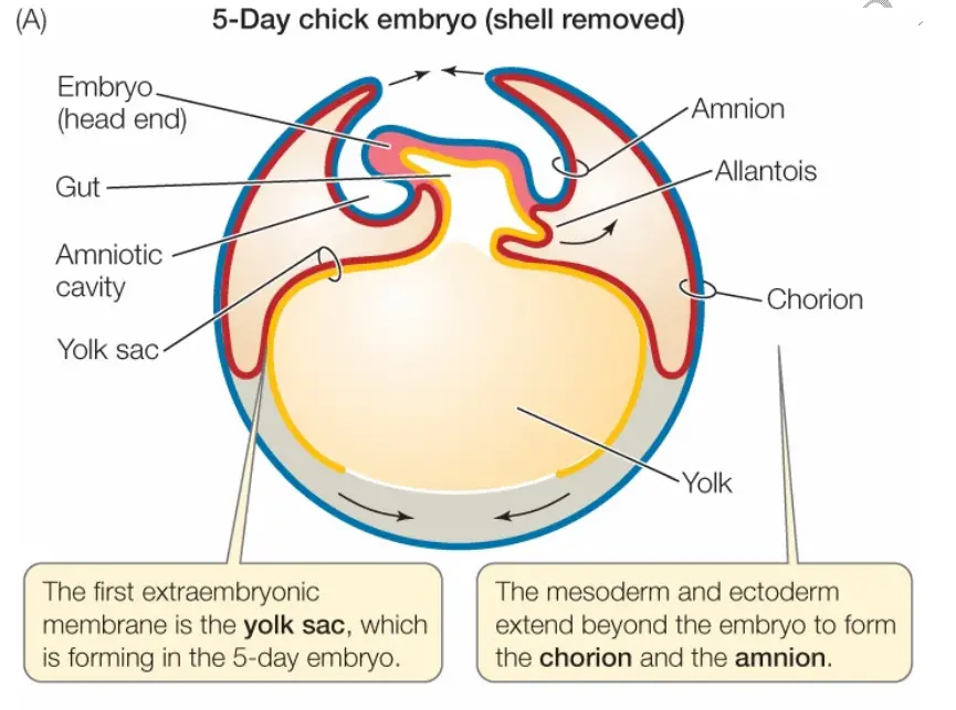
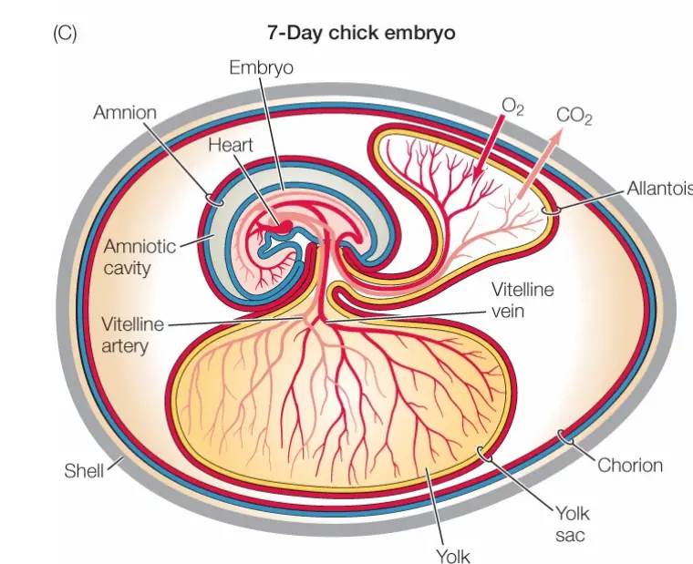
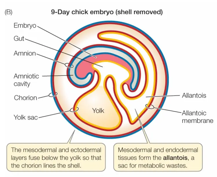
- In birds, reptiles, and mammals, there are four main extra-embryonic membranes: the yolk sac, the allantois, the amnion, and the chorion or serosa. These membranes play crucial roles in providing nutrition, gas exchange, waste removal, and protection for the developing embryo.
- The yolk sac surrounds and nourishes the yolk, supplying essential nutrients to the developing embryo. In some animals, such as birds, the yolk sac is eventually incorporated into the digestive system of the embryo.
- The allantois is involved in waste management and gas exchange. It stores embryonic waste and facilitates the exchange of gases, allowing for the resorption of carbon dioxide and the uptake of oxygen. In avian species, the allantois also participates in the absorption of calcium from the eggshell.
- The amnion acts as a protective cushion around the embryo, preventing mechanical shocks and providing a stable environment for its development. It contains a fluid-filled cavity known as the amniotic cavity, in which the embryo floats, protecting it from desiccation and concussions.
- The chorion or serosa surrounds the other extra-embryonic membranes and plays a role in gas exchange. In birds, the chorion fuses with the allantois during later stages of egg development, forming the chorioallantois, a combined respiratory and excretory organ.
- These extra-embryonic membranes have evolved to enable vertebrate embryos to develop on land. They provide the necessary support and adaptations for embryonic survival outside of an aquatic environment. While their specific structures and functions may vary across different species, the fundamental purpose of these membranes remains the same.
- It is important to note that in humans and other mammals, these extra-embryonic membranes are commonly referred to as fetal membranes. They play similar roles in supporting and protecting the developing fetus during gestation.
Types of Extra-Embryonic Membranes
During chick development, four types of extra-embryonic membranes are formed:
- Amnion or Amniotic Sac: The amnion surrounds the developing embryo, providing a protective environment similar to a private aquarium. It serves to safeguard the embryo from mechanical shocks, such as pressure, abrasion, and irritation, as well as desiccation (drying out).
- Serosa or Chorion: The serosa or chorion is the outermost membrane that encloses the remaining embryonic structures. It provides additional protection and support to the developing embryo.
- Yolk Sac: The yolk sac contains the yolk of the egg and performs multiple functions. It serves as the first respiratory organ for the embryo, allowing for gas exchange. The yolk sac also functions as a digestive organ, aiding in the absorption of nutrients from the yolk. Additionally, it acts as a site of hematopoiesis, similar to the liver, and produces blood cells. As development progresses, the yolk sac diminishes in size and is eventually incorporated into the chick’s digestive system.
- Allantois: The allantois initially forms as a precursor to the urinary bladder. It accumulates the embryonic nitrogenous waste, mainly in the form of non-toxic uric acid. Additionally, the allantois acts as an embryonic respiratory surface, facilitating gas exchange. In avian species, the allantois plays a significant role in calcium resorption from the eggshell.
Each of these extra-embryonic membranes is composed of two germ layers. The amnion and chorion consist of the extra-embryonic ectoderm and the somatic layer of mesoderm, collectively known as the somatopleure. On the other hand, the yolk sac and allantois are composed of the extra-embryonic endoderm and the splanchnic layer of mesoderm, collectively referred to as the splanchnopleure.
These extra-embryonic membranes provide essential support and functions necessary for the successful development of the chick embryo. They ensure protection, gas exchange, waste management, and nutritional supply, allowing the embryo to develop and grow until hatching.
1. Development of The Yolk Sac
The yolk sac plays a crucial role in the early development of the chick embryo. Here is an overview of the development of the yolk sac:

- Early Appearance: The yolk sac is the first extra-embryonic membrane to emerge in the chick embryo. Initially, the floor of the gut is formed by the yolk mass located below it.
- Formation of a Sac-like Investment: The splanchnopleure, the layer of mesoderm associated with the yolk sac, grows over the surface of the yolk, forming a sac-like structure that envelops and protects the yolk instead of forming a complete gut.
- Establishment of the Gut: Up to approximately 16 hours of incubation, the gut in the chick embryo appears as a round cavity beneath the primitive streak. As the extra-embryonic splanchnopleure spreads around the yolk, changes occur in the intra-embryonic splanchnopleure, resulting in the formation of a completely enclosed gut within the body of the embryo.
- Differentiation into Foregut, Midgut, and Hindgut: Around 24 hours of incubation, a small enteric pocket develops into the developing head, forming the foregut. At approximately 48 hours of incubation, a sac-like hindgut develops with the formation of the tail. The undifferentiated part of the gut located between the foregut and hindgut is referred to as the midgut.
- Reduction of Midgut and Connection with Yolk Sac: The foregut and hindgut expand in size while the midgut diminishes in extent. Eventually, the midgut is almost completely reduced, except for a point where it remains connected to the yolk sac through an inverted funnel-like duct called the yolk stalk. The opening of the midgut into the yolk sac is known as the umbilicus.
- Gradual Reduction of Yolk Sac: Over time, the yolk sac gradually reduces in size due to the spreading of the blastoderm, which encompasses the yolk. As the chick embryo develops, the yolk sac becomes incorporated with the tissues of the small intestine.
- Final Stage: Approximately two to three days before hatching, the yolk sac is greatly reduced and is represented as a protuberance from the small intestine. At this point, its function is almost complete, and the chick is prepared for hatching.
The development of the yolk sac is a crucial process in the early stages of chick embryo development, providing essential nutrients and serving as a site for early organogenesis and hematopoiesis. As development progresses, the yolk sac gradually diminishes in size as the embryo becomes more self-sufficient.
Functinon Of Yolk Sac
The yolk sac plays a crucial role in the early development of embryos, including in the chick. Here are some key functions of the yolk sac:
- Nutrient Source: The yolk sac serves as a source of nourishment for the developing embryo. The yolk, which contains various nutrients such as proteins, lipids, and carbohydrates, directly provides essential food for the early stages of embryonic development.
- Enzymatic Breakdown: The endoderm of the yolk sac secretes enzymes that break down the yolk into diffusible substances. These enzymes help to break down complex nutrients present in the yolk into simpler, more easily absorbable forms.
- Absorption and Transport: Once the yolk is broken down into diffusible substances, they are absorbed by the vitelline veins present in the yolk sac. These veins collect the nutrients and carry them to the developing embryo.
- Distribution to Embryo and Extra-Embryonic Structures: The nutrients collected by the vitelline veins are transported to the heart, which then pumps them to different parts of the embryo and extra-embryonic structures. This distribution ensures that the developing embryo and its associated structures receive the necessary nutrients for growth and development.
By utilizing the yolk as a source of nutrients, the embryo can sustain its early development until it becomes capable of obtaining nutrients from external sources, such as the alimentary canal, after hatching. The yolk sac plays a crucial role in facilitating this early nourishment, ensuring the embryo’s growth and development during the initial stages of embryogenesis.
2. Development of The Amnion and The Chorion
The development of the amnion and the chorion, two important extra-embryonic membranes, occurs in conjunction with each other. Here are the key points regarding their formation:

- Derived from the Extra-embryonic Somatopleure: Both the amnion and the chorion are derived from the extra-embryonic somatopleure, which is a combination of the ectoderm and the somatic mesoderm.
- The Amnion: The amnion forms a sac-like investment that encloses the embryo. It surrounds the embryo and creates the amniotic cavity, which remains filled with a saline solution called amniotic fluid. The amnion plays a crucial role in suspending the embryo within the amniotic fluid, allowing it to change shape and position.
- Development of the Amnion: The development of the amnion begins around 30 hours of incubation in the chick embryo. Initially, the head of the embryo starts sinking into the underlying yolk mass and is pushed forward. The somatopleure lying anterior to the head forms a fold called the head fold of the amnion, which bends backward and covers the head like a hood. Lateral amniotic folds also form and fuse above the embryo.
- The Chorion: The chorion represents the outer half of the amniotic folds. It consists of somatic mesoderm on the inner side and ectoderm on the outer side. The chorion develops as the somatopleure continues to grow and envelops the yolk sac. The formation of the chorion involves the doubling of the somatopleure itself.
- Sero-amniotic Connection: The union of the amniotic folds is marked by a scar called the sero-amniotic connection. The sero-amniotic cavity, located between the amnion and the chorion, constitutes the extra-embryonic coelom. It remains connected to the intra-embryonic coelom through the yolk stalk region until later stages of development.
- Completion of Formation: The amnion, composed of inner ectoderm and outer somatic mesoderm, represents the inner half of the amniotic fold. The chorion, composed of somatic mesoderm on the inner side and ectoderm on the outer side, represents the outer half of the amniotic fold. The formation of the chorion is completed at the end of the second week of development.
The development of the amnion and the chorion is crucial for protecting and supporting the embryo during its development. The amnion provides a protective fluid-filled environment, while the chorion forms a protective layer around the embryo and other extra-embryonic structures.
Functions Of Amnion And Chorion
The amnion and chorion, two important extra-embryonic membranes, serve several functions during embryonic development. Here are the key functions of the amnion and chorion:
- Protection from Desiccation: The amnion and amniotic fluid work together to protect the developing embryo from desiccation or drying out. The amnion surrounds the embryo in a sac-like structure, creating a sealed environment that prevents excessive loss of moisture. The amniotic fluid within the amnion keeps the embryo moist and helps maintain an optimal environment for development.
- Shock Absorption: The amnion and amniotic fluid provide a cushioning effect, protecting the developing embryo from mechanical shocks caused by physical forces. They act as a shock absorber, reducing the impact of external pressure or movement on the embryo. This function helps safeguard the delicate structures and tissues of the developing embryo.
- Pressure Equalization: The amnion and amniotic fluid also play a role in equalizing the pressure acting on the embryo. By distributing and balancing the pressure evenly, they help prevent localized pressure points that could potentially harm the developing embryo. This function ensures that the pressure exerted on the embryo is evenly distributed, minimizing the risk of deformities or damage.
- Freedom of Movement: The presence of the amnion allows for the free movement of the developing embryo within the amniotic fluid. The amnion provides a spacious and flexible environment that enables the embryo to move, change shape, and assume different positions as it develops. This freedom of movement is essential for the proper development of the musculoskeletal system and facilitates the unfolding of various developmental processes.
- Rhythmic Movement: The amnion exhibits rhythmic movement, which further contributes to the overall well-being of the developing embryo. These movements can be observed as gentle contractions and expansions of the amniotic sac. The rhythmic movement of the amnion helps prevent adhesion of the embryo to the surrounding tissues, ensuring that the embryo remains free and mobile within the amniotic fluid.
3. Development of The Allantois
The allantois is an important extra-embryonic structure that develops from the intra-embryonic region during chick development. Here is an overview of the key stages in the development of the allantois:

- Initiation: The development of the allantois begins around 72 hours of chick embryo development. It originates as a ventral diverticulum or outpouching from the hindgut, which is a region of the developing digestive system.
- Rapid Growth: Following its initiation, the allantois undergoes rapid growth. It extends and expands, eventually invading the extra-embryonic coelom, which is the space surrounding the developing embryo.
- Tissue Composition: The allantois is composed of two primary germ layers. The outer layer consists of splanchnic mesoderm, which is derived from the embryonic mesoderm. The inner layer is made up of endoderm, which is one of the primary germ layers of the embryo.
- Allantoic Stalk and Vesicle: The allantois has two distinct parts. The proximal part, known as the allantoic stalk, connects the allantois to the hindgut from which it originated. The distal part is called the allantoic vesicle, which lies in the space between the yolk sac, amnion, and serosa. The allantoic vesicle gradually expands as the allantois grows.
- Fusion with Chorion: As the allantois continues to grow, it replaces the extra-embryonic coelom and eventually fuses with the chorion. The chorion is the outermost extra-embryonic membrane that surrounds the rest of the embryonic system. The fusion of the allantois with the chorion establishes a connection between the developing embryo and the outside environment.
The allantois serves several important functions during embryonic development. It acts as a precursor to the umbilical cord in mammals, allowing for the exchange of gases and nutrients between the embryo and the maternal blood supply. In avian development, the allantois accumulates nitrogenous wastes, such as non-toxic uric acid, produced by the developing embryo. It also functions as an embryonic respiratory surface, facilitating gas exchange.
Functions Of Allantois
The allantois plays several important functions during embryonic development in birds. Here are the key functions of the allantois:
- Excretory Function: The allantois serves as a storage site for excretory wastes produced by the developing embryo. These wastes include uric acid and other nitrogenous compounds. The allantois accumulates these waste products, preventing their accumulation within the embryo itself, and helps to maintain the internal environment of the embryo free from toxic substances.
- Respiratory Function: The allantois also acts as a respiratory surface, facilitating the exchange of gases, particularly oxygen, between the embryo and the external environment. Blood vessels within the allantois allow for the absorption of oxygen from the surrounding environment and the elimination of carbon dioxide, thereby ensuring a sufficient oxygen supply for the developing embryo.
- Absorption of Albumen: The allantois plays a role in the absorption of a significant quantity of albumen, which is the nutrient-rich substance surrounding the developing embryo within the egg. The blood vessels in the allantois aid in the absorption of nutrients from the albumen, providing nourishment to the growing embryo.
- Calcium Absorption: The circulation of the allantois, in contact with the eggshell via the chorion (the outermost extra-embryonic membrane), enables the absorption of calcium from the eggshell. This absorbed calcium is utilized by the developing embryo for the formation of bones. As a result, the eggshell gradually loses calcium, making it weaker. This weakening of the eggshell facilitates the hatching process, allowing the embryo to break through the shell when it is fully developed.
FAQ
What are extra-embryonic membranes in a chick?
Extra-embryonic membranes in a chick are specialized structures that develop outside the embryo during embryonic development. They include the amnion, chorion, yolk sac, and allantois.
What is the function of the amnion in a chick?
The amnion surrounds the embryo and is filled with amniotic fluid. Its main function is to protect the developing embryo from desiccation, mechanical shocks, and equalize pressure.
What is the role of the chorion in a chick embryo?
The chorion is the outermost membrane that surrounds the rest of the embryonic system. It plays a role in gas exchange, nutrient absorption, and the fusion with the shell membrane.
What is the function of the yolk sac in a chick embryo?
The yolk sac functions as the first respiratory organ and a digestive organ for the embryo. It also serves as a site for blood cell formation and plays a role in the absorption of nutrients from the yolk.
What is the allantois in a chick embryo?
The allantois is a membrane that develops from the hindgut of the chick embryo. It functions in excretion, respiration, absorption of albumen, and the absorption of calcium from the eggshell.
How do the extra-embryonic membranes develop in a chick embryo?
The extra-embryonic membranes develop concurrently during chick embryonic development from different germ layers, forming specialized structures to support the growing embryo.
What is the purpose of the amniotic fluid in a chick embryo?
The amniotic fluid in the amnion provides a protective environment for the developing chick embryo. It helps cushion the embryo from mechanical shocks, allows freedom of movement, and aids in the exchange of respiratory gases.
How do the extra-embryonic membranes in a chick facilitate nutrient absorption?
The extra-embryonic membranes, such as the yolk sac and allantois, play a role in absorbing nutrients from the yolk and albumen. Blood vessels within these membranes allow for the transport of nutrients to the developing embryo.
What is the relationship between the extra-embryonic membranes and the chick’s eggshell?
The chorion, one of the extra-embryonic membranes, is in contact with the eggshell. This allows for gas exchange between the embryo and the external environment. The allantois also absorbs calcium from the eggshell for bone development.
When do the extra-embryonic membranes in a chick start to develop?
The extra-embryonic membranes begin to develop early in chick embryonic development. The amnion starts forming around 30 hours of incubation, while the yolk sac and allantois develop around 72 hours of incubation. The chorion forms as the amniotic folds fuse.