Whta is Dirt and Soil?
- The terms “dirt” and “soil” are often used interchangeably, but they have distinct differences that make one preferable for agricultural and scientific purposes. Dirt is a fine-grained, unconsolidated mixture found on the ground, primarily composed of sand, silt, and clay, and may contain rocky fragments. However, it lacks the essential organic and living material found in soil. It is commonly accumulated under shoes or fingernails and lacks the cohesive properties of soil.
- To differentiate between dirt and soil, one can simply scoop a small amount of each with their hand. Soil will form loose balls or clumps even without adding water, whereas dirt does not compact even when water is added. Additionally, dirt should not be confused with sediments, which are granular materials eroded by natural forces like wind and water.
- Although dirt and soil are not the same, they share connections in various fields. Earth science and archaeology utilize the study of soil structures and geographical formations to gain insights into the Earth’s history. Agriculture benefits from analyzing soil samples to identify different soil types and determine the best composition for farming purposes. Construction and mining industries also rely on soil studies to ensure stability and appropriate use of materials.
- Microscopy has played a significant role in understanding soil characteristics and composition. The pioneering work of Antonie van Leeuwenhoek in the 1670s involved studying soil particles and chalk under the microscope, describing them as composed of globules. In the 1930s, major investigations of soil using microscopic techniques were conducted by researchers Harrison and Walter Kubiena. Their work laid the foundation for determining the micromorphology of soil in great detail.
- Micromorphology, which involves the use of microscopic techniques to study soil components, continues to be a valuable tool in soil science. Advanced microscopic techniques allow scientists to characterize different types of soil and classify them based on their respective components. This level of analysis provides valuable insights into soil properties, nutrient content, and overall fertility, essential for agricultural practices and land management.
- In conclusion, while dirt and soil may seem similar, their differences are critical in various fields, especially in agriculture and scientific study. The study of soil through microscopy and other techniques has provided a wealth of knowledge about Earth’s history and the composition of soil, contributing to sustainable agricultural practices and informed decision-making in various industries. Understanding the importance of soil in different environments continues to be a key focus for researchers and professionals worldwide.
Direct Observation of the Samples

Requirements
- Stereo Microscope: This type of microscope provides a three-dimensional view of the sample and is suitable for macroscopic observation.
- Compound Microscope: For more detailed observations at the cellular or molecular level, a compound microscope is used, which offers higher magnification.
- Sample Collection: Gather samples of garden soil and dirt for comparison. Both samples should be collected using clean tools, such as a spatula, to prevent contamination.
- Petri Dishes: Several Petri dishes are needed to hold the samples during observation. These dishes help keep the samples contained and prevent them from spilling.
- Microscope Glass Slides: Clean glass slides are essential for mounting the samples and placing them under the microscope. Ensure that the slides are free of dust and debris.
- Water: Having water available is crucial, especially for observing living organisms. Adding a drop of water to the sample can provide a suitable medium for observing cells and other structures.
- Pair of Gloves: To maintain aseptic conditions and avoid contamination, wear gloves when handling the samples and during the preparation process.
- Spatula: Use a spatula to transfer the samples onto the microscope slides carefully. Avoid touching the samples directly to prevent contamination.
- Pipette/Dropper: A pipette or dropper is necessary to add water or specific reagents to the samples if required for further analysis or observation.
Procedure
Step 1: Wear Protective Gloves Before handling soil and dirt samples, put on a pair of gloves to maintain aseptic conditions and prevent contamination.
Step 2: Preparation of Samples Using a spatula, scoop a small amount of dirt and place it onto a clean Petri dish. Repeat this step with a small amount of soil and place it into another clean Petri dish. Ensure that the samples are not mixed to maintain their individual characteristics.
Step 3: Optional Water Addition If desired, use a dropper or pipette to add a few drops of water onto the samples in each Petri dish. This step is optional but can create a suitable medium for observing any living organisms that may be present in the soil. Allow the water to distribute evenly through the soil and dirt samples.
Note: If water is not added, take care not to observe the samples as clumps. Instead, try to spread them out slightly for better inspection.
Step 4: Preparing Microscope Slides Take clean microscope glass slides and carefully transfer a small portion of each sample onto separate slides. If water was added to the samples, ensure the water is also transferred along with the soil and dirt.
Step 5: Mounting the Slides Place the prepared microscope slides under the compound microscope. Adjust the magnification and focus knobs to obtain a clear and detailed view of the samples.
Step 6: Observation With the microscope properly set up, begin the observation process. Take note of any structures, organisms, or other characteristics present in both the soil and dirt samples.
Step 7: Recording Observations Record your observations in a notebook or on a computer, detailing any findings or notable features observed in the samples.
Step 8: Clean-up After completing the observations, dispose of the samples appropriately. Clean the microscope slides and Petri dishes thoroughly to prevent cross-contamination.
Direct observation of soil and dirt samples under a microscope provides valuable insights into the microcosmic world, shedding light on the diverse components and interactions within these materials. By following this procedure, researchers and students can conduct accurate and informative observations, contributing to a deeper understanding of the fascinating world of soil and dirt.
Microscopy of Dirt and Soil
Microscopy of dirt and soil offers a fascinating opportunity to explore the hidden microcosmic world that exists beneath our feet. By using both stereo and compound microscopes, researchers can gain valuable insights into the composition and structure of these essential natural resources. Here is a step-by-step guide for conducting microscopy of dirt and soil:
Using a Stereo Microscope:
- Set Up the Microscope: Begin by turning the turret to set the low-power objective in place. This will provide a broader view of the sample.
- Place the Petri Dish: Position the Petri dish containing the dirt sample on the microscope stage. Try to center the sample under the low-power objective for the best viewing.
- Focus the Image: Look through the eyepieces and gently turn the coarse and fine adjustment knobs to bring the image into focus. Take your time to ensure a clear and sharp view of the sample.
- Observe and Compare: Make observations of the dirt sample under low magnification. Take note of any particles, textures, or organisms present. Afterward, switch to high magnification to examine the sample in greater detail. Repeat the process for the soil sample in the second Petri dish and compare your observations.
Using a Compound Microscope:
- Prepare Glass Slides: Instead of using Petri dishes, prepare clean microscope glass slides for mounting the samples. Place a small amount of each sample (dirt and soil) on separate slides.
- Lower the Microscope Stage: Before mounting the slide, lower the microscope stage to provide enough space for the slide to fit without damage.
- Set Low-Power Objective: Turn the turret to set the low-power objective in place. This will provide an initial overview of the samples.
- Center the Slide: Position the slide on the microscope stage and try to center it under the objective for optimal viewing.
- Focus the Image: Look through the eyepieces and gently turn the coarse and fine adjustment knobs to bring the image into focus. Take your time to achieve a clear and detailed view.
- Observe and Record: Make observations of the dirt and soil samples under low magnification. Note any structures, microorganisms, or other intriguing features. Afterward, lower the stage and switch to the high-power objective for closer inspection. Record your findings for further analysis.
Preparation of Samples:
For a more detailed examination, consider mixing the soil and dirt samples with water in separate vials before placing a small amount of each on the slides or Petri dishes for observation. The addition of water can create a suitable medium for observing living organisms and enhance the observation process.
Microscopy of dirt and soil is a captivating endeavor that allows researchers and students to appreciate the rich complexity of the microcosmic world. By following these steps and taking the time to observe and record their findings, scientists can gain a deeper understanding of the essential roles played by soil and dirt in the ecosystem.
Preparing a Thin Section
Requirements
Preparing a thin section is a valuable technique used in various scientific fields to study the intricate details of materials at a microscopic level. This process involves transforming a solid sample, such as a dirt sample, into a thin slice that can be mounted on a microscope slide for observation. To successfully create a thin section, specific requirements and equipment are essential. Here are the key requirements for preparing a thin section:
- Dirt Sample: Begin with a representative dirt sample that you wish to examine in detail. Ensure the sample is free of debris and contaminants to obtain accurate observations.
- Polyester Resin or Epoxy Resin: Polyester resin or epoxy resin acts as a binding material to encase the dirt sample and create a stable block for further processing. Choose a resin suitable for your specific sample and the subsequent analysis.
- Vacuum Pump: A vacuum pump is necessary to remove air bubbles from the resin as it encases the dirt sample. This ensures a uniform and bubble-free block, which is crucial for obtaining clear and precise thin sections.
- Clean Glass Slide and Coverslip: Once the thin section is ready, it needs to be mounted on a clean glass slide. A coverslip is placed over the thin section to protect it and facilitate easy observation under a microscope.
- Diamond Saw: A diamond saw is used to cut a small and precise slice from the resin-encased dirt sample. This slice will serve as the thin section that will be mounted on the glass slide.
- Grinding Wheel: After cutting the initial slice, a grinding wheel is used to further reduce the thickness of the section. This process gradually grinds away material from the slice, creating an ultra-thin section suitable for microscopy.
Procedure
- Preparation of Sample: Air dry or freeze dry the dirt sample to remove any water content. Resins used for fixing the samples are hydrophobic, so it’s crucial to eliminate water from the sample before proceeding.
- Fixation of Sample: Place a small amount of the dry dirt sample into a small glass or plastic container. Gently add the chosen resin to the container. If using unsaturated polyester resin, mix it with acetone to reduce viscosity. Conduct this step under vacuum using a vacuum pump to ensure thorough penetration of the resin into the sample.
- Curing of Resin: Allow the resin to harden by placing the container in an oven at approximately 40°C. Curing will ensure the resin solidifies, creating a stable block with the embedded dirt sample.
- Cutting and Polishing: Once the resin has dried and hardened, carefully cut the hardened block using a diamond saw to obtain a flat surface. Polish the cut surface to achieve a very smooth and even surface.
- Mounting the Thin Section: Use an isotropic cementing agent, such as colorless polyester resin, to glue the polished surface of the sample onto a clean glass slide. Allow the sample to properly attach to the slide.
- Obtaining the Thin Section: Cut the mounted sample again using the diamond saw to obtain a very thin slice. Take care to achieve the thinnest possible section.
- Grinding the Thin Section: Using a grinding wheel with fine abrasive, carefully grind the thin slice to further trim it and make it even thinner. Aim to trim the slice down to about 30 microns.
- Final Steps: Clean the polished surface of the thin section and cover it with a clean coverslip. Place a drop of the chosen resin on the thin slice and gently place the coverslip on top to protect the sample and facilitate easy microscopic observation.
- Repeat for Comparison (Optional): If time permits, you can repeat the entire process with a sample of garden soil for comparison and further insights.
Petrographic Microscope Procedure
A petrographic microscope is an invaluable tool for geologists, mineralogists, and researchers to examine and analyze the intricate structures and properties of rock and mineral samples at a microscopic level. To make the most of this powerful instrument, a systematic procedure is followed to ensure accurate and detailed observations. Here is a step-by-step guide for conducting a petrographic microscope procedure:
- Set Up the Microscope: With the microscope stage lowered, begin by turning the rotating turret to set the low-power objective in place. This provides a broader view of the sample.
- Prepare the Slide: Place the prepared microscope slide containing the rock or mineral sample on the microscope stage. Try to center the slide so that the sample is directly below the objective, ensuring optimal viewing.
- Focus the Image: While looking through the microscope eyepiece, gently turn the coarse and fine adjustment knobs to bring the image into focus. Take your time to achieve a clear and sharp view of the specimen.
- Explore Different Areas: As you observe the sample under low power, you can gradually rotate the stage to explore different parts of the specimen. This allows you to examine various features and structures within the sample.
- Record Your Observations: During your observation, record any notable characteristics, such as mineral composition, texture, grain size, and any visible geological features. Detailed observations are crucial for accurate analysis and interpretation.
- Switch to High-Power Objective: After recording your observations under low power, switch to the high-power objective to examine the sample in greater detail. This higher magnification allows for closer inspection of fine structures and mineral associations.
- Record High-Power Observations: As you examine the sample under high power, continue to record your observations, paying attention to any minute details that may not have been visible at lower magnifications.
- Analyze and Interpret: Once you have completed your observations under both low and high power, analyze the data and interpret the findings. Compare your observations with known mineralogical and geological characteristics to identify the rock or mineral type and gain insights into its origin and history.
- Note Special Features: Be sure to note any special features, such as mineral inclusions, cleavage planes, or crystal twinning, as these may provide valuable information about the sample’s formation and geological history.
Petrographic microscopy is a meticulous process that requires careful attention to detail and keen observation skills. By following this step-by-step procedure, scientists can unlock the hidden secrets of rock and mineral samples, providing essential information for geological research, mineral exploration, and understanding Earth’s complex geological history.
Observations
The observations made while examining a dirt sample under different microscopes reveal a fascinating and intricate world of components and living organisms. Each magnification level provides unique insights into the composition and characteristics of the sample. Here are the observations at various magnifications:
1. Stereo Microscope (40X Magnification):
- Large rock fragments and sand grains are easily identifiable in the dirt sample.
- Grains/particles of silt and clay appear as a matrix, embedded among the larger sand grains and rock fragments.
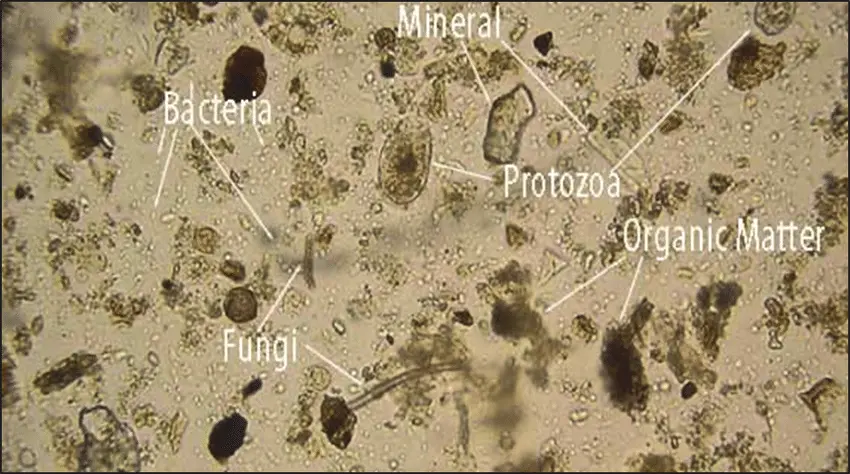
- Some movements in the sample are noticeable, indicating the presence of tiny organisms, possibly nematodes.
2. Compound Microscope (100X Magnification):
- Nematodes can be clearly identified, exhibiting their characteristic wriggling movements.
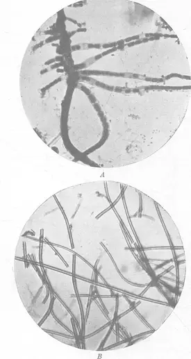
- Elongated fungal hyphae are visible, forming intricate networks within the field of view.
- Clusters of bacteria can be spotted, contributing to the microbial diversity in the dirt sample.
3. Petrographic Microscope (Higher Magnification):
- Under the petrographic microscope, the components of the dirt sample become more distinguishable.
- Sand grains exhibit a white coloration, allowing easy differentiation from other components.
- The clay matrix appears brownish in color, providing a contrasting background for the sand grains.
- Rock fragments can be identified based on their larger size and various colors, ranging from brown to dark brown, adding further diversity to the sample.
Overall, the observations under different microscopes offer a comprehensive understanding of the dirt sample’s complex composition. The stereo microscope provides an initial glimpse of the larger components, while the compound microscope reveals the presence of living organisms such as nematodes and bacteria. Finally, the petrographic microscope offers detailed insights into the distinct characteristics of sand grains, clay matrix, and rock fragments.
It is essential to note that the observations in the dirt sample are intriguing but relatively limited in terms of living organisms when compared to the garden soil. The garden soil, known for its rich microbial life, exhibits a broader range of tiny living organisms that play vital roles in nutrient cycling and soil health. The observations made through the various microscopes shed light on the hidden world of dirt, revealing the complexity and diversity that exists even in seemingly ordinary soil samples.

FAQ
What can I observe in dirt and soil under a microscope?
Under a microscope, you can observe various components of dirt and soil, such as sand grains, silt, clay, organic matter, living organisms like nematodes, bacteria, and fungal hyphae.
How do sand grains appear under a microscope?
Sand grains appear as distinct, granular structures with a defined shape and color, which can vary depending on the mineral composition.
What are the differences between silt and clay particles in a soil sample?
Under a microscope, silt particles appear smaller than sand grains but larger than clay particles. Clay particles are tiny and have a smooth texture.
Can I identify living organisms in dirt and soil samples using a microscope?
Yes, a microscope allows you to identify living organisms like nematodes, bacteria, and fungal hyphae, which are essential components of soil ecosystems.
What is the significance of observing nematodes in soil samples?
Observing nematodes indicates a healthy soil ecosystem, as these microscopic worms play crucial roles in nutrient recycling and soil fertility.
How do bacterial clusters appear under a microscope?
Bacterial clusters appear as dense aggregations of tiny cells, often forming intricate patterns or colonies.
Can I distinguish rock fragments from other components in soil using a microscope?
Yes, under higher magnification, rock fragments may exhibit distinct sizes and colors, allowing differentiation from other soil components.
What does the clay matrix look like under a microscope?
The clay matrix appears as a fine, brownish material that surrounds and embeds other soil particles, providing a cohesive structure.
What are the applications of observing dirt and soil under a microscope?
Observing dirt and soil microscopically is valuable for understanding soil composition, assessing soil health, and studying soil-related environmental processes.
How does the petrographic microscope enhance observations of soil samples?
The petrographic microscope offers higher magnification and resolution, enabling more detailed observations of soil components, mineralogy, and complex soil structures.
- Text Highlighting: Select any text in the post content to highlight it
- Text Annotation: Select text and add comments with annotations
- Comment Management: Edit or delete your own comments
- Highlight Management: Remove your own highlights
How to use: Simply select any text in the post content above, and you'll see annotation options. Login here or create an account to get started.