Insects exhibit a remarkable diversity in their feeding habits, leading to significant adaptations in their digestive systems. These adaptations can be classified based on their dietary preferences, which include phytophagous (plant-eating), entomophagous (insect-eating), wood-boring, wool-feeding, and saprophytic behaviors. Each of these feeding strategies necessitates specific structural modifications within the digestive system to efficiently process the ingested food.
Insects that primarily consume solid food, such as plant parts or host tissues, possess specialized mouthparts designed for biting and chewing. These mouthparts enable them to mechanically break down tough plant fibers and other solid materials. Additionally, these insects often have a well-developed gizzard, a muscular structure that further aids in the grinding of food particles, facilitating the enzymatic digestion that follows.
Conversely, insects that feed on sap or other liquid nutrients utilize sucking mouthparts. These mouthparts are adapted to extract fluids efficiently, minimizing energy expenditure while maximizing nutrient intake. Within the alimentary canal of these sap-feeding insects, a filter chamber is often present. This structure plays a critical role in separating the ingested sap from solid debris, allowing for more efficient nutrient absorption.
The digestive system of insects is thus intricately linked to their feeding habits. The anatomical variations, such as the presence of a gizzard in solid feeders and a filter chamber in sap feeders, highlight the evolutionary adaptations that enhance their ability to extract nutrients from various food sources. Therefore, understanding these structural modifications provides insight into the ecological roles that different insect species play within their respective environments.
Alimentary Canal
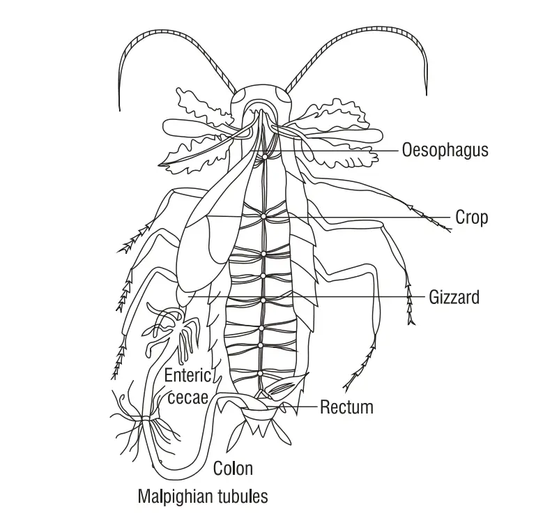
The alimentary canal in insects is a complex structure that varies significantly in length and morphology across different species. This tubular organ extends from the mouth to the anus and plays a vital role in digestion and nutrient absorption. Its length can range from 6 to 7 centimeters and may even extend to match the overall length of the insect body in certain species. The alimentary canal is systematically divided into three main regions: the foregut, midgut, and hindgut.
- Foregut (Stomodaeum):
- The foregut is the anterior section of the alimentary canal, originating from ectodermal tissue.
- It is primarily responsible for the ingestion of food and initial processing.
- This region often includes specialized structures, such as the mouthparts for mechanical breakdown and the crop for temporary food storage.
- Midgut (Mesenteron or Ventriculus):
- The midgut represents the central portion of the alimentary canal and is derived from endodermal tissue.
- It functions primarily in the enzymatic digestion and absorption of nutrients.
- The midgut contains numerous gastric ceca, which are finger-like projections that increase the surface area for nutrient absorption and contribute digestive enzymes.
- Hindgut (Proctodaeum):
- The hindgut is the posterior section of the alimentary canal, also ectodermal in origin.
- Its main functions include the absorption of water and the formation of feces.
- This section includes the rectum, which stores waste until excretion.
The overall structure and length of the alimentary canal can vary significantly among different feeding groups of insects. For instance, phytophagous (plant-eating) insects typically have a longer alimentary canal compared to carnivorous or sap-sucking insects. This adaptation allows them to process the fibrous and complex carbohydrates found in plant material more efficiently.

(a) Fore Intestine (Stomodaeum)
The fore intestine, or stomodaeum, is a crucial component of the insect alimentary canal, serving as the primary site for food intake and initial processing. It connects the mouth to the midgut and is designed to accommodate various feeding strategies. The structure of the fore intestine consists of several specialized regions, each fulfilling specific roles in the digestive process.
- Intima:
- The innermost layer of the fore intestine is a cuticular lining known as the intima.
- This layer provides a protective barrier and assists in the movement of food through the alimentary canal.
- Epithelial Layer:
- Continuous with the hypodermis, the epithelial layer is composed of cells that facilitate absorption and secretion.
- These cells play a vital role in the overall digestive function.
- Basement Membrane:
- The basement membrane bounds the outer surface of the epithelium, providing structural support and anchoring the epithelial layer.
- Peritoneal Membrane:
- This membrane supports the entire structure of the fore intestine and is involved in the overall functioning of the alimentary canal.
- Preoral Food Cavity:
- This cavity lies between the mouthparts and the labrum.
- Although not a true part of the intestine, it plays a critical role in the manipulation of food prior to ingestion.
- In insects with manipulative mouthparts, the hypopharynx divides this space into an anterior (dorsal) cibarium and a posterior (ventral) diversion.
- Pharynx:
- Located between the mouth and esophagus, the pharynx contains muscles that facilitate the movement of food from the mouth into the esophagus.
- This region plays a significant role in swallowing.
- Esophagus:
- The esophagus is a simple, straight tube that extends from the back of the head into the thorax.
- Its walls are longitudinally folded, which aids in the passage of food.
- The length of the esophagus can vary significantly among different insect species.
- Crop:
- The crop is a dilation of the esophagus and serves as a food reservoir.
- Its structure is highly variable; in some insects, such as grasshoppers and cockroaches, it can be quite capacious, forming a substantial part of the fore intestine.
- The walls of the crop are thin, and its muscular coat is weakly developed, allowing for the temporary storage of food.
- Gizzard (Preventriculus):
- Situated behind the crop, the gizzard is well developed in certain insects like grasshoppers.
- Its most distinctive feature is the cuticular lining, which is heavily developed into prominent denticles.
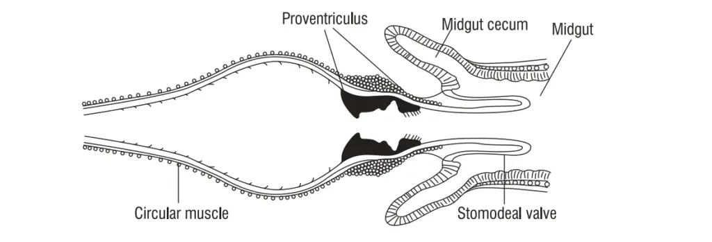
- At the junction of the fore and mid intestines, many insects possess a cardiac or esophageal valve, accompanied by six gastric ceca that assist in digestion.
- Muscle Layers:
- The foregut is also composed of muscle layers, including longitudinal and circular muscles.
- These muscle layers facilitate the movement of food through the foregut, ensuring efficient transport to the midgut.

(b) Mid Intestine (mesenteron or stomach or ventriculus)
The mid intestine, also referred to as the mesenteron, stomach, or ventriculus, is a crucial segment of the insect digestive system. It serves as the primary site for enzymatic digestion and nutrient absorption. The morphology of the mid intestine exhibits considerable variability among insect species, with shapes ranging from sac-like to tubular, and it can even be divided into multiple regions depending on the specific insect’s dietary needs and evolutionary adaptations.
- Structure of the Mid Intestine:
- Internally, the mid intestine is lined with enteric epithelium, with the outer ends of these epithelial cells resting on a basement membrane.
- Beneath the epithelium, there exists an inner layer of circular muscles and an outer layer of longitudinal muscles, which facilitate the movement of food through peristalsis.
- Peritoneal Membrane:
- The outermost layer of the mid intestine consists of a thin peritoneal membrane, providing additional structural support.
- Gastric Ceca:
- In many insects, the surface area of the mid intestine is enhanced by the presence of sac-like diverticula known as enteric or gastric ceca.
- These structures are typically located at the esophageal end of the stomach and their number can vary widely among different insect species. They play a critical role in increasing the surface area available for nutrient absorption.
- Cardia:
- The anterior portion of the mid intestine is known as the cardia, which surrounds the stomodaeal valve.
- From the cardia, eight small, tubular, finger-like blind processes extend freely into the hemocoel, contributing to the digestive process.
- Lining and Membrane:
- The internal lining of the mid intestine consists of endodermal epithelium made up of columnar cells.
- This epithelium features small, villi-like folds that increase the surface area for absorption.
- Unlike the foregut, the mid intestine is covered by a thin, transparent peritrophic membrane composed of chitin and proteins, which serves to protect the epithelium from abrasive food particles and facilitate digestion.
- Malpighian Tubules:
- At the hind end of the mid intestine, long, slender, yellowish structures known as Malpighian tubules project into the hemocoel.
- While these tubules are associated with the digestive system, their primary function is excretion, helping to remove waste products from the insect’s body.
- Functional Role:
- The mid intestine plays a vital role in the overall digestive process, where enzymatic breakdown of food occurs, and nutrients are absorbed into the insect’s circulatory system.
- The combination of muscular layers and specialized structures, such as gastric ceca and the peritrophic membrane, underscores the efficiency of this digestive region in processing various food types.
(c) Hind Intestine (proctodaeum)
The hind intestine, also known as the proctodaeum, is a significant section of the insect alimentary canal, primarily responsible for the final stages of digestion and the excretion of waste. It exhibits several structural features that facilitate these functions, and it is composed of three main regions: the ileum, colon, and rectum. The hind intestine is essential for the absorption of residual nutrients and the formation of feces.
- Structure and Layers:
- The hind intestine comprises similar layers to those found in the fore intestine. It includes an inner layer of circular muscles that may vary in development and an outer layer of longitudinal muscles.
- The hind intestine typically begins at the pyloric valve, which serves as a critical junction between the midgut and hindgut. Additionally, this region marks the insertion point of the Malpighian tubules, which play a key role in excretion.
- Regions of the Hind Intestine:
- Ileum: The small intestine or ileum often features a cuticular lining that is folded and equipped with hair-like projections. These adaptations increase the surface area for absorption, facilitating the uptake of nutrients that may have escaped earlier stages of digestion.
- Colon: Following the ileum, the large intestine or colon continues the absorption process. Similar to the ileum, it is also characterized by folds and projections that enhance nutrient uptake.
- Rectum: The rectum functions as a storage chamber for waste material before excretion. It is generally globular or pyriform in shape and is lined with inwardly projecting papillae, which aid in the reabsorption of water and electrolytes from the waste, contributing to more efficient waste management.
- Salivary Glands:
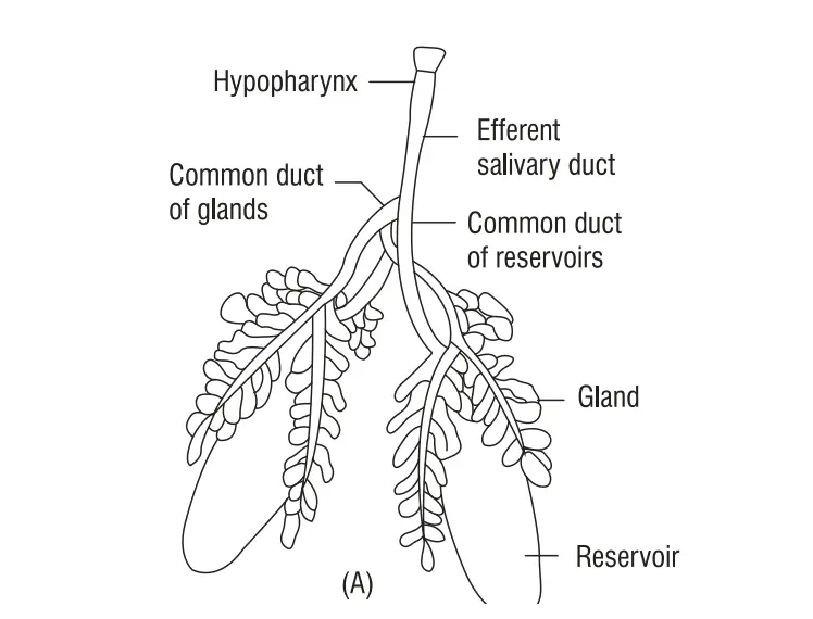
- In proximity to the hind intestine, a pair of prominent, whitish salivary glands is located on each side of the thorax, wrapping around the esophagus and the anterior section of the crop. These glands have multiple components that contribute to the digestive process.
- Each gland consists of a flattened glandular portion divided into several lobules or acini, organized in grape-like clusters. These lobules produce saliva, which is essential for enzymatic digestion.
- The fine ductules from various lobules converge to form a single glandular duct, leading to a thin-walled, semi-transparent sac-like reservoir that stores the saliva. A separate reservoir duct connects the reservoir to the glandular duct.
- The glandular ducts from both salivary glands merge to form a common glandular duct. Similarly, the reservoir ducts unite to create a common reservoir duct, ultimately leading to a common efferent salivary duct.
- This duct traverses through the neck and head, opening at the hypopharynx in the pre-oral space, allowing saliva to mix with food as it enters the digestive tract.
- Functionality and Importance:
- The hind intestine is vital for completing the digestive process, where the absorption of nutrients and water occurs, and fecal matter is formed. The muscular layers of the hind intestine aid in peristalsis, facilitating the movement of contents through the digestive system.
- The salivary glands support the digestive process by secreting enzymes that initiate the breakdown of food as it enters the alimentary canal. This secretion is crucial for ensuring efficient digestion and nutrient assimilation.

Functions of insects Digestive System
Below are the primary functions of the insect digestive system:
- Ingestion: The process begins with the intake of food through specialized mouthparts, which vary depending on the insect’s feeding habits. For example, chewing mouthparts are used by phytophagous insects, while sucking mouthparts are characteristic of sap-feeding insects.
- Mechanical Digestion: Once ingested, food is mechanically broken down in the foregut, particularly in the gizzard (or ventriculus), where it is subjected to physical grinding. This mechanical action is crucial for reducing food particle size, facilitating subsequent chemical digestion.
- Chemical Digestion: The midgut (mesenteron) is the primary site for chemical digestion. It is lined with enteric epithelium that secretes digestive enzymes, breaking down complex food substances into simpler molecules. Enzymatic action is essential for digesting carbohydrates, proteins, and fats, allowing nutrients to be readily absorbed.
- Nutrient Absorption: The midgut is also where most nutrient absorption occurs. The presence of villi-like structures increases the surface area for absorption, enabling the efficient uptake of amino acids, sugars, fatty acids, vitamins, and minerals into the hemolymph (the insect equivalent of blood).
- Water Reabsorption: The hindgut (proctodaeum) plays a key role in reabsorbing water and electrolytes from waste material. This process is vital for maintaining osmotic balance and conserving water, especially in insects inhabiting arid environments.
- Waste Formation and Excretion: The hindgut is responsible for forming and storing fecal matter. Waste products, along with undigested food particles, are compacted into feces and ultimately expelled from the body through the anus. The excretion of waste ensures the removal of harmful byproducts of metabolism.
- Symbiotic Relationships: In some insects, the digestive system houses symbiotic microorganisms that assist in breaking down complex food substances, particularly cellulose in herbivorous insects. These microbes can help in nutrient acquisition that the insect cannot obtain independently.
- Storage: Some insects have specialized structures, such as the crop, which serves as a temporary storage site for food before it moves to the midgut for digestion. This allows insects to consume larger quantities of food when it is available and digest it gradually.
- http://jnkvv.org/PDF/04042020094238Digestive%20system%20AK.pdf
- http://eagri.org/eagri50/ENTO231/lec08.pdf
- https://www.ndsu.edu/pubweb/~rider/Pentatomoidea/Teaching%20Structure/Lecture%20Notes/Week%2011a%20Digestive%20System.pdf
- https://faculty.ksu.edu.sa/sites/default/files/digestion_in_insects.pdf
- Text Highlighting: Select any text in the post content to highlight it
- Text Annotation: Select text and add comments with annotations
- Comment Management: Edit or delete your own comments
- Highlight Management: Remove your own highlights
How to use: Simply select any text in the post content above, and you'll see annotation options. Login here or create an account to get started.