- The digestive system in vertebrates is a complex network of organs responsible for breaking down food, absorbing nutrients, and eliminating waste. It consists of the alimentary canal, which serves as the primary pathway for food to travel, and the associated glands, which aid in digestion by secreting necessary enzymes and fluids.
- Although the basic structure of the digestive system remains consistent across all vertebrates, there are significant variations in specific components depending on the species. These variations are typically adaptations to the dietary needs of the animal, whether herbivorous, carnivorous, or omnivorous.
- The alimentary canal, which is formed during early embryonic development, originates from the endoderm, except for the mouth and rectum, which are lined with ectoderm. This canal begins at the mouth, where food enters, and ends at the anus, where undigested food is expelled. Major organs within the canal include the esophagus, stomach, small intestine, and large intestine, each performing specific roles in the digestive process.
- In addition to the alimentary canal, several glands—such as the liver, pancreas, and salivary glands—play crucial roles in producing digestive enzymes and other substances that facilitate the breakdown of food into absorbable molecules. These enzymes are critical for the chemical digestion of carbohydrates, proteins, and fats.
- The overall function of the digestive system in vertebrates is to ensure that essential nutrients are extracted from food and absorbed into the bloodstream, while non-digestible materials are efficiently expelled from the body. Thus, despite variations in structure and function across species, the vertebrate digestive system follows a fundamental blueprint that serves the vital purpose of maintaining life.

The digestive tract of vertebrates
The digestive tract of vertebrates consists of several distinct parts, each playing a crucial role in the overall process of digestion. From the mouth to the glands associated with digestion, this system is designed to break down food, absorb nutrients, and expel waste. Below is a detailed breakdown of the various components of the vertebrate digestive tract.
- Mouth and Oral Cavity
- Teeth: The teeth are specialized structures used for breaking down food mechanically. Depending on the dietary habits of the vertebrate, teeth can be adapted for cutting, grinding, or tearing food. Herbivores tend to have flat teeth for grinding plants, while carnivores have sharp teeth for tearing meat.
- Tongue: The tongue is a muscular organ that aids in the manipulation of food, facilitating chewing and swallowing. In many vertebrates, the tongue also plays a role in taste perception and in mixing food with saliva.
- Oral Glands: The oral glands, including the salivary glands, secrete saliva that contains enzymes, such as amylase, which initiate the chemical breakdown of carbohydrates in the mouth. Saliva also helps moisten food for easier swallowing.
- Pharynx
- The pharynx is a muscular passage that connects the mouth to the esophagus. It serves as a common pathway for both food and air. During swallowing, the epiglottis closes off the airway to ensure that food is directed into the esophagus rather than the respiratory tract.
- Oesophagus
- The esophagus is a tube-like structure that transports food from the pharynx to the stomach through a series of coordinated muscular contractions known as peristalsis. This smooth, wave-like motion ensures that food moves efficiently down the esophagus regardless of the vertebrate’s body position.
- Stomach
- The stomach is a hollow organ where both mechanical and chemical digestion occur. The stomach secretes gastric juices, including hydrochloric acid and digestive enzymes like pepsin, which help break down proteins. The muscular walls of the stomach also churn food, mixing it with digestive fluids to form a semi-liquid mixture known as chyme.
- Intestine
- The intestine is divided into two main sections: the small intestine and the large intestine.
- Small Intestine: The small intestine is where the majority of nutrient absorption takes place. It is lined with villi and microvilli that increase the surface area for absorbing nutrients into the bloodstream. Enzymes from the pancreas and bile from the liver aid in the digestion of fats, carbohydrates, and proteins here.
- Large Intestine: The large intestine primarily functions to absorb water and electrolytes from the remaining indigestible food matter. It also compacts this material into feces, which will be expelled from the body through the rectum and anus.
- The intestine is divided into two main sections: the small intestine and the large intestine.
- Glands Associated with the Digestive System
- Liver: The liver is a large, vital organ that produces bile, which is stored in the gallbladder. Bile helps emulsify fats, making them easier to digest. The liver also plays a role in detoxifying the blood and metabolizing nutrients.
- Pancreas: The pancreas secretes digestive enzymes into the small intestine, which assist in the breakdown of carbohydrates, proteins, and fats. Additionally, the pancreas produces insulin and glucagon, hormones that regulate blood sugar levels.
- Gall Bladder and Bile Duct: The gallbladder stores bile produced by the liver and releases it into the small intestine through the bile duct when needed for fat digestion.

1. Mouth
The mouth serves as the opening that leads into the buccal cavity, playing a critical role in the digestive process across various vertebrate species. Its structure and function exhibit remarkable diversity among different vertebrate groups, reflecting their evolutionary adaptations to dietary habits and feeding strategies.
- General Structure:
- The mouth acts as the anterior opening leading into the oral cavity.
- In lampreys, classified as cyclostomes, the mouth is circular and located at the base of the buccal funnel. It remains permanently open due to the absence of jaws.
- In gnathostomes, which include jawed vertebrates, the mouth is typically terminal, though in certain species like elasmobranchs and sturgeons, it is positioned ventrally.
- Mouth Characteristics in Different Vertebrate Groups:
- Cyclostomes:
- The mouth is circular in shape, suited to the feeding habits of these jawless fishes.
- The continuous opening allows for the suction of food and other materials from their environment.
- Gnathostomes:
- In this group, the mouth is generally terminal, providing a strategic location for food intake.
- The mouth margins in fishes, amphibians, and most reptiles are often accompanied by small lips, aiding in food manipulation.
- Specific Adaptations:
- Turtles, Birds, and Some Mammals:
- In these species, lips have evolved into a hard, beak-like structure, which assists in grasping and processing food.
- This adaptation reflects their particular feeding strategies, enabling them to consume a variety of food sources.
- Mammals:
- Mammals have more complex mouth structures, characterized by fleshy and muscular lips. These muscular lips facilitate suckling in infants and mastication in adults.
- Additionally, the presence of muscular cheeks supports these functions, enhancing the efficiency of food manipulation during feeding.
- Turtles, Birds, and Some Mammals:
- Cyclostomes:
2. Oral (Buccal) Cavity
The buccal cavity, an essential component of the digestive system, serves as the space between the mouth and the pharynx, facilitating various functions crucial for food intake and processing. This cavity is characterized by a distinct structure that varies among different vertebrate groups, reflecting their evolutionary adaptations to dietary habits and feeding mechanisms.
- Anatomy and Structure:
- The buccal cavity is situated posteriorly to the vestibule, which is the space bounded by the lips and jaws. The vestibule is present only in mammals and is absent in fishes, amphibians, reptiles, and birds.
- The buccal cavity is lined with stratified epithelium and is bounded by several structures:
- Palate: Divided into two parts:
- The hard palate contains transverse ridges known as rugae, which help grip food.
- The soft palate is smooth and fleshy, terminating in the uvula, which plays a role in closing the internal nostrils during swallowing.
- Tongue: Positioned below the buccal cavity, it is composed of muscle and connective tissue and plays a significant role in manipulating food.
- Jaw Cheeks: Located on the sides, they contribute to the overall structure of the buccal cavity.
- Palate: Divided into two parts:
- Nasal and Buccal Cavities:
- In fishes, the nasal cavities do not connect to the oral cavity through internal nares, resulting in a shorter oral cavity.
- In tetrapods (amphibians, reptiles, and mammals), the oral cavity is comparatively longer. Crossopterygians and amphibians possess internal nares that open into the oral cavity.
- In reptiles and birds, palatal folds separate the oral cavity from the nasal passage, while in mammals, a bony secondary palate completely separates the nasal passage from the oral cavity.
- Oral Components:
- Teeth:
- Teeth in vertebrates are categorized into two main types:
- Epidermal Teeth: Present in cyclostomes and are hard, conical structures derived from the stratum corneum. They are found on the walls of the buccal funnel and on the tongue of lampreys.
- True Teeth: Most vertebrates possess true teeth, which differ in structure and replacement patterns across species. For instance:
- In lower vertebrates (fishes, amphibians, and most reptiles), teeth are acrodont, homodont, and polyphyodont, meaning they are attached to the jawbone’s surface, similar in shape, and can be replaced throughout life.
- In mammals, teeth are thecodont, heterodont, and diphyodont, allowing for variation in shape and limited replacement.
- The structure of teeth includes three sections: the crown (exposed part above the gums), the neck (middle section covered by gums), and the root (lower part attached to the jawbone).
- Teeth in vertebrates are categorized into two main types:
- Tongue:
- The tongue serves multiple functions, including gustation, food manipulation, and swallowing. Its structure varies among vertebrates:
- In cyclostomes, the tongue is muscular and rasping, adapted for feeding.
- In fishes, the tongue lacks musculature and glands but may have receptors.
- In amphibians, the tongue may be immovable or highly protrusible for capturing prey.
- In reptiles and birds, the tongue often has unique adaptations, such as bifid or sticky tips for capturing food.
- In mammals, the tongue is well-developed, containing taste buds and various types of papillae (fungiform, circumvallate, and filiform), allowing for complex food manipulation.
- The tongue serves multiple functions, including gustation, food manipulation, and swallowing. Its structure varies among vertebrates:
- Oral Glands:
- The buccal cavity contains various glands that secrete substances essential for digestion:
- Mucous Glands: Present in fishes and amphibians, these glands produce mucus that lubricates food.
- Salivary Glands: In mammals, several salivary glands (parotid, sublingual, and submaxillary) secrete saliva containing enzymes like ptyalin, which aid in starch digestion. Salivary glands are absent in fishes and amphibians.
- Specialized glands in certain species, such as venom glands in snakes, further illustrate the diversity of oral gland functions across vertebrates.
- The buccal cavity contains various glands that secrete substances essential for digestion:
- Teeth:
3. Pharynx
The pharynx is a crucial region within the digestive and respiratory systems of vertebrates, acting as the transitional space between the oral cavity and the esophagus. Its structure and function vary across species, but its role as a crossroad for food and air is consistent, especially in tetrapods. Below is a detailed and organized explanation of the pharynx, based on specific anatomical divisions and functions:
- General Structure and Location
- The pharynx is situated in the foregut, positioned between the oral cavity and the esophagus.
- It is separated from the buccal cavity by the fauces, a constricted opening that marks the transition between these two regions.
- The lining of the pharynx is derived from the endoderm, the innermost of the three primary germ layers.
- Divisions of the Pharynx
- The pharynx is divided into three main regions:
- Nasopharynx:
- This is the uppermost part of the pharynx, located directly behind the uvula.
- It contains the openings of the internal nares on its roof and the Eustachian tubes on the sides. These Eustachian tubes connect the pharynx to the middle ear, helping to equalize air pressure in the ear.
- Oropharynx:
- Located behind the buccal cavity, this middle portion of the pharynx serves as a common passage for both food and air.
- The dual function of this region makes it a crucial component for both the respiratory and digestive systems.
- Laryngopharynx:
- Positioned posterior to the tongue, the laryngopharynx forms the lower portion of the pharynx.
- This region contains two important openings:
- The glottis, which leads into the trachea and allows the passage of air to the lungs.
- The gullet, which leads to the esophagus, directing food toward the digestive tract.
- The glottis is protected by a cartilaginous valve called the epiglottis. This valve functions during swallowing, covering the glottis to prevent food from entering the trachea and instead directing it toward the esophagus.
- Nasopharynx:
- The pharynx is divided into three main regions:
- Functionality in Different Vertebrates
- In fishes, the pharynx acts primarily as a respiratory organ, facilitating the flow of water over the gills where gas exchange occurs.
- In tetrapods (four-limbed vertebrates), the pharynx serves as a crossroad between the digestive and respiratory systems. It allows for the proper routing of food to the esophagus and air to the lungs.
- The pharynx in tetrapods also contains the openings of the auditory (Eustachian) tubes, which connect the middle ear to the pharynx and play a role in maintaining equilibrium of air pressure.
4. Oesophagus
The esophagus is a distensible, muscular tube that connects the pharynx to the stomach, playing a vital role in the passage of food during digestion. Though it does not participate directly in the digestive process itself, the esophagus is essential for transporting food from the mouth to the stomach for further breakdown. Below is an organized explanation of the esophagus based on its structure and function across different vertebrates, highlighting key details relevant to human anatomy and comparative anatomy.
- General Structure and Description
- The esophagus is a muscular tube, approximately 25 cm long in humans, extending from the gullet down to the stomach.
- It runs parallel to the trachea, though it functions exclusively in food transportation, without contributing to digestion.
- In vertebrates with short necks, such as fishes and amphibians, the esophagus is relatively short, reflecting the minimal distance between the pharynx and stomach.
- In sharks and Latimeria (a genus of coelacanth fish), the esophagus is notably large, whereas in Polypterus (bichirs), a primitive fish, it is long.
- Among amniotes (reptiles, birds, and mammals), the esophagus is generally longer, reaching its greatest length in species like birds and giraffes, whose long necks necessitate an extended esophagus to bridge the gap between the head and stomach.
- Special Adaptations
- In birds, the esophagus contains a distensible sac known as the crop. This structure serves as a temporary storage site for food, allowing birds to gather food quickly and digest it later. The crop is particularly useful for birds that need to process food over time or regurgitate it for feeding young.
- Human Esophagus
- In humans, the esophagus begins at the upper esophageal sphincter (UES), a region composed of skeletal muscle that helps control the entry of food into the esophagus. At the other end, the lower esophageal sphincter (LES), composed of smooth muscle, regulates the passage of food into the stomach and prevents reflux, ensuring that stomach acids do not flow back into the esophagus.
- The esophagus does not produce digestive enzymes or carry out absorption; its primary function is to transport food to the stomach.
- Along the esophagus, cardiac glands secrete mucus, which helps lubricate the passage of food, facilitating smooth movement toward the stomach.
5. Stomach
The stomach is a vital muscular chamber located at the end of the esophagus, serving as a primary site for food storage and digestion. Positioned in the upper left portion of the abdominal cavity, the stomach plays a crucial role in processing ingested food through mechanical and chemical means. Below is a detailed exploration of the structure, variations, and functions of the stomach across different animal groups.

- Basic Structure and Location
- The stomach is a pouch-like organ that opens from the esophagus via the cardiac sphincter, which regulates the entry of food into the stomach.
- Its shape varies significantly among different animal species and can also change within the same animal depending on whether the stomach is full or empty.
- Absence in Certain Animals
- Notably, the stomach is absent in certain vertebrates such as cyclostomes, chimeras, lung fishes, and some teleosts (bony fishes).
- In the embryos of vertebrates, the stomach initially forms as a straight tube-like structure.
- Variations Across Species
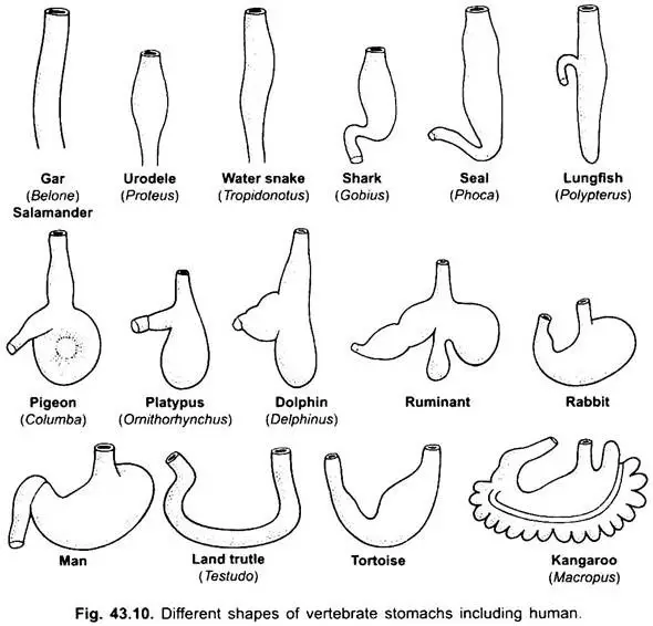
- In some fishes, long-bodied amphibians, lizards, and snakes, the stomach can be described as cigar-shaped. Conversely, in most fishes, it takes on an I-shaped configuration.
- Certain deep-sea fishes possess a highly distensible stomach that enables them to consume prey larger than their own body size.
- In amphibians, the stomach is long and notably lacks a fundus, a part commonly found in other vertebrates.
- For example, the stomach of Uromastix (a type of lizard) is U-shaped, characterized by thick muscular walls and gastric glands within its mucosal lining.
- Stomach in Birds
- Birds exhibit a unique stomach structure that is generally short with no storage capacity. Their stomach consists of two main regions: the proventriculus, which is glandular, and the gizzard, which is muscular and assists in grinding food.
- Stomach in Mammals
- Mammals possess a well-developed stomach, which is divided into four distinct regions: the cardiac, fundus, body, and pylorus.
- The cardiac region is the uppermost part of the stomach.
- The fundus is the thickest section, situated to the left of the cardiac area.
- The body constitutes the main portion, located centrally.
- The pylorus is the lower segment that opens into the duodenum, the first part of the small intestine.
- The inner lining of the stomach contains gastric glands, which house three types of secretory cells:
- Chief cells, responsible for producing pepsinogen and prorennin.
- Oxyntic cells, which secrete hydrochloric acid (HCl).
- Mucus cells, that produce mucus for protection and lubrication.
- Mammals possess a well-developed stomach, which is divided into four distinct regions: the cardiac, fundus, body, and pylorus.
- Stomach in Monotremes and Ruminants
- In monotremes (egg-laying mammals), the stomach takes the form of a sac-like structure and lacks gastric glands.
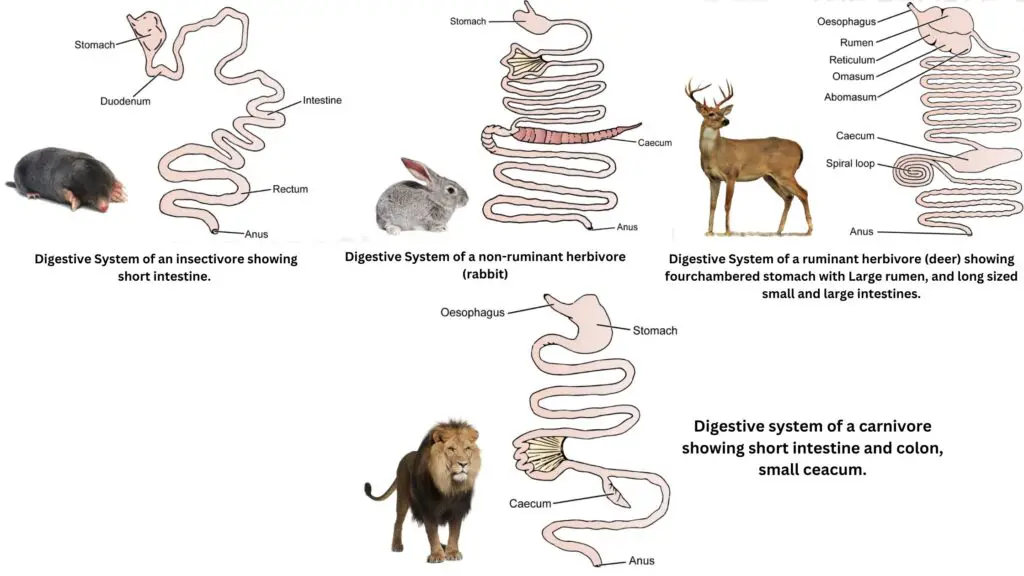
- Ruminants possess a specialized stomach consisting of four chambers: the rumen, reticulum, omasum, and abomasum.
- The rumen is the largest chamber, while the reticulum serves as a small accessory chamber featuring a crisscross ridge on its inner surface. In both the rumen and reticulum, food is reduced to a pulp-like consistency.
- The omasum further triturates the food, and the abomasum acts as the true stomach, where enzymatic digestion occurs.
- It is important to note that in camels, the omasum is absent.
- Functions of the Stomach
- The gastric glands in the stomach secrete gastric juice, which plays an essential role in food digestion in an acidic medium.
- The stomach serves as a temporary storage site, holding food for approximately four hours.
- Furthermore, it facilitates the churning and mixing of food with gastric juices, ensuring thorough breakdown and preparation for subsequent digestion in the small intestine.
6. Intestine
The intestine is a critical component of the digestive system, located between the stomach and the cloaca or anus. It plays a vital role in the digestion and absorption of nutrients, and its structure varies significantly among different vertebrate groups. This variation reflects the evolutionary adaptations to dietary needs and digestive strategies.
- General Structure and Location:
- The intestine is positioned strategically between the stomach and the cloaca or anus.
- In many vertebrates, it is divided into a small intestine and a large intestine, although the extent of this differentiation varies across species.
- Intestinal Characteristics in Different Groups:
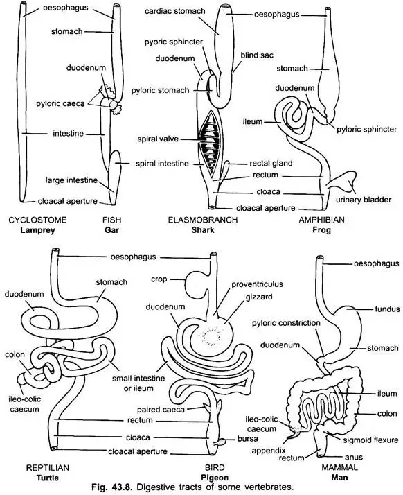
- Cartilaginous and Primitive Bony Fishes:
- In these species, the intestine is typically straight and short, lacking distinct differentiation.
- Cartilaginous fishes feature a spiral valve that enhances nutrient absorption.
- The distal end forms a short, narrow rectum that opens into the cloaca. A caecum is absent, and a tubular rectal gland, whose function remains unknown, may be present.
- Amphibians:
- Amphibians possess a coiled small intestine and a short, straight large intestine.
- The duodenum is a straight segment forming a “U” shape with the stomach and receives bile and pancreatic juice from the hepato-pancreatic duct.
- True villi are absent in the ileum, and amphibians lack both caecum and rectal glands, though urinogenital apertures and Bursa Fabricii are present.
- Reptiles and Birds:
- Both reptiles and birds exhibit a coiled small intestine and a relatively short large intestine that empties into the cloaca.
- In birds, the Bursa Fabricii is well-developed, while it is absent in reptiles.
- Mammals:
- In mammals, the small intestine is significantly longer and more coiled, divided into the duodenum, jejunum, and ileum.
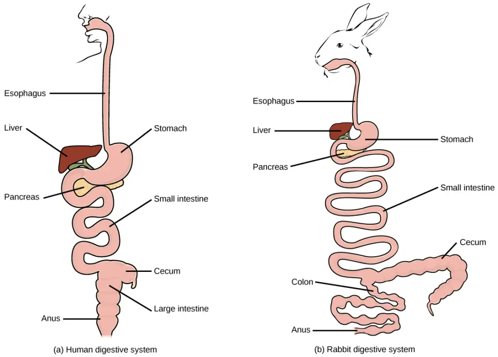
- The large intestine is often lengthy but generally not as long as the small intestine. A caecum is commonly found at the junction of the small and large intestines, particularly in herbivorous mammals.
- In many bony fishes and mammals (excluding monotremes), the urinogenital and anal openings are distinct. The vermiform appendix is absent in fishes, amphibians, reptiles, and birds, but present in mammals.
- Cartilaginous and Primitive Bony Fishes:
- Small Intestine:
- The small intestine is a long, narrow, and coiled tube extending from the pylorus, functioning as the primary site for digestion and absorption.
- In cyclostomes, it is a short, straight tube with a spirally arranged longitudinal flap.
- In elasmobranchs, the small intestine is divided into small and large portions, with a spiral valve that greatly increases the absorptive surface area. This spiral valve is also found in some primitive bony fishes, but it is absent in higher forms, which have long, coiled intestines.
- Caecilians have a less coiled intestine that is not differentiated into small and large tracts, while frogs and toads feature a relatively long and coiled small intestine.
- Reptiles exhibit a more coiled small intestine than amphibians, and for the first time in vertebrates, a caecum arises at the junction of the small and large intestines, though this is not permanent in all reptiles.
- In birds, the small intestine is coiled or looped, often accompanied by one or two colic caeca at the junction with the large intestine. Most mammals also have a proportionately long and coiled small intestine, with its length correlating to dietary habits; herbivores typically possess longer intestines compared to insectivores and carnivores.
- The first part of the small intestine is the duodenum, which is short and begins at the pylorus, terminating beyond the entrance of pancreatic and hepatic ducts.
- It contains many folded villi and branching Brunner’s glands in the submucosa that secrete mucus, alkaline fluids, and some enzymes. The duodenum also produces two hormones, secretin and cholecystokinin, which stimulate the pancreas and gall bladder to release their juices.
- The ileum follows the duodenum, with mammals differentiating it into an anterior smaller jejunum and a posterior longer ileum. Numerous small digestive glands, known as crypts of Lieberkuhn, are present throughout the length of the small intestine, secreting mucus and a digestive fluid known as succus entericus that contains several enzymes.
- The lining of the small intestine is folded to form villi, which significantly increase the surface area for secretion and absorption. These villi are further covered by minute finger-like projections called microvilli, which enhance absorption into the villi. In mammals, lymphoid tissue nodules known as Peyer’s patches are located in the ileum.
- Large Intestine:
- The large intestine has a larger diameter than the small intestine and is generally shorter in fishes, amphibians, reptiles, and birds, but longer in mammals.
- In lower vertebrate forms, the large intestine forms a rectum; in tetrapods, it consists of a colon and a terminal rectum. In most fishes and amphibians, the rectum leads into a cloaca formed by the proctodaeum, where the rectum, excretory ducts, and genital ducts converge before opening to the exterior via a cloacal aperture.
- However, in many bony fishes and all mammals (except monotremes), the rectum and urinogenital ducts have separate openings; in mammals, the anus serves as the opening for the rectum.
- The rectum in mammals is not homologous with that of other vertebrates, as it is derived from the partitioning of the embryonic cloaca. In most vertebrate embryos, a postanal gut extends the intestine into the tail, but this structure typically disappears later in development.
- In elasmobranchs, the large intestine features a pair of rectal glands that secrete mucus and sodium chloride.
- In amniotes, an ileocolic valve is present at the junction of the small and large intestines, a feature absent in fishes, preventing bacteria from entering the ileum from the colon.
- An ileocolic caecum arises from this junction in amniotes; birds possess two caeca, which house cellulose-digesting bacteria. The caecum is particularly long in herbivorous mammals (e.g., rabbits, horses, cows), while in primates, it is relatively small, containing a vestigial vermiform appendix.
Glands Associated with the Digestive System
The glands associated with the digestive system are vital components that aid in the breakdown, digestion, and absorption of nutrients. These glands, including the liver, pancreas, salivary glands, and gastric glands, contribute to various digestive processes. The following outlines their structures, functions, and interconnections.
- Liver
- The liver develops as a single or double outgrowth from the ventral wall of the embryonic archenteron, forming a hollow structure called the hepatic diverticulum. This structure differentiates into two parts:
- An anterior part proliferates to become the liver and its bile ducts.
- A posterior part develops into the gall bladder and cystic duct.
- The bile ducts merge to create a hepatic duct, which unites with the cystic duct, leading to the formation of the common bile duct (ductus choledochus). The area from which the liver arises transforms into the duodenum.
- The liver, recognized as the largest lobed gland in the body, is suspended by a double layer of peritoneum from the transverse septum.
- The gall bladder, situated within the liver, serves as a storage organ for bile secreted by hepatic cells. Bile drains into the duodenum through the common bile duct, formed by the union of the cystic duct and hepatic duct. Although the gall bladder is present in many species, it is not essential and may be absent in several birds and mammals.
- The liver is present in all vertebrates:
- In cyclostomes (like lampreys), it is small and single-lobed, while hagfishes exhibit a two-lobed liver.
- In elasmobranchs (cartilaginous fishes), the liver is bilobed, whereas bony fishes, amphibians, reptiles, and birds possess two to three lobes.
- In mammals, the liver can be multi-lobed and takes on different shapes depending on the species; for instance, it is long and narrow in fishes, urodeles, and snakes, while it appears short, broad, and flattened in birds and mammals.
- Gall bladders and bile ducts are observed in larval cyclostomes, though these structures are absent in adults. Most fishes, amphibians, and reptiles have gall bladders; however, many birds do not. Most mammals have gall bladders, but cetaceans (whales and dolphins) and ungulates (hoofed mammals) typically lack them.
- The liver secretes an alkaline bile, which, while devoid of enzymes, neutralizes the acidity of food entering the duodenum and aids in fat digestion.
- The liver develops as a single or double outgrowth from the ventral wall of the embryonic archenteron, forming a hollow structure called the hepatic diverticulum. This structure differentiates into two parts:
- Pancreas
- The pancreas originates from the endoderm of the embryonic archenteron. Its formation involves:
- A dorsal diverticulum from the embryonic duodenum and one or two ventral outgrowths from the liver, collectively forming pancreatic diverticula.
- The proximal segments of these diverticula evolve into pancreatic ducts, which may undergo reduction or fusion, resulting in one or two pancreatic ducts in the adult. These ducts open into the duodenum either independently or after merging with the common bile duct.
- The distal sections of the diverticula bud to create the main mass of pancreatic cells, enhanced by mesodermal derivatives, resulting in a single gland that can have either a diffuse or compact structure.
- The pancreas serves as both an exocrine and endocrine gland, interconnected by delicate connective tissue strands. The exocrine component secretes digestive enzymes that enter the duodenum through pancreatic ducts, while the endocrine component releases hormones such as insulin and glucagon.
- The pancreas is found in all vertebrates:
- In lampreys, some bony fishes, lungfishes, and lower tetrapods, it appears as a diffuse organ embedded within the liver, mesenteries, and intestinal wall.
- Hagfishes possess a small pancreas, while elasmobranchs have a well-defined bilobed pancreas.
- In higher tetrapoda, the pancreas typically takes on a compact structure, with one or two pancreatic ducts opening into the duodenum.
- The pancreas originates from the endoderm of the embryonic archenteron. Its formation involves:
- Salivary Glands
- Salivary glands secrete saliva, a viscous, colorless, and opalescent liquid primarily consisting of salivary amylase. Saliva is secreted in small amounts throughout the buccal cavity, keeping it moist.
- The primary functions of saliva include:
- Breaking down starch into maltose.
- Maintaining oral hygiene by keeping the mouth and teeth healthy.
- Enhancing food flavor by acting as a solvent and increasing taste sensations.
- Acting as a lubricant, moistening dry food, which facilitates easier swallowing.
- Gastric Glands
- Gastric glands consist of simple or branching tubular structures. In adults, these glands produce approximately 2-3 liters of gastric juice daily, regulated by both neurological and hormonal mechanisms.
- The stomach’s mucosa features three primary types of gastric glands:
- Parietal cells, which secrete hydrochloric acid (HCl). This acid is crucial for protein breakdown as it activates pepsinogen, the inactive precursor of pepsin.
- Chief cells, located in the gastric glands and the stomach’s antrum, secrete pepsinogen. Pepsin is instrumental in digesting proteins into smaller peptides or amino acids, which can then be absorbed in the small intestine.
- Mucous cells, which secrete mucin, contribute to the protective mucosal barrier of the stomach.
Feeding Mechanisms
Feeding mechanisms are critical for the survival of animals, as they directly impact their ability to obtain essential nutrients. Different species have evolved various strategies to secure food, adapt to their environments, and avoid predation. The complexity of these feeding mechanisms reflects the evolutionary pressures faced by different groups of vertebrates. Below is an overview of the feeding mechanisms employed by various vertebrate groups, including fishes, amphibians, reptiles, birds, and mammals.
- Fishes
- Fishes exhibit a wide variety of feeding mechanisms, including adaptations among cyclostomes, elasmobranchs, and teleosts.
- They possess pointed teeth mounted on jaws or palates that assist in holding, tearing, and swallowing prey.
- Most fishes are carnivorous, feeding on a diverse array of animal food, ranging from zooplankton and insect larvae to larger vertebrates.
- Some deep-sea fishes can consume prey nearly twice their own size, which is an adaptation to the scarcity of meals in their environment.
- Many advanced ray-finned fishes cannot masticate their food; however, species like the wolf eel have molar-like teeth for crushing hard-bodied prey, including crustaceans.
- Other fish utilize powerful pharyngeal teeth to grind food. The incompressibility of water facilitates suction feeding, a method used by large-mouthed predators to create negative pressure in their mouths, which draws prey inside.
- Additionally, ram feeding is a technique where predators move past prey with open mouths, engulfing them along with surrounding water.
- Filter feeding is a prevalent and successful method among many fishes, which involves using ciliated surfaces to create currents that draw food particles into their mouths.
- Free-swimming filter feeders, such as herring and basking sharks, can actively swim through food, allowing for more selective feeding.
- Herbivorous fishes consume flowering plants, algae, and grasses, playing a crucial role in freshwater ecosystems.
- Omnivorous fishes eat both plant and animal foods, while scavengers feed on organic debris and parasites from other fish.
- Amphibians
- Adult amphibians exhibit varied feeding habits. Anurans (frogs and toads) and salamanders primarily consume insects and other arthropods, while caecilians feed mainly on earthworms and small bugs.
- Aquatic salamanders employ a lunging technique, using their expanded buccal cavities to suck in prey.
- Frogs, as carnivores, utilize their protrusible tongues to capture prey, which is attached to the front of the mouth and free at the back. The free end produces a sticky secretion that adheres to captured insects, spiders, and other small moving organisms.
- Teeth on the premaxillae and vomers help prevent the escape of prey but are not used for biting or chewing.
- The larval stages of anurans, known as tadpoles, are herbivorous, feeding on pond algae and other plant matter.
- Reptiles
- Reptiles primarily exhibit carnivorous feeding habits, although some, like land turtles and tortoises, are herbivorous, consuming grass and other vegetation. Green iguanas and Uromastyx are also herbivorous, while marine iguanas feed on seaweed.
- Chameleons utilize their long tongues to catch insects from a distance.
- Reptilian jaws and palates contain pointed teeth that aid in holding, tearing, or swallowing prey. Generally, non-mammalian teeth are less differentiated.
- However, some venomous snakes, such as vipers, cobras, and rattlesnakes, have modified fangs that inject venom. These fangs are either hollow or have grooves that allow for venom delivery, functioning similarly to syringe needles.
- Snakes cannot chew their food; instead, they swallow their prey whole, often capturing prey larger than themselves due to the flexibility of their jaws and skull structure. The lower jaw can detach and stretch, and the absence of a sternum allows the ribs to move freely, facilitating the passage of large prey through the digestive system.
- Birds
- Birds lack teeth; instead, they have evolved beaks that exhibit adaptive radiation suited to their feeding lifestyles.
- They consume a wide variety of foods, including seeds, fruits, insects, worms, mollusks, crustaceans, fish, and other vertebrates.
- Many birds feed on nectar, while some species, such as crows and bluejays, are generalists that adapt to seasonal food availability. Others, known as stenophagous species, focus on specific food types.
- The beak structure in birds is adapted to their feeding strategies, ranging from the strong, pointed beaks of crows to the specialized beaks of flamingos and toucans.
- Seed-eating birds have short, stout beaks, while those like the crossbill possess crossed beaks that enable them to open pine cones. Seed eaters often use a muscular gizzard containing stones (gastroliths) to grind their food.
- Insectivorous birds, such as woodpeckers, feature straight, hard beaks for chiseling into wood to access insects. Their long, barbed tongues assist in extracting insects from their burrows.
- Water feeders, including herons and kingfishers, have sharp, pointed beaks suited for catching fish, while ducks and geese possess flat beaks adapted for straining aquatic plants from mud and water.
- Hummingbirds have long, slender beaks that serve as protective coverings for their specialized tongues, which use capillary action to draw up nectar.
- Birds of prey, such as hawks and eagles, possess strong hooked beaks for tearing flesh, while their feet feature long, curved talons for grasping prey.
- Mammals
- Mammals display a wide variety of feeding strategies, with some requiring specialized diets while others are opportunistic feeders.
- The relationship between feeding habits and physical structure is evident in mammals, as adaptations for finding, capturing, and digesting food significantly influence their shapes and behaviors.
- Teeth are the most revealing physical characteristics regarding a mammal’s dietary habits. While most mammals possess teeth, some, like certain whales, monotremes, and anteaters, have evolved modifications that impact their feeding strategies.
- As mammals evolved, significant changes in tooth and jaw structure occurred during the Mesozoic era. Mammalian teeth became heterodont, exhibiting differentiation for various functions, such as cutting, gnawing, tearing, and grinding.
- The primitive mammalian tooth formula includes a combination of incisors, canines, premolars, and molars. Unlike reptiles, mammals typically do not replace their teeth continuously throughout their lives. Instead, they develop two sets of teeth: deciduous (milk) teeth, which are later replaced by a permanent set as the skull grows.
- Only the incisors, canines, and premolars are deciduous; molars remain fixed throughout an individual’s lifetime.
Comparative Anatomy of Digestive Systems in Vertebrates
The digestive systems of vertebrates exhibit remarkable diversity, shaped by evolutionary adaptations that cater to their varied diets and lifestyles. This comparative analysis focuses on the digestive systems of five major vertebrate groups: fish, amphibians, reptiles, birds, and mammals. Each group’s unique structural and functional traits reflect their ecological niches and feeding habits.
- Fish Digestive System:
- Fish represent the oldest vertebrate group and possess relatively simple digestive systems.
- Their digestive tract typically consists of a mouth, esophagus, stomach, and intestine. Notably, many species lack a true stomach.
- Digestion occurs primarily in the intestine, where nutrient absorption takes place.
- The dietary habits of fish are diverse, ranging from carnivorous to herbivorous. This variation influences the length and functionality of their digestive tracts.
- Some cartilaginous fish have specialized structures, such as the spiral valve, which enhances nutrient absorption.
- Amphibians Digestive System:
- Amphibians undergo metamorphosis from aquatic larvae to terrestrial adults, which significantly impacts their digestive adaptations.
- Their digestive systems are relatively straightforward, featuring a stomach and short intestines.
- Larval amphibians primarily consume small prey and aquatic plants, while adult amphibians transition to a more terrestrial diet, leading to the development of longer intestines to improve nutrient absorption.
- Reptiles Digestive System:
- Reptiles exhibit a wide range of digestive adaptations across different orders, showcasing their dietary versatility.
- Snakes possess highly specialized digestive systems to accommodate large prey, featuring elongated intestines that facilitate efficient nutrient extraction.
- Crocodilians have powerful stomachs capable of digesting hard substances like bones and tough tissues.
- Many reptiles exhibit fermentation processes in specialized regions of their digestive tracts, aiding in the breakdown of complex plant materials.
- Birds Digestive System:
- Birds have evolved some of the most specialized and efficient digestive systems among vertebrates.
- Their digestive tract includes a crop, which serves as a food storage organ, and a muscular gizzard that performs mechanical digestion.
- Lacking teeth, birds utilize their gizzard to grind food with ingested stones.
- The crop and proventriculus secrete digestive enzymes, while the highly developed small intestine is adapted for maximum nutrient absorption.
- Mammals Digestive System:
- Mammals, including humans, possess intricate and highly specialized digestive systems.
- They feature stomachs with multiple compartments, particularly evident in ruminants, which have a rumen for fermentation.
- Mammals exhibit efficient dentition for grinding and chewing, contributing to effective food processing.
- The lengthy small intestine is crucial for nutrient absorption, while herbivorous mammals have evolved complex fermentation chambers that allow them to extract nutrients from plant materials through microbial action.

- Finegan, E.J., & Stevens, C.E. (2010). THE DIGESTIVE SYSTEM OF VERTEBRATES.
- https://www.academia.edu/5248174/THE_DIGESTIVE_SYSTEM_OF_VERTEBRATES?auto=download
- https://byjus.com/question-answer/name-the-glands-associated-with-human-digestive-system-and-mention-their-secretion-and-function/
- https://www.slideshare.net/slideshow/comparative-anatomy-of-digestive-system-of-vertebrates/232624777
- https://content.patnawomenscollege.in/zoology/COMPARITIVE%20ANATOMY%20OF%20DIGESTIVE%20SYSTEM%20IN%20VERTEBRATES.pdf
- https://www.notesonzoology.com/vertebrates/digestive-system-of-vertebrates-with-diagram-chordata-zoology/8977
- https://opened.cuny.edu/courseware/lesson/780/student/?section=4
- https://www.zoologytalks.com/comparative-anatomy-of-digestive-systems-in-vertebrates/
- Text Highlighting: Select any text in the post content to highlight it
- Text Annotation: Select text and add comments with annotations
- Comment Management: Edit or delete your own comments
- Highlight Management: Remove your own highlights
How to use: Simply select any text in the post content above, and you'll see annotation options. Login here or create an account to get started.