A cell is like the tiny building block that makes up all living things, from plants and animals to bacteria. Imagine it as a microscopic bag filled with stuff that keeps life going. Every cell has a thin outer layer called a membrane, which holds everything inside and controls what goes in or out. Inside, there’s a jelly-like substance called cytoplasm, where all the tiny structures—like the nucleus—float around. The nucleus acts like the cell’s brain, holding DNA, the instructions that tell the cell how to grow, work, and make more cells.
Cells aren’t all the same, though. They come in different shapes and sizes depending on what they do. For example, muscle cells are stretchy to help you move, while nerve cells are long and thin to send signals across your body. Some organisms, like bacteria, are just a single cell doing everything on its own. Others, like humans, have trillions of cells working together.
What’s wild is that every cell comes from another cell. When a cell splits, it copies its DNA so the new cell knows what to do. This keeps life ticking, whether it’s healing a scrape or growing a whole new person. Cells might be small, but without them, nothing alive would exist. They’re the unsung heroes keeping the show running, quietly doing their jobs so you can breathe, think, and live.
What is the cell?
- The cell is the basic unit of life that makes up all organisms, whether unicellular or multicellular.
- This is a sophisticated, self-contained system capability of carrying out all required life functions.
- Prokaryotic cells, which lack a defined nucleus and comprise bacteria and archaea, and eukaryotic cells, which have a membrane-bound nucleus and comprise plants, animals, fungi, and protists, are two generally accepted categories for cells.
- In eukaryotes, organelles include mitochondria, chloroplasts (in plants), and the endoplasmic reticulum serve specialised purposes; fundamental components within a cell include the plasma membrane, cytoplasm, and genetic material.
- Scientists such as Matthias Schleiden, Theodor Schwann, and Rudolf Virchow developed cell theory in the 19th century, proving that all living entities are made of cells and that new cells are produced only by division of pre-existing cells.
- Comprehensive investigations of cellular mechanisms have helped us to grasp metabolism, energy generation, reproduction, and genetic inheritance.
- Modern research explores cell processes using cutting-edge tools including high-resolution microscopy, molecular biology tests, and genomic analysis, therefore affecting knowledge of illnesses, technologies, and medicine.
- Constant progress in gene editing (e.g., CRISpen-Cas systems) and stem cell research helps to further understanding of biological processes and create fresh therapeutic intervention paths.
Cell Theory
- A fundamental tenet of biology, the cell hypothesis holds that one or more cells—which form the fundamental units of structure and function—make up every living entity.
- According to the hypothesis, the cell is the smallest unit able to perform all basic functions including metabolism, development, and reproduction.
- Its formulation cleared the path for contemporary study in genetics, embryology, and molecular biology as well as a uniting framework for biology.
- Historical events include the 1665 observation of cells in cork by Robert Hooke and the 1830s understanding by Matthias Jakob Schleiden and Theodor Schwann that plants and animals are made of cells.
- Emphasising that cell division is fundamental to life, Rudolf Virchow later polished the theory in 1855 by adding the idea that all cells develop from pre-existing cells.
- With contemporary findings, the classical cell hypothesis has changed to now include DNA in heredity and show that all cells have identical biochemical characteristics.
- By exposing the complex complexities of cellular processes and the dynamic character of cell membranes, organelles, and cytoskeletal components, advances in microscopy and molecular methods have bolstered cell theory.
- By offering essential insights into cellular mechanisms underpinning health, sickness, and developmental processes, the cell theory supports several fields including medicine and industry.
- Focussing on cellular communication, energy transfer, and the integration of cellular activities within multicellular organisms, continuous research is extending the idea.
Cell Organelles/Cell Structure
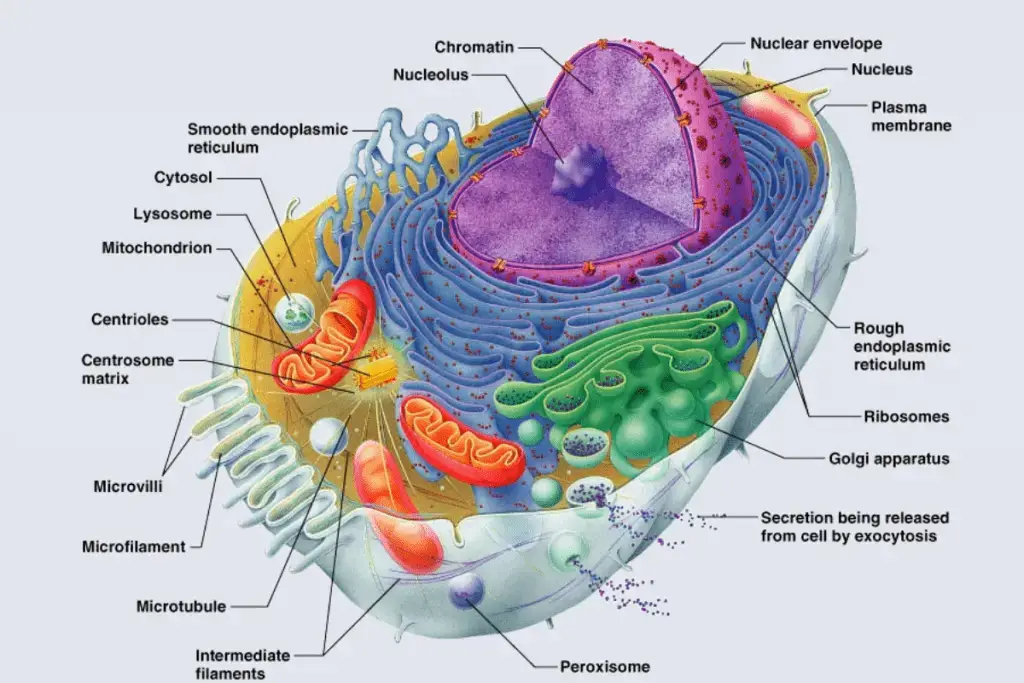
1. Cell Membrane or plasma membrane
- The outermost barrier of a cell separating the internal cytoplasm from the external world is the cell membrane, sometimes referred to as the plasma membrane.
- Its main component is a phospholipid bilayer, in which hydrophilic (water-attracting) heads face outward and hydrophobic (water-repelling) tails face inside to form a semi-permeable barrier.
- Whether peripheral or integral, embedded proteins have important roles including signal transmission, chemical transport, and cell identification and adhesion assistance.
- Especially at different temperatures, cholesterol molecules scattered inside the bilayer assist control membrane fluidity and stability.
- Key functions in cell-to—cell communication and immune recognition are glycoproteins and glycolipids formed on the extracellular surface by carbohydrate chains coupled to proteins and lipids.
- Emphasising that lipids and proteins can migrate laterally inside the layer, the fluid mosaic model explains the dynamic character of the cell membrane and helps the membrane to change with the surroundings.
- Its selective permeability helps the cell to control waste products, nutrients, and ion movement, thereby preserving cellular homeostasis.
- Specialised microdomains, including lipid rafts, crucial in cell signalling and trafficking, have been found using advanced imaging and biochemical methods.
- The cell membrane is essentially essential for preserving the integrity and activity of cells; its intricate structure is therefore vital for functions such endocytosis, exocytosis, and intercellular communication.
Structure and Chemical composition of Membrane
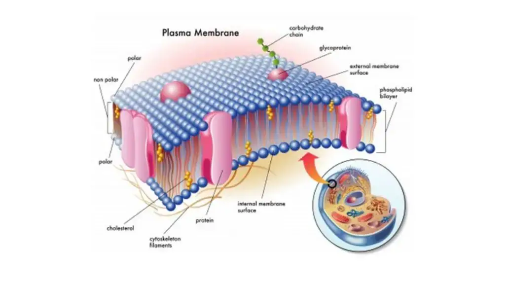
- A phospholipid bilayer makes up the main component of the plasma membrane; each phospholipid molecule contains a hydrophilic (water-attracting) head and two hydrophobic (water-repelling) fatty acid tails, therefore forming a selective barrier separating the internal from the exterior environment.
- Underlying the membrane’s capacity to self-assemble into a bilayer and preserve its integrity in an aqueous environment, phospholipids are amphipathic—that is, they have both hydrophilic and hydrophobic areas.
- Integral and peripheral proteins abound in the lipid bilayer; integral proteins span the membrane and act as channels, transporters, or receptors; peripheral proteins are loosely bound to the membrane surface and help in structural support and signalling.
- Distributed among the phospholipids of animal cell membranes, cholesterol molecules assist control membrane fluidity and stability by preventing too stiff or fluidity, particularly in relation to temperature changes.
- On the extracellular side, carbohydrate chains covalently bind proteins (forming glycoproteins) and lipids (forming glycolipids), therefore facilitating important roles in cell identification, adhesion, and immune response.
- According to the fluid mosaic concept, the plasma membrane is a dynamic and flexible structure in which lipids and proteins may migrate laterally within the layer, allowing the cell to adapt to environmental changes and participate in sophisticated activities including signal transduction and endocytosis.
- Specialised microdomains including lipid rafts enhanced in cholesterol and sphingolipids provide platforms for signalling molecule organisation and enhancement of intracellular communication.
- The chemical makeup of the plasma membrane—phospholipids, cholesterol, proteins, and carbohydrates—ensures it operates efficiently as a barrier, a venue for biochemical activities, and a mediator of cell-to–cell connections generally.
Functions of Cell Membrane
- The cell membrane functions as a physical barrier that mechanically encloses the cell’s contents and preserves its structural integrity
- It controls the movement of particles, such as ions and molecules, across the membrane to maintain the proper internal environment
- Small molecules like oxygen and carbon dioxide can move passively through the membrane via diffusion and osmosis along concentration gradients
- The membrane regulates the exchange of materials between the cell and its surroundings, contributing to cellular homeostasis
- It facilitates bulk transport processes, including exocytosis, where secretory vesicle contents are expelled from the cell, and endocytosis, where materials are internalized into the cell
2. Cell Wall
- Providing structural support and protection, the cell wall is a stiff outer covering around the cells of plants, fungi, bacteria, and certain protists.
- Along with hemicelluloses and pectin, which help preserve cell structure and control water transport, the cell wall of plants is mostly made of cellulose.
- Whereas bacterial cell walls usually consist of peptidoglycan, indicating adaptations to various conditions, fungal cell walls are mostly composed of chitin.
- The cell wall acts as a physical barrier shielding the cell from mechanical strain, infections, and facilitates the regulation of material movement into and out of the cell.
- In multicellular organisms, it is absolutely important for cell development, turgor pressure regulation, and intercellular communication facilitation.
- Understanding the development and variety of life as well as in uses like agriculture and biotechnology depend on the fundamental knowledge of cell walls.
- various species have various structural compositions of their cell walls, which reflect evolutionary adaptations to certain ecological niches and functional needs.
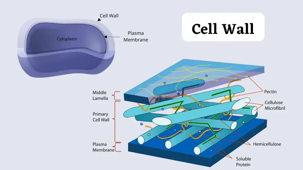
Structure and composition of Cell wall
- Found outside the plasma membrane in plants, fungi, bacteria, and certain protists, the hard, multilayered cell wall gives structural support, protection, and helps to define cell form.
- Embedded in a matrix of hemicelluloses and pectins, which govern porosity and water retention, the main cell wall of plant cells is mostly made of cellulose—a long-chain polymer of β-1,4-linked glucose units that assembles into microfibrils.
- Found in specialised cells like xylem vessels, secondary cell walls include extra polymers like lignin, which boosts stiffness and water resistance.
- Mostly composed of chitin, a polymer of N-acetylglucosamine, coupled with glucans and glycoproteins taken together to offer mechanical strength and osmotic stress resistance, fungal cell walls
- Gram-positive (thick, multilayered peptidoglycan) and Gram-negative (thin peptidoglycan layer accompanied by an outer membrane) bacterial cell walls mostly consist of peptidoglycan, a complex polymer made of alternating sugars (N-acetylglucosamine and N-acetylmuramic acid) cross-linked by peptide chains—with structural differences.
- Structural proteins and enzymes are among the extra elements included into the cell wall that help to enable intercellular communication, defence against pathogens, and remodelling throughout development.
- Maintaining tissue integrity, controlling cell growth, and mediating interactions between cells and their external environment all depend on the general form and chemical makeup of cell walls.
Functions of the Cell Wall
- Maintaining the cell’s predetermined form and shielding it from outside physical pressures, the cell wall gives mechanical strength and structural support.
- It serves as a strong barrier protecting the cell from environmental threats and pathogen invasion, therefore supporting its whole defence systems.
- Controlling turgor pressure helps the cell wall of plant cells to regulate water absorption and retention, therefore enabling plant stiffness and development by means of this mechanism.
- It serves as a selective filter controlling the flow of molecules, letting in necessary nutrients and signalling chemicals while rejecting toxins.
- Key for the organisation and structural integrity of multicellular tissues, cell-to—cell adhesion and communication are facilitated by the cell wall.
- By letting the cell expand in a controlled way throughout development and growth, it helps to govern cell morphogenesis.
- Maintaining cell form and avoiding lysis in bacteria depends on the cell wall, which also helps to control osmotic pressure variations in different environmental situations.
3. Cytoskeleton
- It is a complex, dynamic network that interlinks protein filaments within the cytoplasm of all cells, such as bacteria and archaea.
- The cytoskeleton is extended from the cell membrane to the cell nucleus.
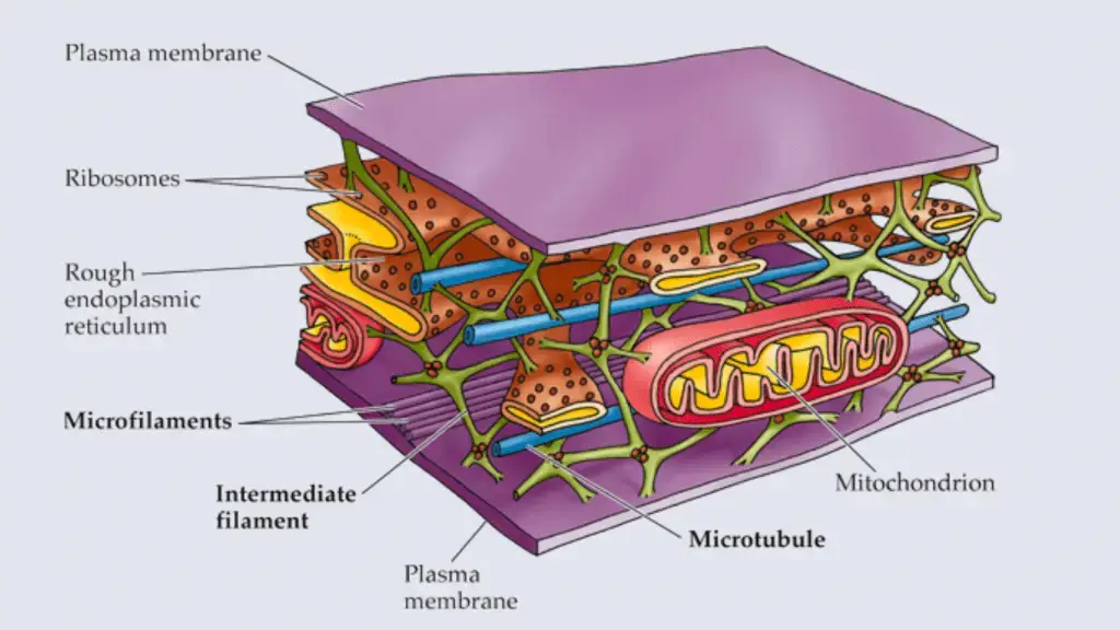
Structure and composition of Cytoskeleton
- In Eukaryotic cells, the Cytoskeleton is composed of microfilaments, microtubules, and intermediate filaments.
- The diameter of a microfilament is about 7 nm which is made up of many linked monomers of a protein known as actin These actions are combined and formed a double helix structure.
- The Intermediate filaments are composed of multiple strands of fibrous proteins that are wound together. They have an average diameter of 8 to 10 nm.
- The Microtubules have a diameter of about 25 nm which is composed of tubulin proteins (tubulin protein consists of two subunits, α-tubulin, and β-tubulin). These proteins are arranged to form a hollow, straw-like tube.
Functions of Cytoskeleton
- The cytoskeleton maintains the shape of the cell.
- It helps in cell locomotion.
- It organizes the structures and activities of the cell.
4. Capsule
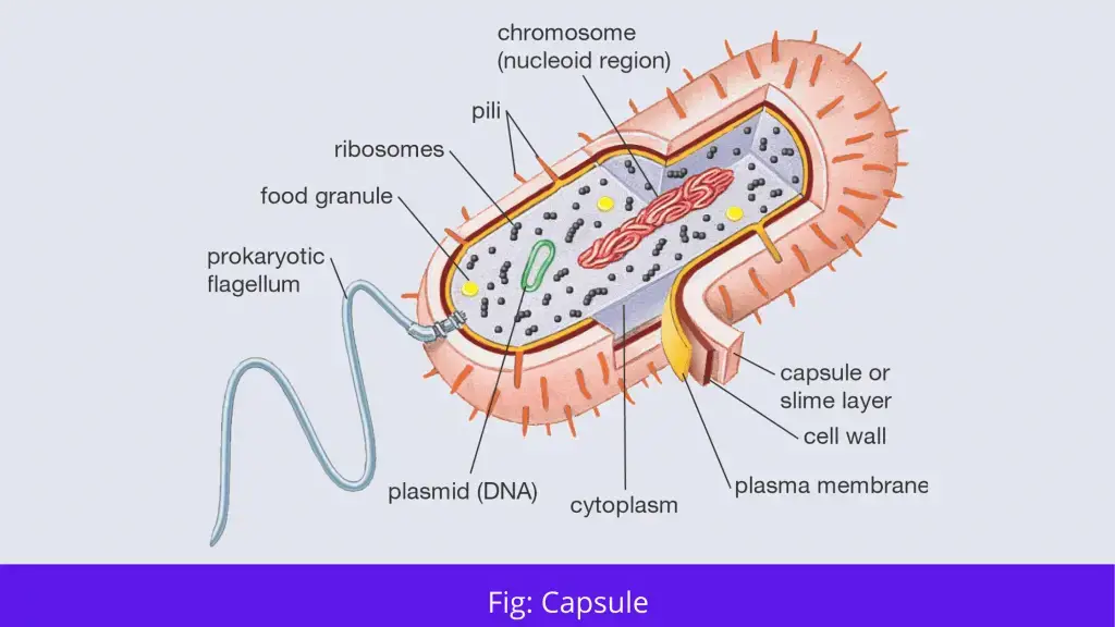
- The capsule is present at the outside of the cell membrane and cell wall in some bacteria.
- The capsule can be detected by staining them with India ink or methyl blue. The standard stains are not used to stain them because they cannot penetrate the capsule.
Structure and composition of Capsule
- The Capsule in bacteria cells consist of polysaccharide except Bacillus anthracis, because their capsule consists of poly-D-glutamic acid.
Function of Capsule
- Protecting the cell wall from different antibacterial agents, thus it is considered a virulence determinant.
- It protects the bacteria from desiccation.
- Allow in adhesion of bacteria to surface.
- It prevents phagocytosis.
5. Flagella
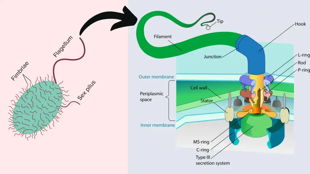
- The bacterial flagellum are long and thick thread-like appendages which are stretched from cytoplasm through the cell membrane(s) and extrude through the cell wall.
- There are present different types of flagella based on their number and location such as Monotrichous, Peritrichous, Lophotrichous, and Amphitrichous.
Structure and composition
- Bacterial flagella is about 20-nanometer-thick and composed of the protein flagellin.
- The flagella has three important parts such as Basal body, Hook, and Filament.
- The basal body consists of four rings such as P-ring, M-S-ring, L-ring and C-ring.
- Filaments are thin hair like structure and Hook is the broader area present at the base of the filament.

Function
- Helps in movement of organisms.
- It is also used as a secretary organelle in Chlamydomonas.
- They detect the changes in pH and temperature.
- Some eukaryotes use flagellus to enhance their reproduction rates.
6. Fimbriae
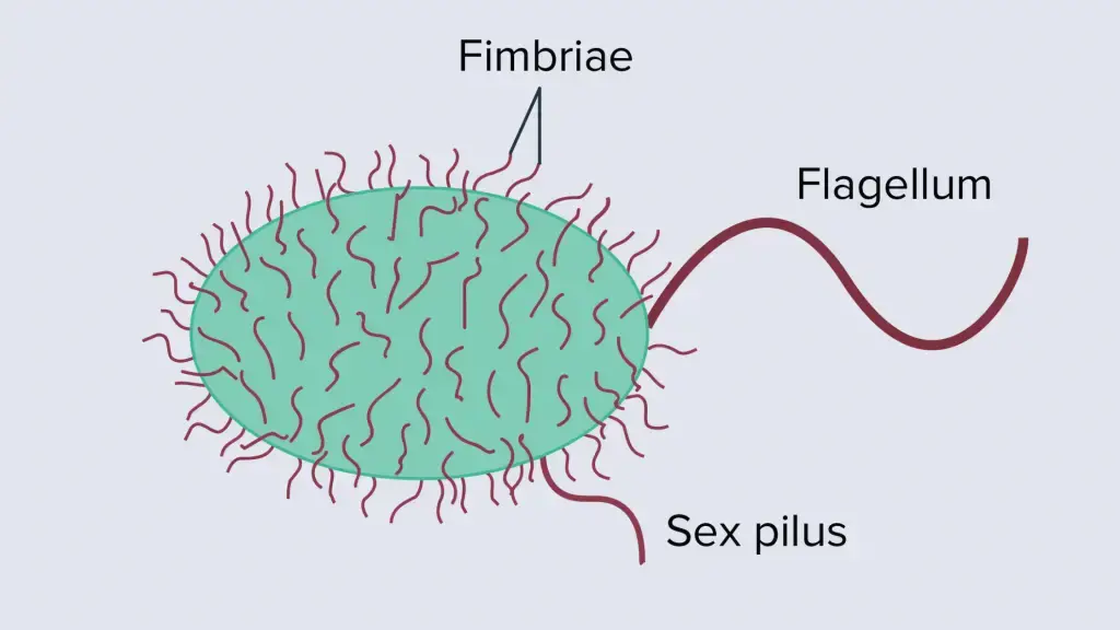
- Fimbriae also known as pilus or pili which is a short, thin, hair-like filament located at the bacterial surface.
- Fimbriae is thinner and shorter than a flagellum.
Structure and composition
- The diameter of fimbriae ranges from 3–10 nanometers and several micrometers long.
- The fimbriae are made up of pilin proteins which are antigenic in nature.
Functions
- It helps in attachment of bacterial cells with the host cells.
- It also determines the virulence properties of several bacteria.
7. Cell Organelles
Cell organelles are referred to when those vital organelles are present within the cell and perform one or more vital functions.
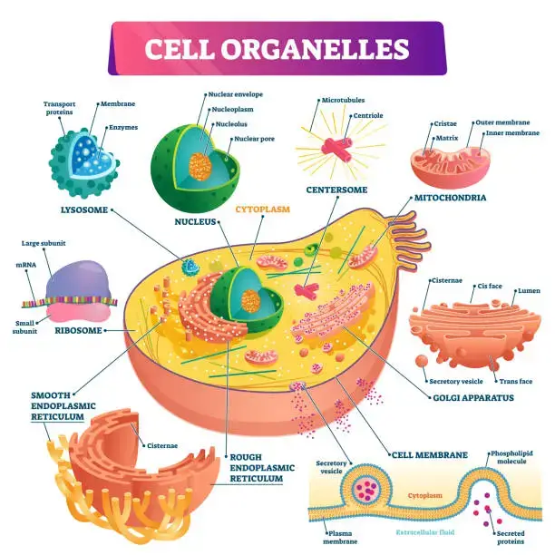
There are present several types of organelles such as nucleus, golgi apparatus, mitochondria, chloroplasts, peroxisomes and lysosomes, etc.
(i). Cell nucleus:
- The nucleus stored the genetic information of the cell that is why it is called the information center of the cell. It is also called “brain of the cell”, because it controls the functions of other organelles.
- In eukaryotic organisms the nucleus is surrounded by a nuclear membrane, and a nuclear envelope, which is separating it from the cell cytoplasm.
- It contains double-stranded, spiral-shaped deoxyribonucleic acid (DNA) molecules within its membrane. In Prokaryotic cells, the nucleus is absent whereas the genetic material distributed in the cytoplasm.
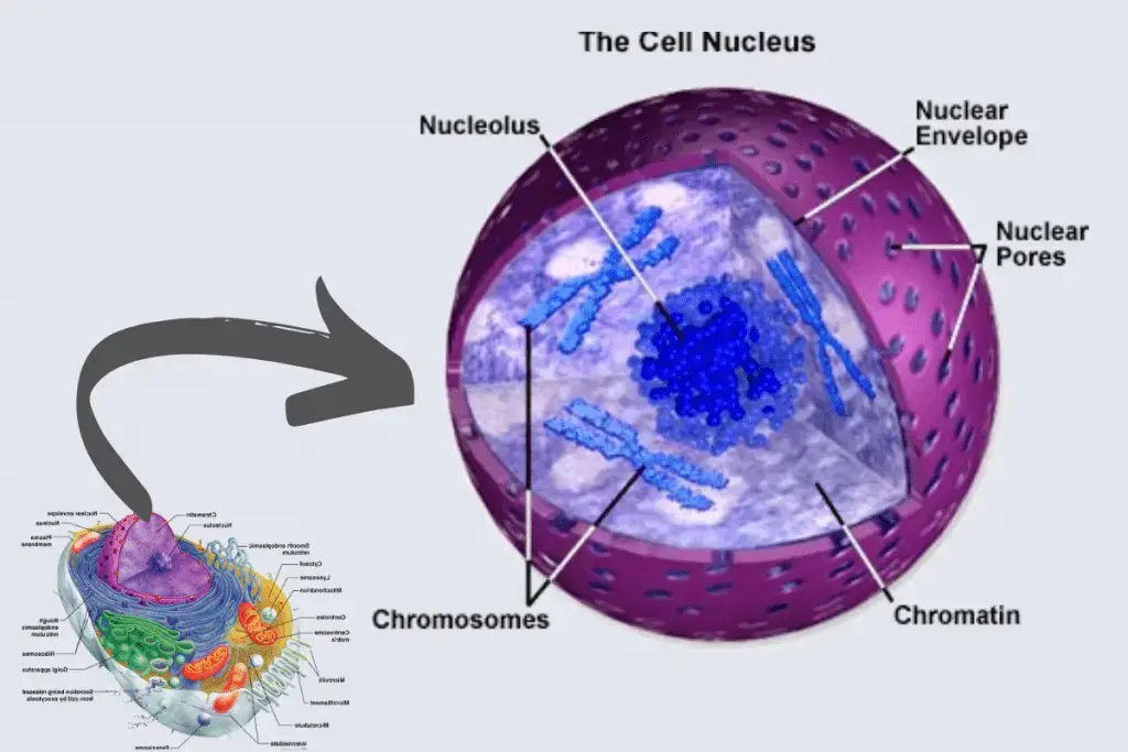
Structure of Nucleus
- The nucleus has a diameter of 6 micrometres (6 × 10⁻⁶ metre), and contains about 1.8 metres of DNA.
- Nucleus has several important parts such as chromatin (Genetic material), nuclear envelope, nucleoplasm and nucleolus.
- The nuclear envelope is a lipid bilayer, which contains two layers such as outer and an inner phospholipid bilayer. This envelope separates the chromatin and nucleolus from cytoplasm. The nuclear envelope contains several pores which helps in import and export of proteins and RNA from cytoplasm.
- Chromatin is a dense, compact fibre which is formed by the combinations of both DNA and proteins.
- The nucleoplasm is referred to the portion which is located within the envelope.
- The nucleolus has four important components such as Fibrillar Centers, Granular Components, Dense Fibrillar Components, and Nucleolar vacuoles. It occupied the 25% volume of the nucleus. It performed the assembling of ribosomes and synthesis of rRNA.
Functions
- Nucleus stored the genetic material and proteins(DNA or RNA).
- It controls the protein synthesis, cell division, growth, and differentiation.
- It produces messenger RNA (mRNA) for protein synthesis.
(ii). Mitochondria
- Mitochondria is referred to as the powerhouse of a cell because it generates energy for the cell.
- It is a self-replicating organelles which is found in both plant and animal cells in various numbers, shapes, and sizes.
- It produces ATP or energy by oxidative phosphorylation of end products which are arising from the cytoplasmic metabolism.
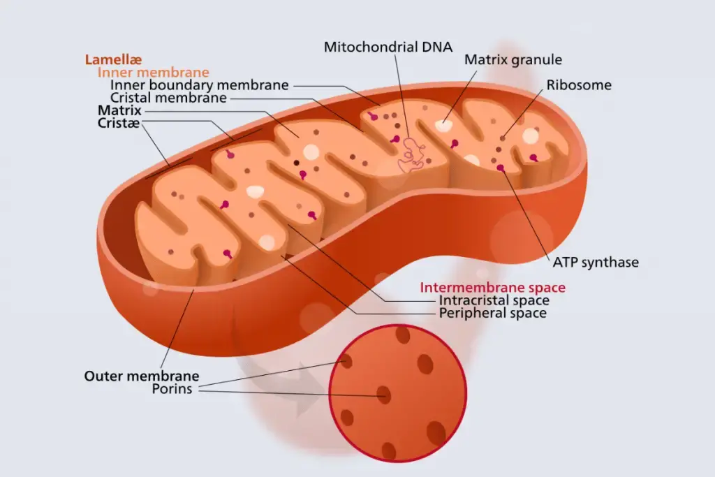
Structure of Mitochondria
- Mitochondria has two distinct membranes such as inner layer and outer layer.
- The outer layer is smooth whereas the inner layer is folded and forms finger-like structures which are called cristae.
- The inner membrane enclosed space is called matrix, this space contains a fluid which is a mixture of metabolic products, enzymes, and ions.
Functions of Mitochondria
- It produces energy in the form of ATP which is required for the proper functioning of cell organelles.
- It helps to detoxify the ammonia.
- Mitochondria produces different types of components of blood and segments of hormones.
- It assists the process of apoptosis.
- It also balanc the amount of Ca+ ions within the cell.
(iii). Chloroplast
- Chloroplasts are a type of plastid which are found in cells of plants and green algae, which help in photosynthesis. In photosynthesis it converts the light energy into chemical energy as a result it produces oxygen and energy-rich organic compounds.
- They contain pigments such as chlorophyll a and chlorophyll b, which helps in photosynthesis. Because of this pigment they appear as green colour which is distinguished from other types of plastids.
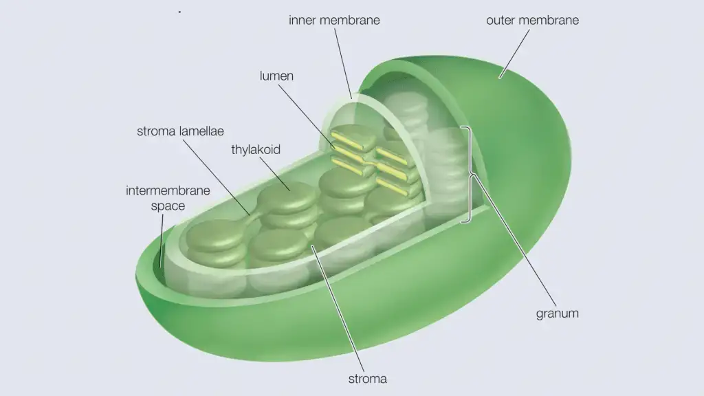
Structure of Chloroplast
- Chloroplasts are round, oval, or disk-shaped with a diameter of 5–7 μm and thickness is about 1–2 μm (1 μm = 0.001 mm).
- The chloroplast is surrounded by a double membrane which has two layers such as the outer and inner layers. There is a gap between the outer and inner layer which is called intermembrane space.
- The porous nature of outer and inner membranes in chloroplast help in transport of different materials.
- Inside the inner membrane it contains thylakoids. There is another internal membrane which is extensively folded and characterized by the presence of thylakoids; it is known as the thylakoid membrane. In some plants the thylakoids are tightly stacked which is called grana.
- Between the inner membrane and thylakoid membrane it contains dissolved enzymes, starch granules, and copies of the chloroplast genome within a matrix which is known as stroma.
Function of Chloroplast
- It performed the light-independent and light-dependent reactions during photosynthesis.
(iv). Centriole
- Centrioles are located near the nucleus but they appear only during cell division, they work in the process of mitosis and meiosis.
- Centrioles are cylindrical in shape and made up of a protein called tubulin. They mainly appear in most eukaryotic cells.
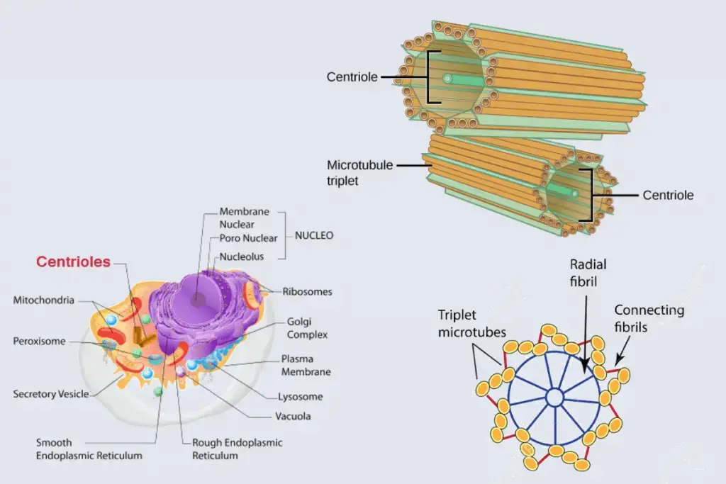
Structure of Centriole
- Centriole consists of nine sets of microtubules, each in groups of three known as triplet microtubules.
- The Triplet microtubules consist of three concentric rings of microtubules which makes it very strong.
- A special protein is bound with each triplet which provides the shape of centriole.
- A amorphous material known as the pericentriolar material is surrounded by the triplet microtubules.
Function of Centriole
- During the cell division centriole helps in the formation of spindle fibers.
- They help in the formations of cilia and flagella.
(v). Cytoplasm
- The semifluid substance which is located between the nucleus and the cellular membrane is called cytoplasm.
- Different types of organelles are suspended within the cytoplasm such as ribosomes, mitochondria, golgi bodies, centriole, lysosomes and peroxisomes, etc.
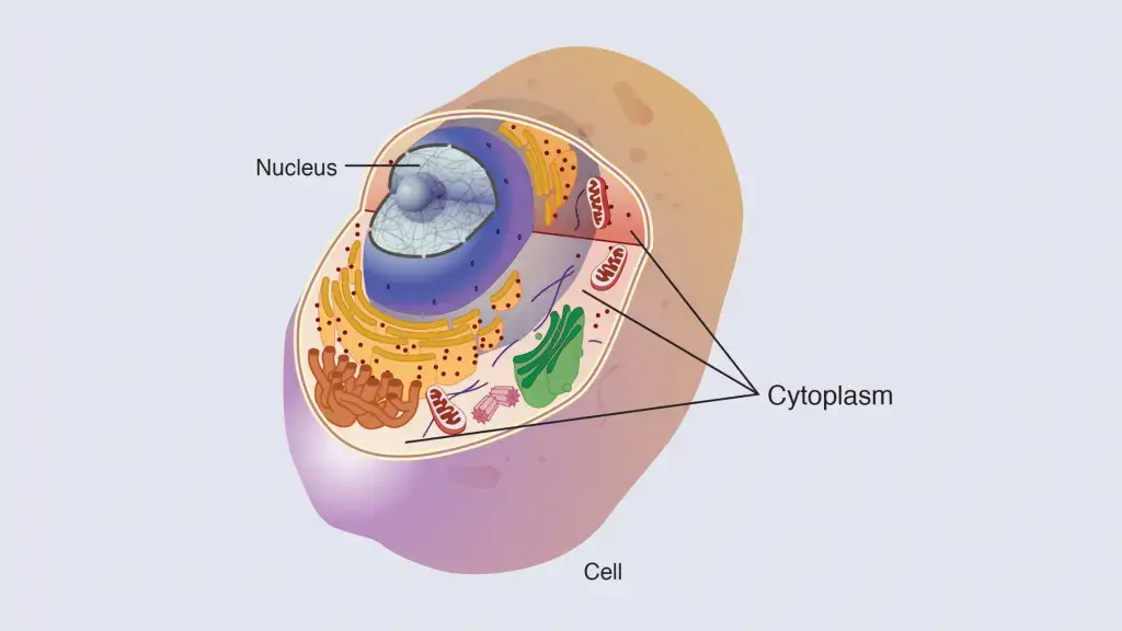
Structure of Cytoplasm
- Cytoplasms consist of three main components such as cytosol, organelles and inclusions.
- Cytosol is a gel-like substance in which other organelles remain suspended. It occupies about 70% of the cell volume. Cytoplasm is a mixture of cytoskeletal filaments, dissolved molecules, and water.
- Organelles are referred to as membrane-bound structures, which perform specific functions and remain suspended within the cytoplasm. Organelles include mitochondria, the endoplasmic reticulum, the Golgi apparatus, vacuoles, lysosomes, etc.
- Cytoplasmic inclusions are insoluble small particles which are suspended in the cytosol.
Function of Cytoplasm
- Most of the Cellular and enzymatic reactions occur within the cytoplasm.
- Cytoplasm protects the other organelles and genetic materials from damage which can occur due to collision or change in the pH.
- Cytoplasm helps in distribution of nutrients and conducting the movement of cell organelles within the cell by a process is called cytoplasmic streaming.
(vi). Endosomes
- It is a membrane-bound vesicle formed by endocytosis.
- Endosomes are found in animal cell cytoplasm.
Structure of Endosomes
- Endosomes are three types including early endosomes, late endosomes, and recycling endosomes.
- The early endosome is composed of tubular-vesicular networks.
- The late endosome has several close-packed intraluminal vesicles but lacks tubules.
- The recycling endosome is made of tubular structures.
Function of Endosomes
- It sorts the material before it reaches the degradative lysosome.
(vii). Golgi Apparatus/ Golgi Complex/ Golgi Body
- Golgi Body mainly found in eukaryotic cells and they modify and transport molecules from endoplasmic reticulum to their destination.
- They are found in cytoplasm next to the endoplasmic reticulum and near the cell nucleus.
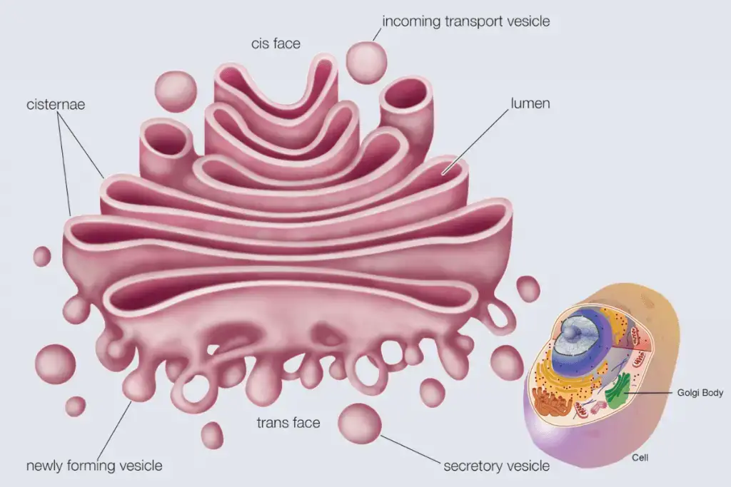
Structure of Golgi Complex
- Golgi Complex is formed by several flattened, stacked pouches called cisternae.
- Generally golgi complexes are made up of four to eight cisternae but in mammalian cells they are made of 40 to 100 stacks of cisternae. In protists they are made of sixty cisternae.
- A matrix protein helps in keeping together the cisternas and a cytoplasmic microtubule supports the golgi complexes.
- The golgi complex has three main part such as cis, which is cisternae nearest the endoplasmic reticulum, medial, central layers of cisternae, and trans, cisternae farthest from the endoplasmic reticulum.
- The cis and trans portion of glogi complex is responsible for sorting proteins and lipids.
Function of Golgi Complex
- It is also called “traffic police” because it directs proteins and lipids to their destination.
- It helps in sulfation of molecules.
- It helps in synthesis of cell membrane, lysosomes and other organelles.
- Helps in exocytosis of zymogen, mucus, lactoprotein, and parts of the thyroid hormone.
(viii). Intermediate filaments
- Intermediate filaments are the main components of the cytoskeleton which is found in vertebrates, and many invertebrates cells.
- There are present six types of intermediate filaments such as Types I and II – acidic and basic keratins (epithelial keratins, trichocytic keratins), trichocytic keratins (Desmin, GFAP, Vimentin, etc), Type IV (Synemin, Neurofilaments, Syncoilin, etc), Type V – nuclear lamins (Lamins), Type VI (Nestin, Filensin, etc)
Structure of Intermediate filaments
- It contains a family of related proteins.
- A pair of two intertwined proteins formed a coiled-coil structure which is preferred as the central building block of an intermediate filament.
Function of Intermediate filaments
- It provides structural integrity of a cell by holding tissues of various organs.
(ix). Lysozyme
- They are membrane bounded organelles and mainly found in cytoplasm of animal cells.
- These organelles degrade various macromolecules with the help of a hydrolytic enzyme.
- There are present two types of lysozyme such as primary and secondary lysozyme.
- The primary lysozyme has several hydrolytic enzymes such as lipases, amylases, proteases, and nucleases.
- Whereas the secondary lysosome is formed by the fusion of primary lysosomes containing engulfed molecules or organelles.
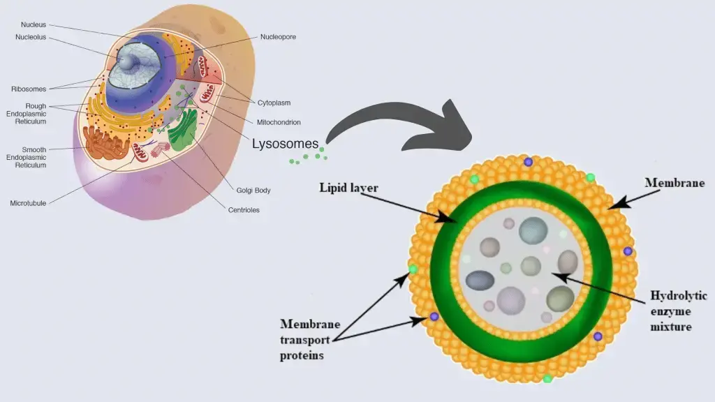
Structure of Lysozyme
- They have an irregular or pleomorphic shape but mostly they appear in spherical or granular structure.
- A lysosomal membrane is present around the Lysosomes which contain different types of hydrolytic enzymes. This membrane protects the other organelles in cytoplasm from harmful enzymes.
Function of Lysozyme
- They help in intracellular digestion of larger macromolecules, where the enzymes which are present in lysozyme degrade the larger macromolecules into smaller molecules.
- They also help in autolysis of unwanted organelles.
- They also help in secretion, plasma membrane repair, cell signaling, and energy metabolism.
(x). Microfilaments
- Microfilaments are also known as the actin filaments, because they are the polymer of a protein called actin.
- It is a network of protein filaments which provides cell structure and keeping organelles in place by extending throughout the cell.
Structure of Microfilaments
- These are referred to as the thinnest filaments of the cytoskeleton by having a diameter of about 6 to 7 nanometers.
- It is made of two strands of actin protein subunit which is wound in a spiral.
- When actin subunits come together and form a microfilament are known as globular actin or G-actin, and when they join together are known as the filamentous actin (F-actin).
- The one end of microfilament is positively charged and the other one is negatively charged.
Function of Microfilaments
- They help in Muscle Contraction.
- They also help in Cell Movement.
- They help in cell division during the mitosis.
(xi). Microtubules
- These are the polymers of tubulin protein which separate them into microfilaments.
- Microtubules are the part of cytoskeleton and provide structure and shape to eukaryotic cells.
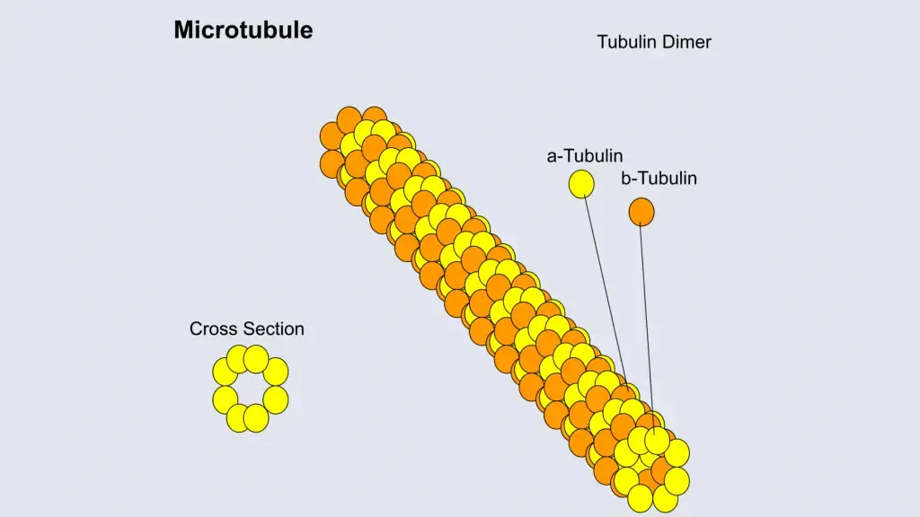
Structure of Microtubules
- Microtubules are 50 micrometres long and the outer diameter is between 23 and 27 nm and inner is between 11 and 15 nm.
- The microtubules are long, hollow cylindrical shapes and are composed of polymerised α- and β-tubulin dimers.
- The inner space of hollow microtubule cylinders is known as lumen.
- Like microtubules the microfilaments have a positively charged and negatively charged end.
Function of Microtubules
- Microtubules help in Cell migration.
- They also help in structure formation of eukaryotic cilia and flagella.
- These are essential for formations of cytoskeleton.
(xii). Microvilli
- The finger-like structures which are projected on or out of the cells are called Microvilli.
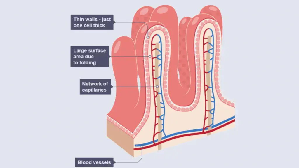
Structure of Microvilli
- These are the bundles of protuberances which are arranged on the surface of the cell.
- They are surrounded by a plasma membrane enclosing cytoplasm and microfilaments.
Function of Microvilli
- In the gastrointestinal tract they help in nutrient absorption.
- They increase the surface area of cells.
- The membrane of Microvilli contains enzymes which break down complex nutrients into simpler compounds.
- During fertilization they act as an anchoring agent in WBCs and in sperms.
(xiii). Peroxisomes
- It is a membrane-bounded organelle found in the cytoplasm of eukaryotic cells.
- They contain enzymes that help in the oxidation of molecules such as fatty acids and amino acids. As a result of the oxidation reaction they produce hydrogen peroxide, that is why they are named peroxisome.
- Hydrogen peroxide is toxic to cells that’s why it contains another enzyme called catalase which converts the Hydrogen peroxide into water and oxygen and neutralizes the toxicity.
Structure of Peroxisomes
- They are made of a single membrane and granular matrix which are scattered in the cytoplasm.
- They appear scattered in the cytoplasm.
- They have different types of enzymes such as urate oxidase, D-amino acid oxidase, and catalase.
Function of Peroxisomes
- They help in oxidation of specific biomolecules.
- They help in biosynthesis of membrane lipids called plasmalogens.
- In plant cells during photorespiration they help in recycling of carbon from phosphoglycolate.
(xiv). Plasmodesmata
- Plasmodesmata is a small passage or channel which is formed in cell walls of plant cells and some algal cells.
- These channels help in transport and communication between two neighboring cells.
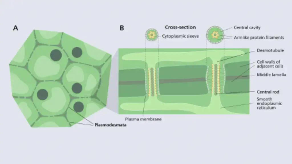
Structure of Plasmodesmata
- A plant cell has approximately 103 – 105 plasmodesmata which connect with the adjacent cells.
- The diameter of a Plasmodesmata is about 50–60 nm.
- Plasmodesmata can traverse up to 90 nm thick cell walls.
- They contain three layers such as the plasma membrane, the cytoplasmic sleeve, and the desmotubule.
- The plasma membrane is a continuous extension of the cell membrane or plasmalemma and has a similar phospholipid bilayer structure.
- The cytoplasmic sleeve is a fluid-filled space enclosed by the plasmalemma and is a continuous extension of the cytosol. Trafficking of molecules and ions through plasmodesmata occurs through this space.
- Desmotubule which is a part of the endoplasmic reticulum that provides a network between two cells and allows the transport of some molecules.
Function of Plasmodesmata
- It helps in transfer of proteins, RNA, and viral genomes between two adjacent cells.
(xv). Plastids
- These are double membrane organelles which are mainly found in plant and algal cells and also in some eukaryotic organisms.
- Sometimes they contain pigments which help in photosynthesis of cells.
- There are present different types of plastids such as; Chloroplasts, Chromoplasts, Gerontoplasts, Leucoplasts.
Structure of Plastids
- These are oval or spherical shapes with a double membrane. The membrane contains two layers such as outer and an inner membrane. The gap between the inner and outer membrane is called an intermembrane space.
- The grana is suspended within the stroma which is enclosed by the inner membrane.
- Grana is made of sac-like thylakoids piled one on the other and connected by stroma lamellae.
- Plastids carry both nucleic acids such as DNA and RNA that allows it to synthesize essential proteins which are required for different processes.
Function of Plastids
- It is the main center for many metabolic activities such as photosynthesis.
- They help to store the foods such as starch.
(xvi). Ribosomes
- These are present in all living cells as free particles or attached to the membranes of the endoplasmic reticulum (eukaryotic cells) and function as the site of protein synthesis.
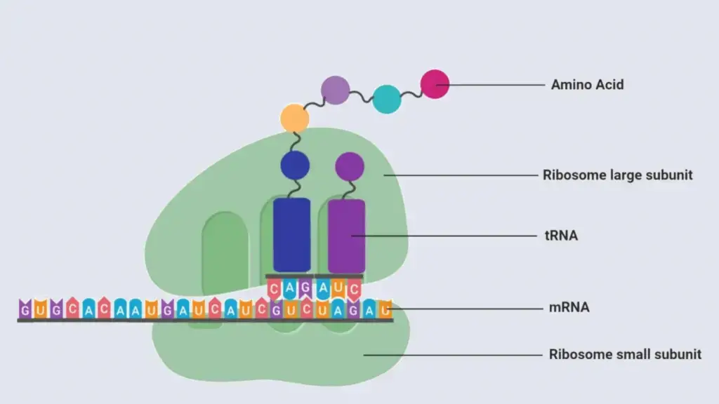
Structure of Ribosomes
- Ribosomes consist of ribosomal RNA and dozens of ribosomal proteins, these are arranged and formed two distinct subunits such as the small subunit and large subunit.
- The eukaryotic cell contains 80s ribosome with 40S smaller subunit and 60S larger subunit.
- The prokaryotic cell contains 70S ribosome where it has a 50S large subunit and 30s smaller subunits.
Function of Ribosomes
- Ribosomes help in protein synthesis.
- They involve protein folding.
- Ribosomes array the amino acids in the order designated by tRNA and assist in protein synthesis.
(xvii). Storage granules
- Storage granules are also known as zymogen granules that store cell’s energy and other metabolites.
Structure of granules
- They contain a lipid bilayer and are made of phosphorus and oxygen.
- The components within these storage granules depend on their location in the body with some even containing degradative enzymes yet to participate in digestive activities.
Function of granules
- It helps to store nutrients and reserves in both prokaryotes and eukaryotes.
- In prokaryotes Sulfur granules utilize hydrogen sulfide as an energy source.
(xviii). Vacuole
- Vacuole refers to an empty space within the cytoplasm which is a membrane-bound structure filled with fluid.
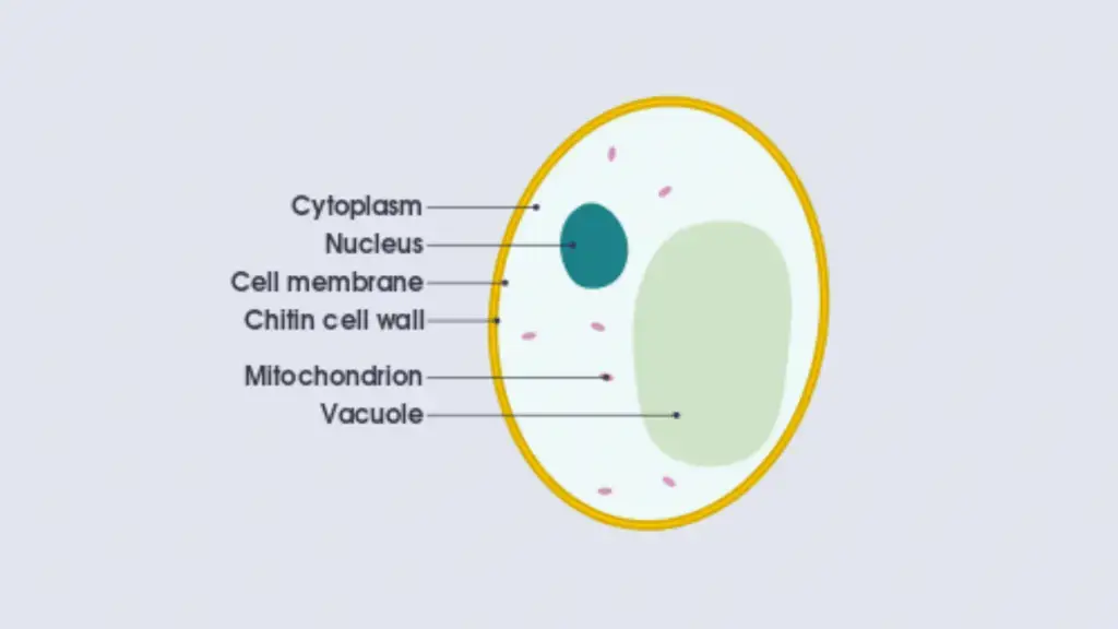
Structure of Vacuole
- It has a membrane called tonoplast, which contains a fluid mixture of inorganic materials and organic materials such as nutrients and even enzymes.
- Vacuoles are similar to vesicles because they form When vesicles are fused.
Function of Vacuole
- They store the nutrients.
- They also store waste materials.
- They involve homeostasis, where they balance the pH of the cell by influx and outflow of H+ ions to the cytoplasm.
- They also contain different enzymes for different metabolic reactions.
(xix). Vesicles
- Vesicle is a structure which is present within or outside a cell.
- They can be formed both naturally (during the processes of secretion (exocytosis), uptake (endocytosis) and transport of materials within the plasma membrane) and artificially. The artificially formed Vesicles are called liposomes.
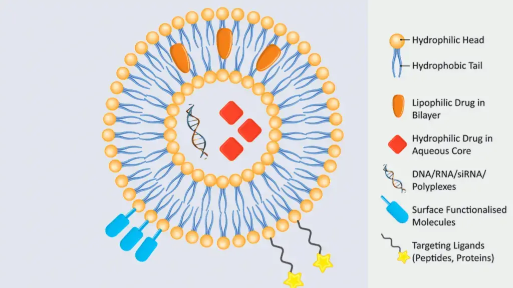
Structure of Vesicles
- They are composed of liquid or cytoplasm enclosed by a lipid bilayer.
- The outer layer of vesicles is similar to plasma membranes, which is enclosing a liquide.
- It has a lipid bilayer with hydrophobic and hydrophilic end.
Function of Vesicles
- It involves the storage and transport of materials.
- It also helps in exchange of molecules between two cells.
- They also function in metabolism and enzyme storage.
Functions of Cell
- Maintains Support and Structure: Cell provides the support and structure to animals, plants, etc.
- Reproduction: Cells help in mitosis and meiosis during the reproduction processes.
- Energy: Cells help in energy production in animals and plants.
- Facilitate Growth Mitosis: Cells facilitate the growth of organisms during the mitosis process by dividing into daughter cells from the parent cells.
- Transport of Substances: Cells transport various nutrients and small substances including oxygen, carbon dioxide, and ethanol, which are essential for chemical processes going on inside the cells.
References
- https://en.wikipedia.org/wiki/Cell_(biology)#Cell_types
- https://www.khanacademy.org/test-prep/mcat/cells/eukaryotic-cells/v/the-nucleus
- https://biologydictionary.net/microfilament/
- https://en.wikipedia.org/wiki/Vesicle_(biology_and_chemistry)
- https://www.britannica.com/science/cell-biology/Actin-filaments#ref37428
- https://byjus.com/biology/cells/
- http://www.biology4kids.com/files/cell_nucleus.html
- https://microbenotes.com/cell-organelles/
Quality content is the secret to invite the visitors to pay a visit the site, that's what this website is providing.
Since the admin of this web site is working, no question very shortly it will be renowned, due to its quality contents.
Hi, after reading this remarkable post i am as well cheerful to share my familiarity here with mates.
thank you
There is perceptibly a bunch to know about this. I suppose you made certain nice points in features also.
Thank you