What is Cell Wall?
- The cell wall is a crucial non-living component that surrounds the outermost layer of a cell. Its composition and characteristics vary across different organisms. The primary function of the cell wall is to provide structure, support, and protection to the cell, while also separating the interior contents from the external environment.
- In plants, the cell wall is predominantly composed of polysaccharides such as cellulose, hemicelluloses, and pectin. These materials confer rigidity and strength, helping the plant maintain its shape and withstand mechanical stress. Additionally, polymers like lignin, suberin, or cutin may be embedded within the plant cell walls, enhancing their structural integrity.
- Fungi, on the other hand, have cell walls made up of chitin, a glucose derivative also found in the exoskeletons of arthropods. This composition provides fungi with similar structural support and protection against desiccation, akin to the role of cell walls in plants.
- Prokaryotic organisms, such as bacteria, also possess cell walls, although their chemical makeup is distinct from that of plants and fungi. Bacterial cell walls are primarily composed of peptidoglycans, large polymers that offer protection and prevent cell lysis. The structure of bacterial cell walls typically includes an inner layer of peptidoglycans and an outer layer comprised of lipoproteins and lipopolysaccharides.
- Eukaryotic cells, which include plants and fungi, possess a defined nucleus and membrane-bound organelles. Notably, the cell wall is absent in most eukaryotic organisms like animals. In contrast, it is a defining feature in plants, fungi, algae, and some prokaryotes, except for certain bacteria like mollicutes.
- The historical discovery and study of the cell wall began with Robert Hooke in 1665, who first observed and named it. Over time, the understanding of cell wall structure and formation evolved, with significant contributions from scientists like Karl Rudolphi, J.H.F. Link, Hugo von Mohl, Carl Nägeli, and Ernst Münch. These studies revealed that cells have independent walls and that the cell wall grows through processes like apposition and intussusception.
Definition of Cell Wall
A cell wall is a rigid, protective layer that surrounds the cell membrane in plants, fungi, algae, and some prokaryotes, providing structural support, shape, and protection.
Bacterial Cell Wall Types
Based on the result of gram staining it is stated that there are two types of cell wall such as
- Gram-positive cell wall (+Ve)
- Gram-negative cell wall (-Ve)
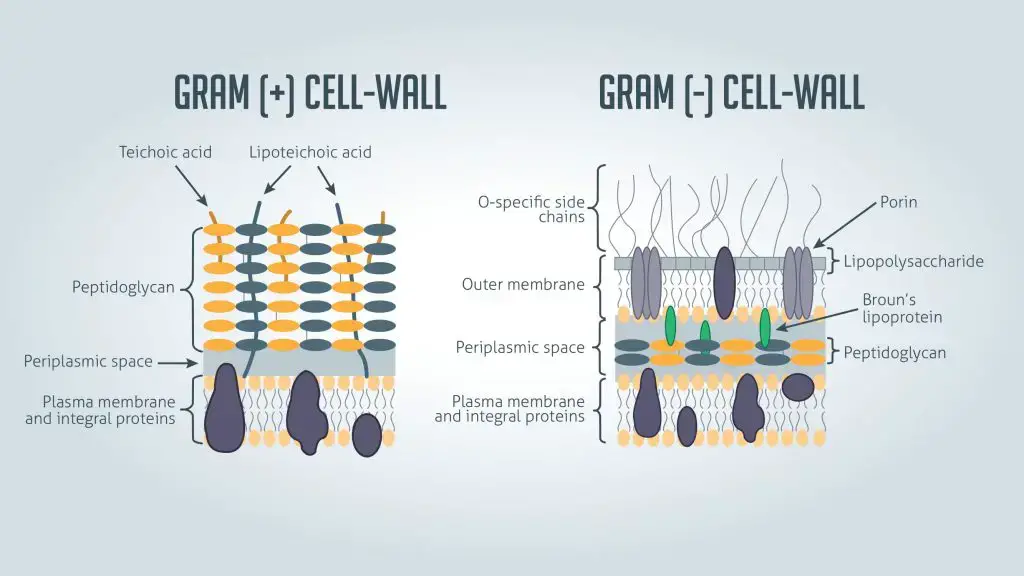
Cell wall structure of gram-positive bacteria
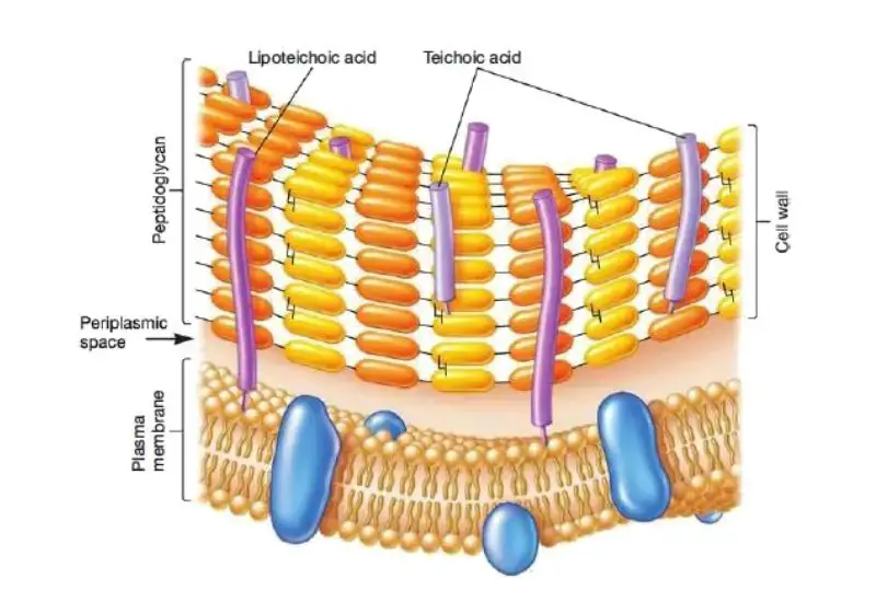
The cell wall of gram-positive bacteria is characterized by a thick peptidoglycan layer, ranging from 20 to 80 nm in thickness. This layer constitutes 40-90% of the cell wall’s dry weight, providing significant rigidity and structural integrity to the cell.
Key Components:
- Peptidoglycan Layer: The primary component of the cell wall, offering mechanical strength and shape to the bacteria. Its thickness can vary among different species.
- Teichoic Acid: Besides peptidoglycan, gram-positive bacteria have another crucial component, teichoic acid, which contains phosphorus. This polymer is embedded within the cell wall and contributes to its overall function and stability.
- Murein: Another term for the cell wall, highlighting its composition and function.
Function and Characteristics:
- Gram Staining: During the Gram staining process, gram-positive bacteria retain the crystal violet dye, resulting in a purple color. This staining method is pivotal for differentiating them from gram-negative bacteria.
- Lysozyme Sensitivity: The cell wall can be completely dissolved by lysozymes, which break the bond between N-acetylmuramic acid and N-acetylglucosamine. However, Staphylococcus aureus is an exception due to the presence of O-acetyl groups on some muramic acid residues, providing resistance to lysozymes.
- Types of Teichoic Acid: The cell wall contains ribitol teichoic acids and glycerol teichoic acids. These polymers, composed of ribitol phosphate and glycerol phosphate, are unique to the surface of gram-positive bacteria.
Cell wall structure of gram-negative bacteria
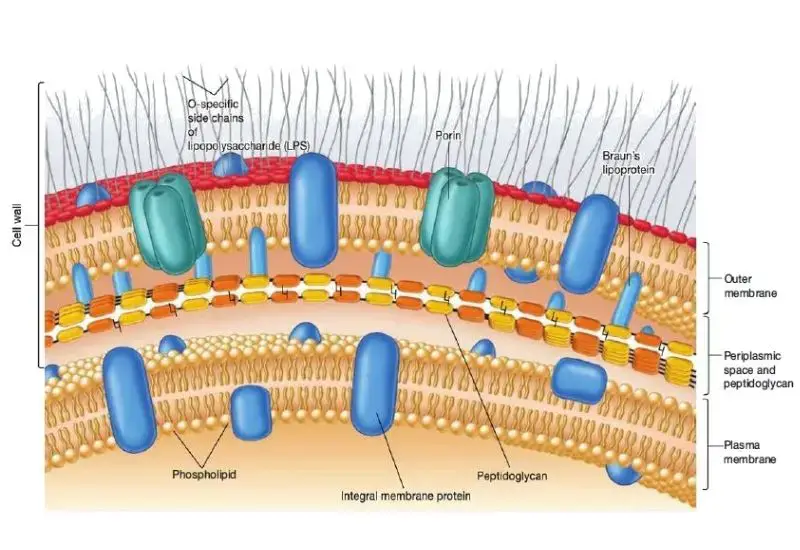
The cell wall of gram-negative bacteria is distinctively thinner compared to that of gram-positive bacteria. Despite its reduced thickness, it has a complex structure with multiple layers that contribute to its unique properties and functions.
Key Components:
- Peptidoglycan Layer: This layer is much thinner in gram-negative bacteria, comprising only 5-10% of the cell wall’s composition. It provides some structural integrity but is not as robust as in gram-positive bacteria.
- Outer Membrane: Superficial to the peptidoglycan layer is a second plasma membrane, known as the outer membrane. This outer layer is a bilayer consisting of proteins and lipopolysaccharides. The presence of this membrane is a defining characteristic of gram-negative bacteria.
- Periplasmic Space: The area between the cytoplasmic membrane and the outer membrane is called the periplasmic space. This space is filled with a loose network of lipoproteins and a significant number of enzymes and transport proteins, facilitating various cellular functions.
Functional Characteristics:
- Gram Staining: During Gram staining, gram-negative bacteria take up the safranin dye, resulting in a pink color. This distinguishes them from gram-positive bacteria, which stain purple.
- Absence of Teichoic Acid: Unlike gram-positive bacteria, gram-negative bacteria lack teichoic acid in their cell wall.
- Antigenic Properties: The lipopolysaccharides present in the outer membrane are responsible for many antigenic properties of gram-negative bacteria, playing a critical role in immune system interactions.
Structure and Function:
- The thin peptidoglycan layer provides essential structural support but is not the primary defensive barrier.
- The outer membrane serves as a formidable barrier against harmful substances, including antibiotics, and plays a crucial role in protecting the bacterial cell.
- The periplasmic space contains various enzymes and transport proteins, facilitating nutrient acquisition and metabolic processes.
Bacterial Cell Wall Structure
The bacterial cell wall is made up of different components which are divided into the following groups
A. Internal Component of Bacterial Cell Wall
1. Peptidoglycan
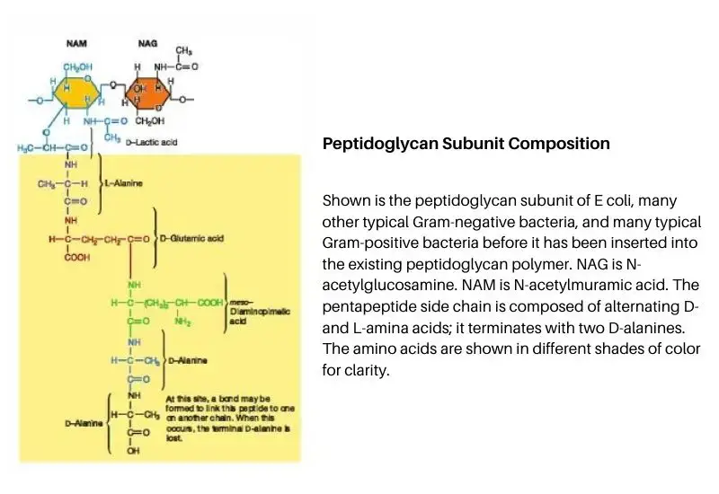
- The general composition of peptidoglycan differ from species to species, but there are basic similarities.
- Peptidoglycan is a very large polymers composed of three kinds of building blocks such as;
- Glycan backbone: 2 sugar derivatives linked by Beta-1,4-glycosidic linkage, called N-acetylglucosamine and N-acetyl muramic acid.
- Tetra-peptide: A tetrapeptide side chain of L-alanine, D-glutamine acid, L-lycine (gram-positive), or Mesodiaminopimelic acid (gram-negative).
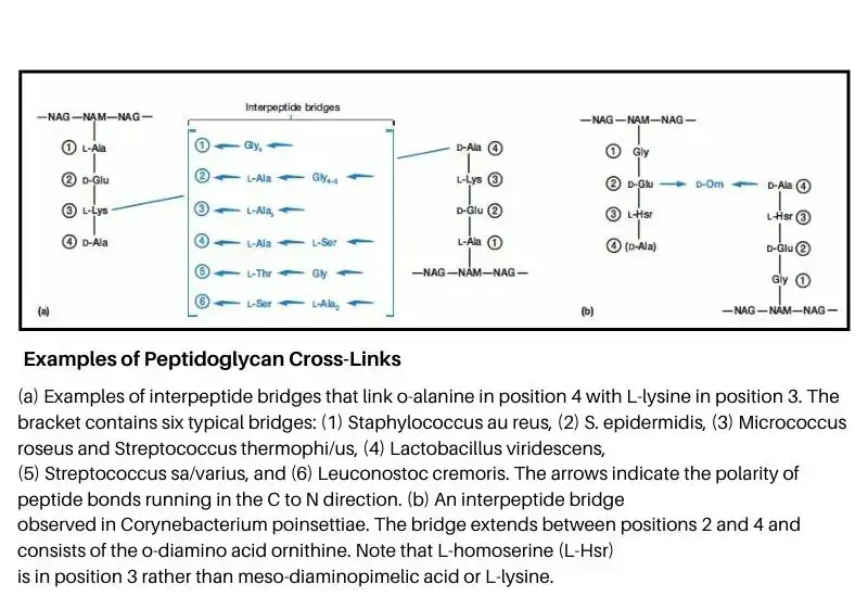
- Peptide cross-linkage: A pentapeptide cross-bridge of glycine (gram-positive) or direct peptide cross bridge.
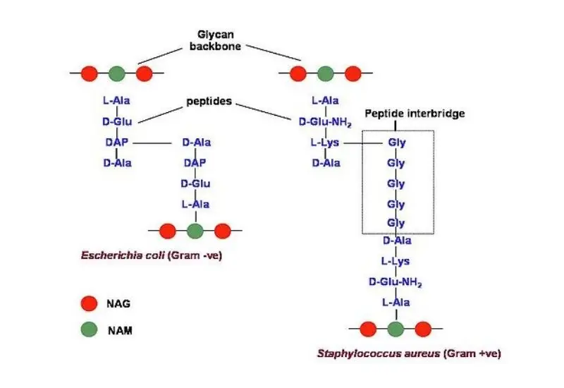
- The sugar derivatives are linked by Beta-1,4 linkage and then four amino acids linked two NAM serially (L-Alanin, D-Glutamic acid, L-Lycine/Mesodiaminopemilic acid, D-alanine).
- The third amino acid varies with different bacteria (In gram-positive bacteria L-lysine and In Gram-negative bacteria mesodiaminopemilic acid).
- In gram-positive bacteria, the parallel tetrapeptide side chains are linked by a pentapeptide (glycine) cross bridge that links the third amino acid of one tetrapeptide fourth amino acid of another tetrapeptide.
- In gram-negative bacteria, the amino acid group of the third amino acid is linked with the carboxyl group of the fourth amino acid of another tetrapeptide side chain by signal peptide bond.
- The rigidity of the cell wall depends on a number of cross-linked molecules.
The members of the phylum Chlamydiae and Planctomycetes both lack peptidoglycan in their cell walls. These are gram-negative strains.
2. Teichoic acid
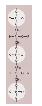
- Many gram-positive bacteria contain acidic polymers of sugar, teichoic acid embedded in their cell wall.
- Teichoic acid consists of alcohol (Ribital or glycerol) and phosphate.
- These poly alcohol are connected to phosphate by ester link and other sugar, alanine is attached to the carboxyl group.
- They are responsible for the negative charge of the cell wall and help the movement of ions through the cell wall, it also prevents the lysis of the cell wall.
- Teichoic acid composes about 50% of the dry weight of the cell wall.
- In gram-positive bacteria, Teichoic acid is the major surface antigen.
Types of Teichoic acid
There are three types of Teichoic acid such as;
- Ribitol teichoic acid (Staphylococcus aureus)
- Glycerol Teichoic acid (Bacillus subtilis)
- Glycerol Ribital teichoic acid (Bacillus licheniformis)
3. Lipo-protein
- Lipo-protein complex is found on the inner side of the outer membrane.
- Lipo-protein has a molecular weight of about 700 daltons and consists of 58 amino acids.
- In lipoprotein of the outer membrane, the protein binds to lipid by non-covalent bond while in lipoprotein of plasma membrane proteins binds to lipid by a covalent bond.
- Lipo-protein together with matrix protein form a complex which contains a channel called porins.
4. Lipopolysaccharide
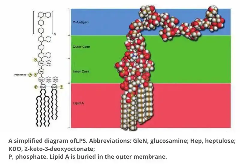
- The outer membrane of gram-negative bacteria is covered by lipo-polysaccharide, which is made up of polysaccharides covalently linked to lipid-A.
a. Lipid-A
- It consists of glucosamine, phosphate, and fatty acids. The Beta 1,6 linkage linked to glucosamine is the carbohydrates component lipid-A.
- The hydroxyl (-OH) and amino groups (-NH2) of these disaccharides are substituted by phosphate, pyro-phospet and fatty acids.
- The fatty acids provide hydrophilic property to lipid-A.
- There are 6 fatty acids – palmitic acid, lauric acid, myristic acid (Ratio 1:1:1), and 3 molecules of 3-hydroxy myristics acid.
b. Polysaccharide
- The polysaccharide portion of LPS of Salmonella cell wall consists of three important components such as inner core, outer core, and ‘O’-antigenic side chain.
- The ‘0’-antigenic side chain is absent in rough bacteria.
- The inner-core consists of two regions, the ket-deoxy-octane region (KDO) and Di Hepto region.
- The outer core consists of two molecules of GLC sandwiching a molecular galactose.
- The ‘O’-antigenic side-chain consists of four sugar Galactose, Rhamnose, mannose, and abequose.
- The O -antigenic side chain may be repeated many times according to species.
Functions
- It is responsible for the negative charge on the bacterial surface due to the presence of charged sugars and phosphate in core polysaccharide.
- It assists to maintain the outer membrane structure because lipid A is a primary component of the outer leaflet of the outer membrane.
- It assists to produce a permeability barrier. The geometry of LPS and communications between adjacent LPS units are thought to prevent the entrance of bile salts, antibiotics, and other toxic substances that might eliminate or injure the bacterium.
- It also protects the pathogenic bacteria front the host defense system. The O side chain of LPS is additionally termed as the O antigen because it evokes an immune response by an infected host. This response includes the formation of antibodies that adhere to the strain-specific form of LPS that evoked the response.
- The lipid A part of LPS can function as a toxin and is known as endotoxin. If LPS or lipid A invades the bloodstream, a pattern of septic shock produces for which there is no primary treatment.
Despite the part of LPS in forming a permeability barrier, the outer membrane is more porous than the plasma membrane and allows the entrance of small particles such as glucose and other monosaccharides. This happens due to the appearance of porin proteins.
B. External Component of Bacterial Cell Wall
The external components of the bacterial cell wall play essential roles in protection, motility, and interaction with the environment. These components vary among bacterial species and are crucial for various cellular functions.
1. Capsules
Capsules are extracellular layers surrounding the bacterial cell wall. They are composed of polysaccharides or proteins.
- Structure and Types:
- Capsule: When the layer is well-organized and firmly attached to the cell wall, it is termed a capsule. This structure is typically dense and resistant to removal.
- Slime Layer: If the extracellular layer is less organized and easily washed away, it is referred to as a slime layer. This layer is often more diffuse and loosely attached.
- Functions: Capsules and slime layers protect bacteria from phagocytosis by immune cells, help in adhering to surfaces, and can also prevent desiccation.
2. Flagella
Flagella are long, hair-like appendages that extend from the bacterial cell wall.
- Structure: Flagella are approximately 20-30 nanometers in diameter and can be up to 15 micrometers long. They are helical in shape and act as motility structures.
- Functions: Flagella enable bacterial motility by rotating and propelling the cell through its environment. This movement is crucial for bacteria to navigate toward favorable conditions or away from harmful substances.
3. Fimbriae and Pili
Fimbriae and pili are short, hair-like appendages that are distinct from flagella.
- Fimbriae: These are fine, straight, and rigid structures. They are generally shorter and thinner compared to flagella and are found in large numbers on the bacterial surface.
- Pili: Sometimes used interchangeably with fimbriae, pili are slightly thicker and can be involved in processes such as conjugation. They are 0.2-20 micrometers long and about 250 angstroms in diameter.
- Functions: Both fimbriae and pili are primarily involved in adherence to surfaces and other cells. They play a crucial role in the formation of biofilms and in the initial stages of infection.
4. Sheath
Sheaths are protective, tube-like structures found in certain aquatic bacteria.
- Composition: Sheaths are composed of polysaccharides and form a hollow, protective covering around the bacterial cell.
- Functions: Sheaths protect bacteria from being engulfed by other organisms and can aid in maintaining cell integrity in aquatic environments.
5. Surface Layer (S-Layer)
Surface Layers (S-Layers) are crystalline layers of proteins or glycoproteins that coat the outer surface of some bacteria.
- Composition: S-layers have a regular array with patterns that can resemble floor tiles or exhibit symmetries such as hexagonal or tetragonal.
- Functions:
- Protection: S-layers protect cells from environmental stresses, such as changes in ion concentration and pH.
- Shape Maintenance: They help maintain the cell’s shape and provide a barrier to external substances.
6. Prosthecae and Stalks
Prosthecae and stalks are tube-like appendages found in certain bacterial genera.
- Prosthecae: These are semi-rigid extensions of the cell wall and cytoplasmic membrane, found in bacteria such as Planctomyces.
- Stalks: Sometimes used interchangeably with prosthecae, stalks are similar appendages excreted by certain bacterial species.
- Functions:
- Nutrient Absorption: Prosthecae increase the surface area available for nutrient uptake.
- Attachment: Stalks assist in adhering to surfaces or substrates.
7. Outer Membrane
Outer Membrane is an additional membrane found in gram-negative bacteria, situated outside the thin peptidoglycan layer.
- Composition: It is composed of a lipid bilayer, proteins, and lipopolysaccharides (LPS). Braun’s lipoprotein connects the outer membrane to the peptidoglycan layer.
- Functions:
- Structural Component: It acts as a structural component, providing protection and maintaining cell integrity.
- Endotoxin Function: The LPS in the outer membrane, particularly lipid-A, can act as an endotoxin, contributing to the pathogenicity of gram-negative bacteria. Lipid-A is antigenic and plays a role in immune responses.
FAQ
What is the function of the bacterial cell wall?
The bacterial cell wall provides structural support to the cell and protects it from osmotic lysis. It also helps maintain the shape of the cell.
What is the composition of the bacterial cell wall?
The bacterial cell wall is primarily composed of peptidoglycan, which is a polymer consisting of alternating units of two sugars and short peptide chains.
How do gram-positive and gram-negative bacteria differ in terms of their cell wall structure?
Gram-positive bacteria have a thick cell wall consisting of multiple layers of peptidoglycan, while gram-negative bacteria have a thinner cell wall consisting of a single layer of peptidoglycan surrounded by an outer membrane.
What is the function of lipopolysaccharides in the outer membrane of gram-negative bacteria?
Lipopolysaccharides are important in the immune response and can cause toxic effects in some animals.
How do antibiotics target the bacterial cell wall?
Many antibiotics work by inhibiting the synthesis or function of peptidoglycan, which makes the cell wall weaker and more susceptible to lysis, ultimately leading to bacterial death.
Can bacteria survive without a cell wall?
Some bacteria, such as Mycoplasma, are able to survive without a cell wall, but they have a specialized cell membrane that provides them with structural support.
How do bacteria build and maintain their cell walls?
Bacteria use enzymes to synthesize and modify peptidoglycan, and they use energy from ATP to transport peptidoglycan precursors across the cell membrane and incorporate them into the cell wall.
Are there any human diseases caused by defects in the bacterial cell wall?
Yes, defects in the bacterial cell wall can lead to diseases such as tuberculosis and leprosy.
How do bacteria evolve resistance to antibiotics that target the cell wall?
Bacteria can evolve resistance to antibiotics that target the cell wall by modifying the enzymes that synthesize peptidoglycan or by developing mechanisms to pump out the antibiotic.
Can the bacterial cell wall be used as a target for new antibiotics?
Yes, the bacterial cell wall can be used as a target for new antibiotics. The cell wall is a unique structure found in bacteria that is essential for their survival and plays an important role in maintaining the structural integrity of the cell.
Many antibiotics target the cell wall by disrupting its synthesis or by damaging its structure, leading to the death of the bacterial cell. For example, penicillin and other beta-lactam antibiotics target the cell wall by inhibiting the enzymes responsible for the synthesis of the peptidoglycan layer, which is a major component of the cell wall in many bacterial species.
Other antibiotics, such as vancomycin and teicoplanin, target the cell wall by binding to and disrupting the structure of the peptidoglycan layer itself. These antibiotics are particularly effective against gram-positive bacteria, which have a thick peptidoglycan layer in their cell walls.
In recent years, there has been a growing interest in developing new antibiotics that target the bacterial cell wall, particularly as resistance to existing antibiotics continues to increase. Researchers are exploring various approaches, including the development of new beta-lactams, as well as non-beta-lactam compounds that target the cell wall.
References
- http://www.textbookofbacteriology.net/structure_5.html
- https://www.onlinebiologynotes.com/bacterial-cell-wall-structure-composition-types/
- https://en.wikipedia.org/wiki/Bacterial_cell_structure#Cell_wall
- https://www.microscopemaster.com/cell-wall.html
- https://byjus.com/biology/cell-wall/
- https://en.wikipedia.org/wiki/Cell_wall