What is Amoeba?
- Amoebae, the extraordinary unicellular organisms, possess a remarkable ability to alter their shape at will. This fascinating characteristic is achieved through the extension and retraction of pseudopods, which are bulges of cytoplasm controlled by actin microfilaments. These pseudopods push out the plasma membrane surrounding the cell, facilitating movement and feeding for the amoeba.
- Amoebae are not restricted to a single taxonomic group; instead, they can be found in various major lineages of eukaryotic organisms. These versatile cells occur among the protozoa, fungi, algae, and animals, making them a diverse and widely distributed group.
- Microbiologists often use the terms “amoeboid” and “amoeba” interchangeably when referring to any organism that exhibits amoeboid movement. However, due to molecular phylogenetic studies, the older classification system that grouped most amoebae into the class Sarcodina has been disproven. As a result, amoeboid organisms are now categorized separately.
- Some of the most well-known amoeboid protists include Chaos carolinense and Amoeba proteus, which have been extensively studied in classrooms and laboratories. Other notable species are the infamous “brain-eating amoeba” Naegleria fowleri, the intestinal parasite Entamoeba histolytica responsible for amoebic dysentery, and the intriguing multicellular “social amoeba” or slime mould Dictyostelium discoideum.
- One key feature distinguishing different groups of amoebae is the appearance and internal structure of their pseudopods. For instance, amoebae in the genus Amoeba typically have bulbous (lobose) pseudopods, while Cercozoan amoeboids like Euglypha and Gromia have slender, thread-like (filose) pseudopods. Foraminifera emit fine, branching pseudopods that merge to form net-like (reticulose) structures. In contrast, some groups like the Radiolaria and Heliozoa have stiff, needle-like, radiating axopodia (actinopoda) supported by microtubule bundles.
- Amoebae showcase a fascinating range of external coverings. They may be “testate,” enclosed within a protective shell made of substances such as calcium, silica, chitin, or agglutinations of found materials like grains of sand and diatom frustules. On the other hand, “naked” amoebae, also known as gymnamoebae, lack any hard covering, revealing their bare form.
- To maintain osmotic pressure and prevent cell rupture, most freshwater amoebae possess a contractile vacuole. This organelle expels excess water from the cell, ensuring that the surrounding hypotonic water does not overwhelm the amoeba’s internal fluids. In contrast, marine amoebae do not usually possess a contractile vacuole, as the solute concentration within the cell is balanced with the tonicity of the surrounding water.
- Amoebae, with their exceptional shape-shifting abilities and diverse habitats, continue to intrigue scientists and inspire awe among those who study these fascinating microorganisms. Their survival strategies and cellular adaptations make them true masters of the microscopic world, and they remain a subject of endless curiosity and exploration in the realms of biology and beyond.
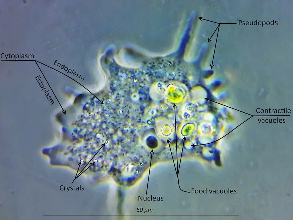
Amoeba of Structure
Amoebas, as eukaryotic organisms, possess a cellular structure that sets them apart in the microscopic world. Under the microscope, the architectural marvels of these single-celled wonders come to life, particularly when observed after staining. Their eukaryotic nature is characterized by a well-defined cell membrane encompassing their cytoplasm, housing the DNA within a central nucleus.
Under microscopic scrutiny, the distinctive components of amoebas become clearly visible, offering valuable insights into their cellular organization. Staining techniques further enhance the visibility of these components, enabling researchers to unlock the secrets of amoeba biology.
Some of the key organelles that stand out when viewing amoebas under the microscope include:
- Nucleus: The nucleus is the control center of the amoeba’s cell. It houses the organism’s genetic material, the DNA, which contains the instructions for essential cellular functions and characteristics. This central nucleus serves as a pivotal hub for regulating amoeba activities.
- Food Vacuole: Food vacuoles are observable within amoebas, highlighting their unique feeding mechanism. Amoebas are known for their phagocytic nature, engulfing and absorbing food particles through the process of endocytosis. Food vacuoles are temporary structures that contain the ingested particles and facilitate the digestion of nutrients.
- Cytoplasm: The cytoplasm fills the interior of the amoeba’s cell, serving as the site of various cellular activities. It houses the organelles and acts as a medium for biochemical reactions, ensuring essential cellular processes take place efficiently. The staining techniques aid in distinguishing the cytoplasm from other cellular components.
- Contractile Vacuole: Another intriguing feature visible under the microscope is the contractile vacuole. This organelle plays a crucial role in regulating the amoeba’s osmotic pressure. It expels excess water from the cell, preventing it from bursting due to osmotic imbalances. The contractile vacuole is particularly prominent in freshwater amoebas.
The Remarkable Resemblance:
Amoebae, in their structural complexity, bear striking similarities to cells found in higher animals, including humans. Despite their single-celled status, amoebas display organizational features that are reminiscent of more complex organisms. This similarity highlights the fundamental nature of cellular organization and function across various forms of life.
In conclusion, the structure of amoebas presents a captivating display of eukaryotic wonders under the microscope. Their cell membrane-enveloped cytoplasm and centrally located nucleus are hallmarks of eukaryotic life. Staining techniques further unveil the intricacies of amoeba organelles, revealing the nucleus, food vacuoles, cytoplasm, and the vital contractile vacuole. As researchers continue to explore the mysteries of these enigmatic microorganisms, the resemblance of amoebae to higher animal cells underscores the unity and interconnectedness of life on a cellular level.
Amoeba Microscopy
In the mesmerizing realm of microscopic life, amoebas reign as captivating single-celled organisms. To catch a glimpse of these elusive creatures, one must turn to the wonders of microscopy. Amoebas, being minuscule, demand the aid of specialized techniques for observation, unlocking their secrets for eager scientists and students alike.
The primary approach to amoeba microscopy is the simplest yet most intriguing one: observing them under the microscope without any staining. This method offers the unique opportunity to witness these living organisms in action as they gracefully move about. Watching amoebas glide and extend their pseudopods, the cytoplasmic projections that enable their shape-shifting movement, is a sight to behold. This technique grants a real-time window into their natural behavior, offering invaluable insights into their life processes.
However, to delve deeper into the intricacies of amoeba structure and uncover the hidden nuances of their organelles, a second method comes into play. This advanced technique involves fixing and staining the amoebas. Fixing refers to preserving the amoebas in a specific state, often by using chemical agents, to halt any further biological activity and preserve their structure. Staining, on the other hand, employs colored dyes or stains that selectively target specific cellular components, making them more visible under the microscope.
By fixing and staining the amoebas, scientists can obtain a clearer and more detailed view of their internal architecture. The stains highlight the various organelles within the amoebas, such as the nucleus, mitochondria, and contractile vacuoles, among others. This method allows for enhanced scrutiny of amoeba morphology, aiding researchers in identifying key cellular components and unraveling the intricate mechanisms that govern their functioning.
Amoeba microscopy plays a pivotal role in educational settings, enabling students to explore the hidden world of these microorganisms. By peering through the microscope lens, students can witness firsthand the dynamic and vibrant nature of amoebas. These observations spark curiosity and encourage a deeper understanding of cellular biology and the intricate processes governing life at the microscale.
Moreover, amoeba microscopy serves as a valuable tool for researchers in diverse fields. The insights gained from studying amoebas can contribute to a broader understanding of cell biology, evolution, ecology, and even medical research. Additionally, amoebas’ versatility and adaptability make them fascinating subjects for investigating various scientific phenomena and cellular behaviors.
1. Simple or Direct Method
Requirement
- A Sample of Water Collected from a Pond with Organic Material: The first step in the direct method of observing amoebas is to obtain a water sample from a pond teeming with life. Amoebas flourish in such environments, and this pond water will serve as the natural habitat for these microscopic organisms. The presence of organic material in the water provides the amoebas with nourishment, making it an ideal place to find these elusive creatures.
- Pondweed from a Pond: In addition to the water sample, acquiring some pondweed from the same environment further enhances the chances of spotting amoebas. Pondweed serves as a valuable source of nutrients and provides an environment that supports the existence of these intriguing microorganisms.
- Petri Dish: A Petri dish acts as a miniature laboratory for microscopic explorations. It provides a controlled environment where the water sample and pondweed can be placed for observation under the microscope. The clear and flat surface of the Petri dish allows for easy positioning of the specimens and ensures a stable viewing platform.
- A Compound Light Microscope: A fundamental tool in any microscopy endeavor, a compound light microscope is indispensable for studying amoebas. This type of microscope employs multiple lenses to magnify the specimen, enabling the observation of tiny details at high resolutions. With the aid of a compound light microscope, the invisible world of amoebas becomes accessible and captivating.
- Water: Maintaining the water sample’s moisture and integrity is crucial during the observation process. Having some additional water on hand ensures that the specimens stay hydrated and that any adjustments to the microscope’s focus can be made without disturbing the amoebas.
- A Dropper: A dropper proves invaluable for transferring the water sample containing amoebas and pondweed to the Petri dish. This delicate tool allows for precise and controlled handling of the specimens, ensuring they are adequately placed for optimal viewing under the microscope.
Procedure for Microscopy
Amoeba Culture Procedure:
- Prepare the Amoeba Culture: To initiate the amoeba culture, carefully place a few pondweeds into a clean and sterile Petri dish. Ensure that the pondweeds are adequately covered with water, creating a suitable environment for amoebas to thrive.
- Incubate the Culture: Allow the Petri dish to rest undisturbed in a dark room for a few days. During this incubation period, nature will work its magic, leading to the formation of a brown scum on the surface of the water. This scum is an indicator that the amoebas have multiplied and flourished in the culture.
Microscopy Procedure:
- Prepare the Microscope Glass Slide: Using a dropper, carefully place a few drops of the sample onto a clean microscope glass slide. The sample can be either a small amount of pond water collected directly or a portion of the brown scum from the amoeba culture.
- Cover and Secure the Specimen: Gently cover the sample on the microscope glass slide with a thin, transparent cover slip. This ensures that the specimen is well-preserved and prevents it from drying out during the observation process. Take care to avoid air bubbles between the sample and the cover slip, as they may interfere with the observation.
- Mount the Slide on the Microscope: Place the prepared slide with the specimen on the microscope stage. Carefully secure it in place, ensuring the slide is positioned correctly to allow for easy adjustment of the microscope’s focus.
- Begin Observation: Start the observation process with the microscope set at low power. Gradually increase the magnification as needed to obtain a clearer and more detailed view of the amoebas. Using the coarse and fine focus adjustments, bring the amoebas into sharp focus, revealing their intricate structures and movements.
As the microscopic world unfolds before your eyes, take the time to marvel at the elegance and complexity of these single-celled organisms. Witness the graceful dance of amoebas as they extend and retract their pseudopods, and uncover the mysteries of their cellular structures through varying magnifications.
The simple or direct method procedure allows for a fascinating journey into the microscopic realm of amoebas, igniting curiosity and passion for the wonders of nature at its smallest scale. Through careful observation and exploration, students and scientists alike gain a deeper appreciation for the intricacies of life hidden within a drop of water.
Observation
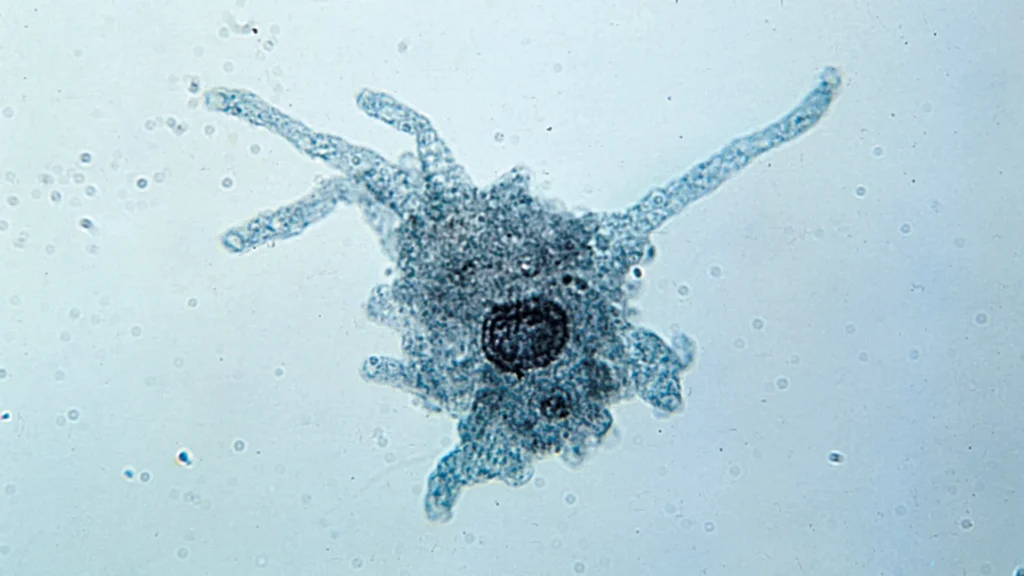
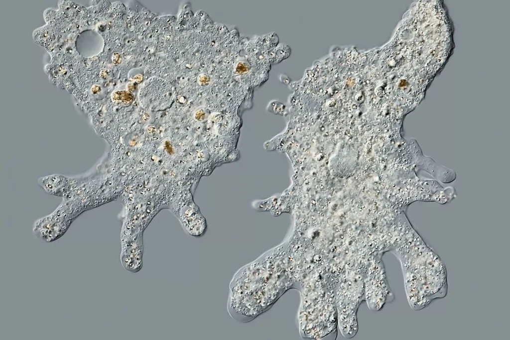
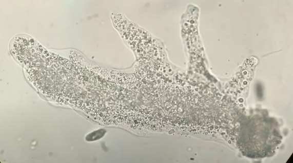
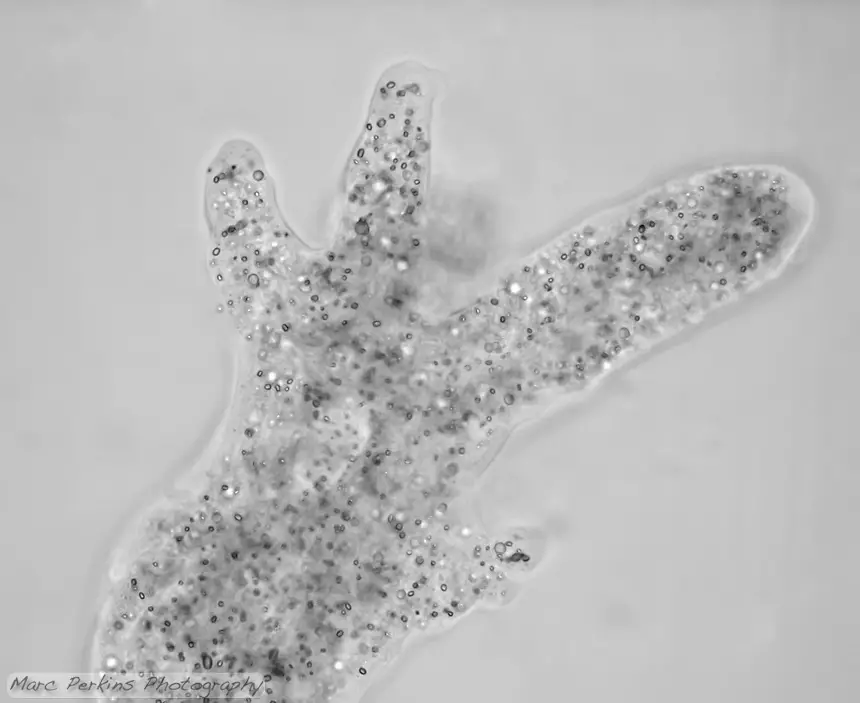
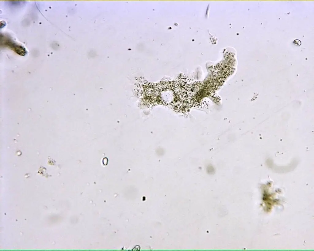
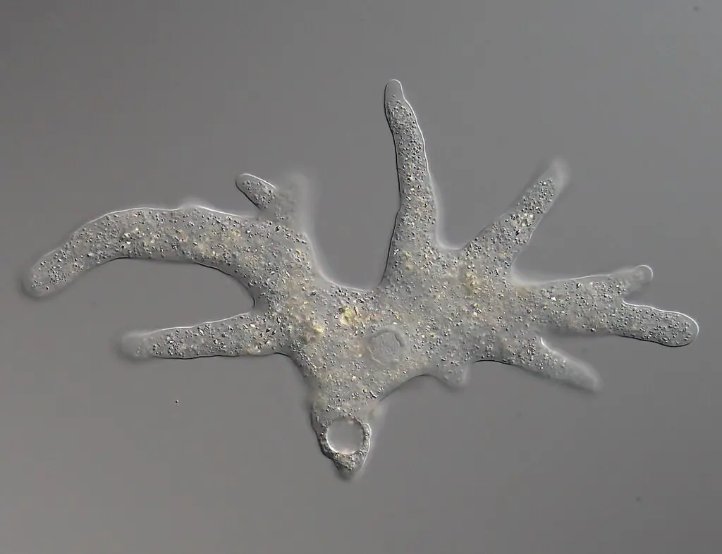
Through the simple or direct method of microscopy, the microscopic world of amoebas unfolds, offering a mesmerizing spectacle of colorless, transparent entities gracefully gliding across the field. Observing amoebas under the microscope reveals a captivating dance of shape-shifting, as they extend and withdraw long, finger-like projections known as pseudopods.
As you focus the microscope, the amoebas come into view, resembling elusive, jelly-like creatures. Their colorless, transparent appearance adds an air of mystery to their existence, as if they are ethereal beings, moving at their own pace in their microscopic realm.
The enchanting aspect of amoebas lies in their unique method of locomotion. As they change shape, the amoebas elegantly protrude and withdraw their pseudopods. These finger-like projections serve as versatile appendages, enabling them to move with grace and precision. Like skilled artists, amoebas craft their movements, molding their form to navigate through the aquatic world they call home.
Observing amoebas under the microscope is an exercise in patience and appreciation for the subtleties of life at the cellular level. Their unhurried pace may seem slow to the impatient observer, but this deliberate movement holds the key to understanding their fascinating biology.
As you continue to watch these captivating microorganisms, you might notice how they respond to changes in their environment. They gracefully adapt their shape and movements to interact with their surroundings, a testament to their resilience and adaptability in the face of ever-changing conditions.
In this realm of the infinitesimal, the simplicity of the direct method of observation reveals the intricacies of amoeba life. Their microscopic world becomes a captivating canvas, painted with the strokes of shape-shifting and propelled by the rhythmic dance of pseudopods.
Observing amoebas through the simple or direct method evokes a sense of wonder and awe at the beauty of life, even at its tiniest scale. These unassuming organisms hold the secrets of survival and adaptation, reminding us of the intricate tapestry of existence that surrounds us.
2. Fixing and Staining Technique for Observation of Amoeba
Requirements
- Amoeba Culture: The process begins with a thriving amoeba culture, providing a stable and controlled environment for these microorganisms to flourish. A well-established amoeba culture ensures a steady supply of specimens for the fixing and staining process.
- A Bench-top MSE Centrifuge: A bench-top MSE centrifuge is a crucial piece of laboratory equipment for separating amoebas from the culture medium. This apparatus employs centrifugal force to separate the amoebas gently and efficiently, ensuring that only the desired specimens are collected for further analysis.
- Neff’s Saline, Sea Water, and Distilled Water: Various solutions play key roles in the fixing and staining process. Neff’s saline, sea water, and distilled water are essential for preparing different fixatives, stains, and washing solutions, ensuring that the amoebas are adequately treated and preserved for observation.
- Moist Chamber: Maintaining the ideal humidity and conditions for the specimens during the fixing and staining procedure is vital. A moist chamber serves this purpose, providing a controlled environment that prevents the amoebas from drying out during the process.
- Cover Slip: Cover slips are thin, transparent pieces of glass that shield the fixed and stained amoebas on the microscope slide. They help protect the specimens from contamination and allow for precise microscopic observation.
- Sodium Hydroxide (NaOH): Sodium hydroxide is a crucial component used in preparing certain fixatives and stains. It aids in the preservation and enhancement of the amoeba specimens, ensuring their structural integrity for accurate analysis.
- Nissenbaum’s Fixative, Lugol’s Iodine, Formalin Seawater, and Carnoy’s Fixative: These specialized fixatives are vital for preserving the amoebas’ cellular structures and preventing degradation during the staining process. Each fixative has unique properties that make it suitable for specific types of amoeba analysis.
- Heidenhain’s Iron Haematoxylin: This specialized stain is employed to enhance the contrast and visibility of amoeba cell structures. Heidenhain’s iron haematoxylin provides invaluable insights into the cellular components, aiding researchers in identifying and studying the finer details of amoebas.
Procedure for Fixing and Staining Technique
The fixing and staining technique provides a powerful method to delve into the microscopic world of amoebas, unraveling the intricacies of their cellular structures and behaviors. To embark on this scientific journey, the following step-by-step procedure is followed:
- Amoeba Culture and Concentration: Begin by culturing the amoebas using a suitable culture medium and saline agar slopes. Once the culture is established, concentrate the specimen by using a centrifuge at 3 Krpm (thousand revolutions per minute) for approximately 10 minutes. This step separates the amoebas from the culture medium, yielding a more concentrated sample.
- Washing the Pellet: Wash the concentrated pellet of amoebas using a suitable solution such as 75 percent seawater, Neff’s saline, or any other appropriate medium. This washing step helps remove unwanted debris and culture medium, leaving behind a purified specimen.
- Settling the Specimen: Allow the washed specimen to settle on a clean cover slip within a moist chamber. Observe the amoebas closely until they adopt a normal locomotionary morphology. Depending on the type of amoeba being studied, the cover slip may be treated with sodium hydroxide to optimize observation conditions.
Fixing the Specimen:
- Applying Nissenbaum’s Fixative: While the specimen settles on the cover slip, carefully pipette freshly prepared Nissenbaum’s Fixative onto the sample. Allow the fixative to stand for approximately 5 minutes, ensuring proper preservation of the amoebas’ cellular structures.
- Acidified HgCl2 Wash: Wash the fixed sample with acidified HgCl2 for about 7 minutes. This step aids in removing excess fixative and prepares the amoebas for subsequent staining.
- Ethanol Washes: Conduct a series of ethanol washes with concentrations of 50%, 35%, and 15% for approximately 5 minutes each. These washes help in gradual dehydration of the specimen, ensuring optimal staining conditions.
- Final Distilled Water Wash: Conclude the fixing process by washing the sample again with distilled water for about 5 minutes. This step removes any residual ethanol and prepares the specimen for the staining phase.
Alternative Fixatives:
In addition to Nissenbaum’s Fixative, other fixatives that can be used include Carnoy’s Fixative, Lugol’s Iodine, and Seawater formalin. Each fixative offers unique advantages for specific amoeba studies, contributing to the diversity of fixation techniques.
Staining the Specimen:
The aim of staining is to enhance the visibility of mitotic figures and cellular structures in the amoebas. Various stains can be used for this purpose, including Heidenhain’s iron Haematoxylin, Kernechtrot (Nuclear Red), Modified Field’s Stain, and Klein’s Silver relief stain.
For Heidenhain’s iron Haematoxylin staining, the following steps are involved:
- Inoculation in Ammonium Ferric Sulphate: Inoculate the sample in 2 percent ammonium ferric sulphate for approximately 1 and a half hours. This step prepares the specimen for the subsequent staining process.
- Distilled Water Rinse: Rinse the sample in distilled water to remove excess ammonium ferric sulphate.
- Inoculation in Haematoxylin Solution: Inoculate the sample in 0.5% haematoxylin and 2% ammonium ferric sulphate for about one and a half hours. This step allows for effective staining of cellular structures.
- Tap Water Wash: Conclude the staining process by washing the sample with tap water, ensuring proper removal of the staining solution.
After the staining process is complete, mount the slide on the microscope stage for observation. The staining technique enhances the visibility of key cellular components, providing valuable insights into amoeba biology and cellular behaviors.
Observation
The fixing and staining technique offers a unique perspective into the microscopic realm of amoebas, providing valuable insights into their cellular structures and behaviors. As students peer through the microscope lens, they are greeted with intriguing observations, each method offering distinct advantages and revealing different aspects of these captivating single-celled organisms.
Direct Observation without Staining:
When viewing amoebas under the microscope without staining, students are presented with a living, motile spectacle. The absence of staining allows the amoebas to retain their natural colorless and transparent appearance. Tiny dark spots become visible in the cytoplasm, hinting at the presence of cellular components within.
The great advantage of direct observation lies in witnessing the dynamic movement of amoebas in real-time. As students observe, they notice the cytoplasm extending and retracting, forming finger-like projections known as pseudopods. These pseudopods elongate and shorten as the amoeba gracefully moves about its micro-environment or engulfs substrates, providing a captivating display of amoeba locomotion.
However, direct observation has its limitations. While the movement is a fascinating sight to behold, it does not grant students a clear view of the amoeba’s organelles or finer cellular structures. These intricate components remain elusive and obscured, requiring further techniques to unlock their secrets.
Fixing and Staining for Enhanced Contrast:
In contrast, fixing and staining the amoebas sacrifices their motility, as the process involves killing the organisms. Though the amoebas are no longer alive and active, this method offers a critical advantage: enhanced contrast. Staining provides students with a clearer view of the organelles within the amoeba’s cell.
With staining, specific cellular structures become more visible, aiding students in identifying and understanding the organelles responsible for various cellular processes. Stains such as Heidenhain’s iron Haematoxylin, Kernechtrot (Nuclear Red), Modified Field’s Stain, and Klein’s Silver relief stain highlight key components, shedding light on the inner workings of these enigmatic microorganisms.
The Trade-off:
As with many scientific techniques, there is a trade-off between the two observation methods. Direct observation allows students to witness the amoebas’ graceful movements and their pseudopods in action but lacks clarity when it comes to organelles. Fixing and staining, on the other hand, provide enhanced visibility of organelles but sacrifice the amoebas’ live movements.
In conclusion, the observation of amoebas using the fixing and staining technique presents a fascinating journey into the microscopic world. Each method, whether direct observation without staining or fixing and staining for enhanced contrast, offers its unique advantages. Witnessing amoebas in motion through direct observation allows for a deeper appreciation of their motility and pseudopod dynamics. On the other hand, fixing and staining provide a clearer view of cellular organelles, offering valuable insights into amoeba biology. By combining both methods, students can explore the multifaceted nature of amoebas, unraveling the hidden secrets that make them truly exceptional organisms in the intricate tapestry of life.
Amoeba Under The Microscope Video
FAQ
What is an amoeba?
An amoeba is a single-celled, eukaryotic organism known for its ability to alter its shape through the extension and retraction of pseudopods. It belongs to the group of organisms called protozoa and can be found in various aquatic environments.
How do I observe an amoeba under the microscope?
To observe an amoeba under the microscope, you can collect a sample of pond water containing these microorganisms. Place a drop of the water on a microscope slide, cover it with a cover slip, and then observe the slide under the microscope using the appropriate magnification.
What can I see when I observe an amoeba under the microscope?
When you observe an amoeba under the microscope, you may see a colorless, transparent organism with constantly changing shape as it extends and retracts its pseudopods. If you use staining techniques, you may also be able to observe specific cellular structures, such as the nucleus and food vacuoles.
What magnification is suitable for observing amoebas?
For observing amoebas, a low to medium magnification (e.g., 100x to 400x) is usually sufficient. Lower magnifications allow you to see the entire amoeba and its movements, while higher magnifications enable you to study cellular structures in more detail.
Can I observe live, motile amoebas under the microscope?
Yes, you can observe live, motile amoebas under the microscope without staining. Direct observation of living amoebas allows you to witness their graceful movements and pseudopod dynamics in real-time.
What is the benefit of fixing and staining amoebas?
Fixing and staining amoebas preserve the specimens for better visualization of cellular structures. Staining enhances contrast, enabling you to see organelles and other microscopic details that may not be as visible during live observation.
What stains are commonly used for staining amoebas?
Some common stains used for staining amoebas include Heidenhain’s iron Haematoxylin, Kernechtrot (Nuclear Red), Modified Field’s Stain, and Klein’s Silver relief stain. Each stain has specific properties that highlight different cellular components.
Can I see the amoeba’s nucleus under the microscope?
Yes, the nucleus of an amoeba is visible under the microscope, especially after staining. The nucleus serves as the control center of the cell and contains the genetic material (DNA) of the organism.
What is the contractile vacuole in amoebas?
The contractile vacuole is a specialized organelle found in certain amoebas, particularly those living in freshwater environments. It helps regulate osmotic pressure by expelling excess water from the cell to prevent it from bursting.
Are amoebas similar to cells found in higher animals?
Yes, despite being single-celled organisms, amoebas have structural similarities to cells found in higher animals, including humans. These similarities highlight the fundamental nature of cellular organization and function across different forms of life.
- Text Highlighting: Select any text in the post content to highlight it
- Text Annotation: Select text and add comments with annotations
- Comment Management: Edit or delete your own comments
- Highlight Management: Remove your own highlights
How to use: Simply select any text in the post content above, and you'll see annotation options. Login here or create an account to get started.