Albert stain is a differential staining technique used to observe the metachromatic granules that is present inside certain bacteria, especially Corynebacterium diphtheriae. It is the granules which is also referred to as volutin granules or Babes-Ernst granules.
These granules is acidic in nature and the basic dyes present in the staining solution get attracted to it. The staining procedure is done with two solutions known as Albert solution A and Albert solution B. Solution A contains basic dyes like Toluidine blue and Malachite green along with glacial acetic acid and alcohol. It is the dyes which help in colouring the granules inside the cytoplasm. Solution B contains iodine and potassium iodide acting as the mordant which fix the stain and prevent the granules from losing colour.
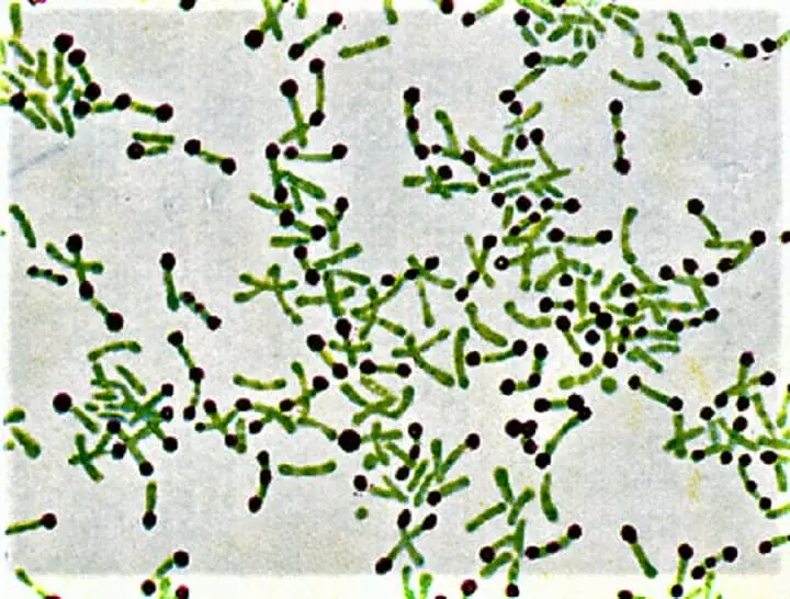
It is the stain which is used mainly for the detection of C. diphtheriae in clinical samples like throat swab or nasal swab. The organisms is seen as green coloured rods arranged in V or L shape (Chinese letter pattern) and the bluish-black granules appears at the poles. This is referred to as a presumptive test for diphtheria because pathogenic diphtheria bacteria possess these granules while non-pathogenic diphtheroids generally lack them. It allows rapid identification which is important for early treatment and prevention of complications.
The history of Albert stain is linked to the early studies of diphtheria when the need was arise to differentiate the Klebs-Loeffler bacillus from other similar organisms. It is named after the scientist who developed or modified the technique. Some sources refer to H. Albert around 1920–1921 while others mention Friedrich von Albert in 1884. The stain became an important tool because it allowed easy visualization of metachromatic granules and helped in tracking the disease outbreaks in the early days of microbiology.
Principle of Albert stain
The principle of Albert stain is based on the differential staining of the metachromatic granules that is present in Corynebacterium diphtheriae. It is the granules which contain polymetaphosphate and these are highly acidic in nature, while the cytoplasm of the cell is relatively neutral. In this method two solutions are used, where Albert solution I contains Toluidine blue, Malachite green and glacial acetic acid. The glacial acetic acid lowers the pH of the stain and creates an acidic medium. It is this acidic condition which allows the basic dye Toluidine blue to get attracted strongly to the acidic granules.
The Malachite green stains the cytoplasm lightly so that the granules can be distinguished clearly from the rest of the cell. After this the Albert solution II which contains iodine is added. This step is referred to as the mordant action. It is the iodine that fixes the dye to the granules and prevents the colour change known as metachromasia. In this way the granules is seen as bluish-black or purple-black structures, while the bacterial body is seen green in colour.
Composition of Albert Stain
Albert stain is used for staining Corynebacterium diphtheriae and it is the technique where two solutions is applied in sequence. It is the process where the cytoplasm and the metachromatic granules is stained differently so the organism can be identified easily.
Albert’s Solution 1 (Staining Solution)
This is the primary staining solution. It is acidic in nature and contains basic dyes which stain the cytoplasm and the granules in different manner. The acidity is maintained by glacial acetic acid so the toluidine blue only goes to the volutin granules while malachite green stains the cytoplasm.
Some of the important ingredients are–
- Toluidine Blue – It is basic dye and it stains the acidic volutin granules. These granules is highly acidic so they take this dye strongly.
- Malachite Green – It stains the cytoplasm light green. It gives a background colour so granules become visible.
- Glacial Acetic Acid – It is used to maintain acidic pH (around 2.8). It is the process where cytoplasm staining by toluidine blue is prevented so only granules take that dye.
- Alcohol (95% Ethanol) – It is used as solvent for dissolving dyes.
- Distilled Water – It is used as diluent.
The usual composition includes–
Toluidine blue (0.15 g), Malachite green (0.20 g), Glacial acetic acid (1 ml sometimes more), Alcohol (2–20 ml), and Distilled water (100 ml).
Preparation of 100ml Albert stain 1
- Add 0.1ml of glacial acetic acids in 100ml of water.
- Incorporate 2ml of ethanol that is 95% into the solution.
- Then dissolve 0.15g of blue toluidine into the solution.
- Then take a moment to dissolve 0.2g of green malachite into the solution
Albert’s Solution 2 (Mordant)
This is the fixing solution. It is used after draining solution 1. It is the process where iodine prevents metachromasia and fixes the granules as blue-black.
Some of the main ingredients are–
- Iodine Crystals – These is the mordanting agent.
- Potassium Iodide – It helps iodine dissolve in water.
- Distilled Water – It acts as solvent.
The usual composition is Iodine (2 g), Potassium iodide (3 g), and Distilled water (300 ml).
Preparation of 300ml of Albert stain 2
- Dissolve 2g of Iodine in 50ml of distillate water
- Add 250ml water to the solution.
- Dissolve 3g Potassium Iodide into the solution.
Functional Insight
Some of the main features of the stain is–
- Selective Staining – Because of the acetic acid, cytoplasm is stained mainly by malachite green, while toluidine blue only goes to the volutin granules. These granules is made of polymetaphosphate so they remain highly acidic.
- Colour Stabilization – Normally toluidine blue can show metachromasia (reddish-purple colour). But when iodine mordant is added it forms a complex and prevents colour shift. The granules is fixed as bluish-black or purple-black while cytoplasm stays green.
This is referred to as a differential staining process which helps in identifying the characteristic granules of Corynebacterium diphtheriae.
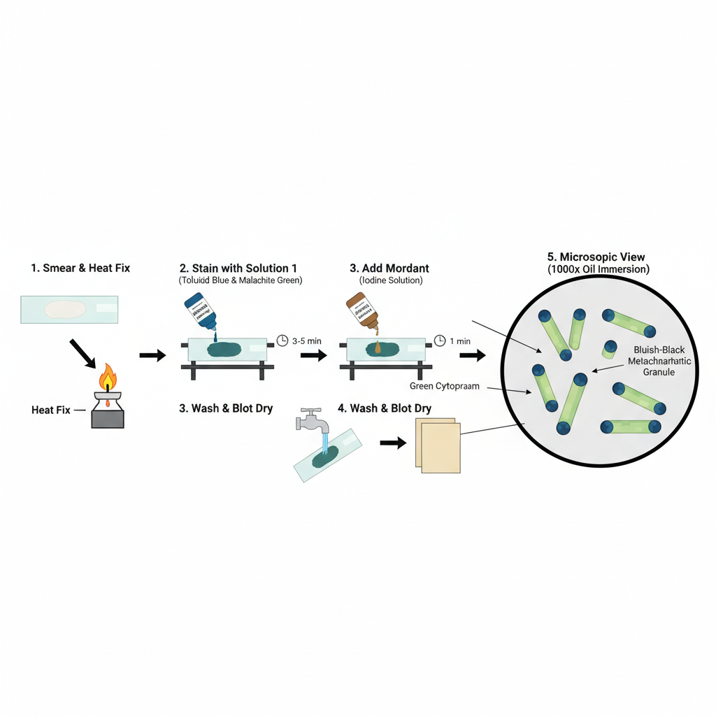
Procedure for Albert Stain

- Preparation of smear
- A loopful culture of Corynebacterium diphtheriae is taken aseptically and a thin smear is made at the center of a clean glass slide. It is allowed to air dry completely.
- The smear is then heat fixed gently by passing over the flame two or three times.
- Staining (Solution–1)
- The fixed smear is placed on a staining rack.
- Albert staining solution 1 is added over the smear so that it is fully covered. It is the solution containing toluidine blue and malachite green.
- It is kept for about 3–5 minutes.
- In this step the cytoplasm is stained and the granules start appearing. The extra stain is drained.
- It is important in this stage that the slide is not washed with water because the malachite green is water soluble and it is lost easily.
- Mordanting (Solution–2)
- Albert staining solution 2 is added on the smear. This solution contains iodine and potassium iodide and it act as mordant.
- It is kept for about 1 minute. It helps in fixing the dye to the granules so that the granules remain bluish black.
- After this step the slide is washed gently with tap water.
- Drying and observation
- The slide is blotted carefully. A drop of cedarwood oil is added on the smear.
- It is observed under the microscope using oil immersion at 1000x.
- The metachromatic granules appear bluish-black while the cytoplasm appears greenish.
Result and Interpretation of Albert Stain

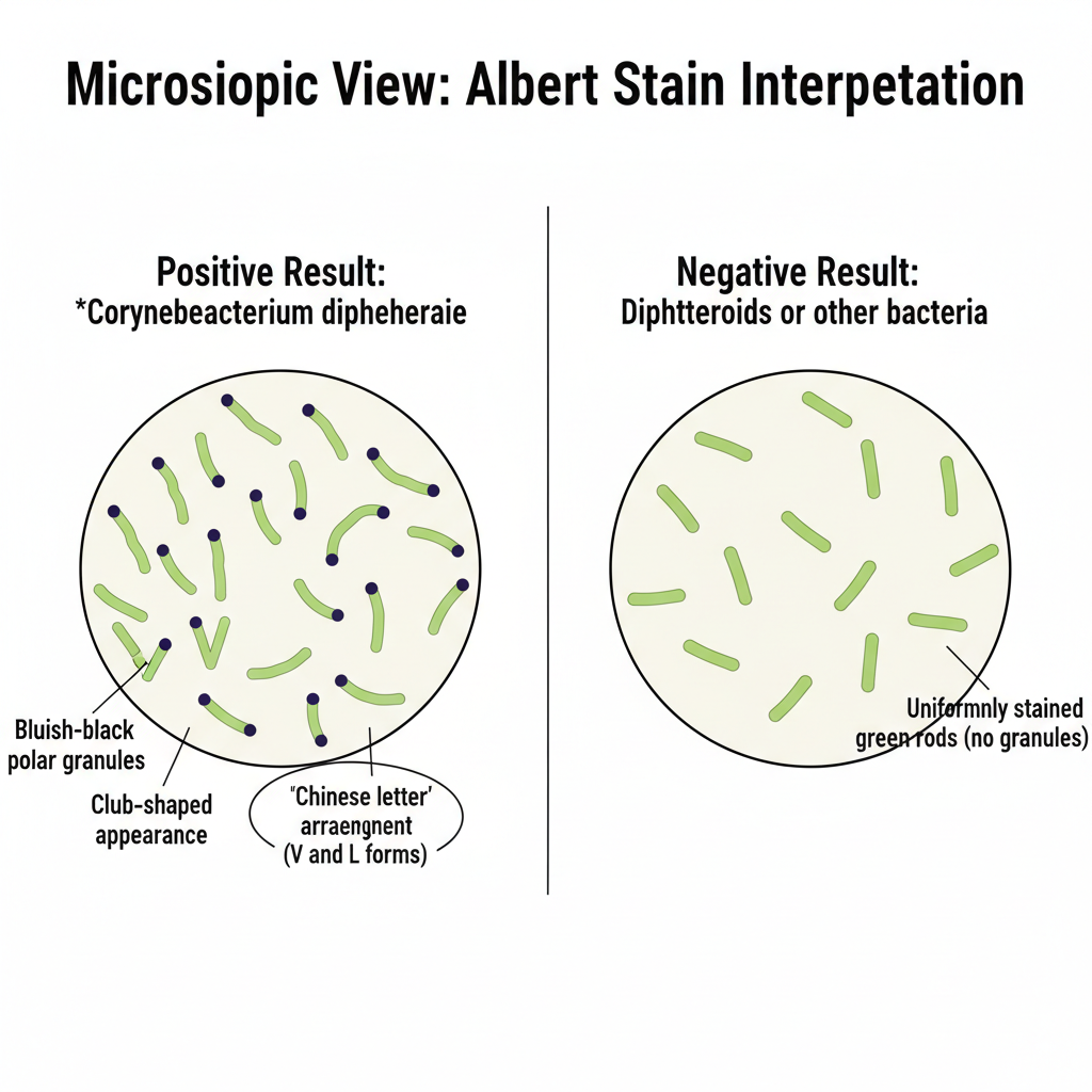
Under oil immersion the organisms is seen as green coloured slender rods. It is the cytoplasm that takes the malachite green stain and appear light green. The metachromatic granules appear as bluish black dots at the two ends of the bacilli. These are polar in position and give the organism a club shaped appearance. In many fields the organisms is arranged at angles forming L, V pattern which is commonly described as Chinese letter arrangement. This is the characteristic appearance of Corynebacterium diphtheriae.
Interpretation
The presence of green bacilli containing dark polar granules is taken as positive finding. It is the indication that granule containing diphtheria bacilli is present in the specimen. These organisms is easily differentiated from other diphtheroids which lack these granules and appear uniformly green. Thus the test helps in presumptive identification of C. diphtheriae.
Limitations
Albert stain is taken as a presumptive test only. It shows the presence of granule containing organisms but it does not confirm its toxigenicity. Some other species may also show similar appearance in rare conditions. So the positive smear needs to be confirmed by culture and by toxigenicity tests like Elek test or PCR. A smear that shows uniformly stained rods without granules is taken as negative slide.

Uses of the Albert Stain
- It is used for detecting Corynebacterium diphtheriae in clinical samples by demonstrating the metachromatic granules.
- It helps in differentiating pathogenic diphtheria bacilli which shows bluish black granules from non-pathogenic diphtheroids that appear uniformly green.
- It is used for rapid presumptive diagnosis because the smear gives immediate information before culture results.
- It helps clinicians in early treatment decision as the presence of granule containing bacilli is taken seriously in suspected diphtheria cases.
- It is used for examining smears taken from throat swab, nasal swab or from growth on Loeffler’s serum slope.
- It is also used in public health laboratories for screening carriers during outbreaks.
- It can show metachromatic granules in some other organisms also though this use is not very common.
Limitations of Albert Staining
- The staining reagents is delicate and the malachite green is easily washed away if water is used after solution–1.
- The slides are not permanent and the smear should be examined immediately because the colour fades with time.
- The test is taken as presumptive only and it cannot confirm the toxigenicity of the organism.
- Some other Corynebacterium species may show similar appearance and this may give misleading result.
- The presence of metachromatic granules depends on proper growth conditions and rich media like Loeffler’s serum slope.
- Old or young cultures may not show granules properly and this may give negative smear even if organism is present.
Advantages of Albert Staining
- It gives rapid presumptive diagnosis because the granules can be seen immediately on the smear.
- It helps the clinician to start early treatment in suspected diphtheria cases without waiting for culture report.
- It shows the metachromatic granules very clearly and helps in differentiating pathogenic C. diphtheriae from non-pathogenic diphtheroids.
- The green cytoplasm and dark granules give good contrast and the characteristic L or V arrangement is easily seen.
- It is simple to perform and the reagents are inexpensive so it is useful in routine laboratories and outbreak screening.
FAQ on Albert Stain
What is Albert stain?
Albert stain is a type of staining technique used in microbiology to identify and differentiate between gram-negative and gram-positive bacteria.
What are the steps involved in Albert stain?
The steps involved in Albert stain include heat fixation, staining with the primary stain, decolorization, staining with the counterstain, and observation under a microscope.
How does Albert stain work?
Albert stain works by using two dyes, a primary stain and a counterstain. The primary stain is used to stain the bacterial cells, while the counterstain is used to provide contrast and make the bacteria more visible under a microscope.
What is the purpose of Albert stain?
The purpose of Albert stain is to identify and differentiate between gram-negative and gram-positive bacteria based on their staining characteristics.
What are the dyes used in Albert stain?
The dyes used in Albert stain are typically crystal violet for the primary stain and safranin for the counterstain.
How does Albert stain compare to other staining techniques?
Albert stain is similar to other differential staining techniques, such as the Gram stain, but it has some key differences. It is less complex and less time-consuming compared to the Gram stain, but it is not as specific and may not provide as much information about the bacteria being studied.
What are the advantages of Albert stain?
The advantages of Albert stain include its simplicity, low cost, and ability to differentiate between gram-negative and gram-positive bacteria.
Can Albert stain be used in food and water quality control?
Yes, Albert stain can be used in food and water quality control to identify the presence of gram-negative and gram-positive bacteria, which can indicate contamination or spoilage.
What are the applications of Albert stain in bacterial research?
The applications of Albert stain in bacterial research include the study of bacterial morphology, classification, and identification, as well as the investigation of bacterial growth and metabolic processes.
What precautions should be taken when performing Albert stain?
Precautions that should be taken when performing Albert stain include wearing gloves and protective clothing, using a fume hood to prevent exposure to toxic chemicals, and properly disposing of used materials to avoid contamination.
- Adarsh College. (n.d.). Unit – IV stains and dyes. https://www.adarshcollege.in/wp-content/uploads/2023/04/UNIT-IV-Stains-and-Dyes.pdf
- Aster Labs. (n.d.). Albert stain test. https://www.asterlabs.in/tests/albert-stain
- Balmiki, J. (n.d.). Staining methods [PowerPoint slides]. SlideShare. https://www.slideshare.net/slideshow/staining-methodspptx/264577243
- Central Drug House (P) Ltd. (2023, December 19). Neisser’s stain A soln. (methylene blue) [Product specification]. https://www.cdhfinechemical.com/images/product/specs/39_457082406_NEISSESSTAINASOLN(METHYLENEBLUE)_868520.pdf
- Diagnopein Diagnostic Centre. (n.d.). What is the Albert stain test and why is it used? https://www.diagnopein.com/BlogDetails/Pathology/What-Is-the-Albert-Stain-Test-and-Why-Is-It-Used
- Fackrell, H. (n.d.). Metachromatic granules stain. University of Windsor. https://web2.uwindsor.ca/courses/biology/fackrell/Methods/Metachromatic_Granules.htm
- HiMedia Laboratories. (2024, February). Albert’s metachromatic stains – kit [Technical data]. https://www.himedialabs.com/media/TD/K002.pdf
- Jamir, I., Ketan, P., Anguraj, S., Jahan, L., Parameswaran, N., Divakarjose, R. R., & Sastry, A. S. (2022). Case report: Bloodstream infection with toxigenic Corynebacterium diphtheriae and Gram-negative sepsis in a child with burns. American Journal of Tropical Medicine and Hygiene, 107(4), 930–933. https://pmc.ncbi.nlm.nih.gov/articles/PMC9651519/
- Kannambath, R., Sistla, S., Jayakar, S., & Pillai, V. (2021). Direct Albert stain from the throat swab showing abundant green coloured bacilli with metachromatic granules arranged in cuneiform pattern [Figure]. ResearchGate. https://www.researchgate.net/figure/Direct-Albert-stain-from-the-throat-swab-showing-abundant-green-coloured-bacilli-with_fig1_353098517
- Kumar, S. (2012). Chapter-87 Staining methods. In Textbook of Microbiology (1st ed.). Jaypee Brothers Medical Publishers. https://www.jaypeedigital.com/eReader/chapter/9789350255100/ch87
- MBBS NAIJA. (n.d.). Albert’s stain (Albert’s solution 1 & 2); Staining technique in microbiology, application, procedure [Video]. YouTube. https://www.youtube.com/watch?v=VXwY6Hayx5w
- MediScan Lab. (n.d.). Albert stain (diphtheria bacilli). https://mediscanlab.com/albert-stain-diphtheria-bacilli
- Micromaster Laboratories. (n.d.). Albert’s stain A (SI 005) Albert’s stain B (SI 006) [Product specification sheet]. https://www.micromasterlab.com/wp-content/uploads/bsk-pdf-manager/SI006-PSS_(1)_1366.pdf
- Microxpress. (n.d.). Albert’s stain B [Technical details]. https://www.microxpress.in/uploads/product/albert%E2%80%99s-stain-b_technicaldetails_3420240313.062852.pdf
- Mistry, Y. (n.d.). Special stains useful in microbiology laboratory [PowerPoint slides]. SlideShare. https://www.slideshare.net/slideshow/special-stains-useful-in-microbiology-laboratory/89068590
- Mokobi, F. (2022, February 1). Albert staining- Principle, reagents, procedure, results, interpretation. Microbe Notes. https://microbenotes.com/albert-staining/
- Pandey, A. (2019, July 26). Alberts stain. myUpchar. https://www.myupchar.com/en/test/albert-stain
- Potadar, R. (n.d.). Volutin granule staining. Scribd. https://www.scribd.com/document/377216830/Volutin-Granule-Staining
- Pro-Lab Diagnostics. (n.d.). Albert’s stains [Instructions for use]. https://www.pro-lab.co.uk/wp-content/uploads/2021/01/Alberts-Stain-IfU-2020-10.pdf
- Rao, T. V. (n.d.). Albert’s staining technique. MicroRao. https://www.microrao.com/alberts_staining.htm
- Sprint Diagnostics. (n.d.). The history of Albert’s stain: A milestone in microbiology. https://www.sprintdiagnostics.in/blog/alberts-stain-microbiology
- The Albert staining technique: A definitive guide to principle, protocol, and clinical utility in diphtheria diagnostics. (n.d.). [Review article].
- Wikipedia. (n.d.). Corynebacterium diphtheriae. https://en.wikipedia.org/wiki/Corynebacterium_diphtheriae
- Yashoda Hospitals. (n.d.). What is Albert’s stain test? https://www.yashodahospitals.com/diagnostics/alberts-stain-test/
- Text Highlighting: Select any text in the post content to highlight it
- Text Annotation: Select text and add comments with annotations
- Comment Management: Edit or delete your own comments
- Highlight Management: Remove your own highlights
How to use: Simply select any text in the post content above, and you'll see annotation options. Login here or create an account to get started.