What is Adrenal Gland?
- The adrenal glands, commonly referred to as the suprarenal glands, are critical components of the endocrine system, located on the superior aspect of each kidney. These triangular-shaped organs, measuring approximately 5 cm by 2 cm and weighing around 4 to 5 grams each, serve multiple vital functions that are essential for maintaining homeostasis in the body.
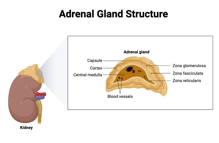
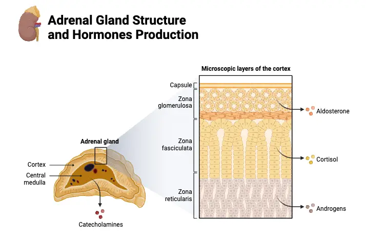
- Structurally, the adrenal glands consist of two main parts: the adrenal cortex and the adrenal medulla. The adrenal cortex is the outer layer and is further subdivided into three zones: the zona glomerulosa, zona fasciculata, and zona reticularis. Each zone is responsible for producing specific types of hormones. The zona glomerulosa primarily secretes mineralocorticoids, such as aldosterone, which play a crucial role in regulating blood pressure and electrolyte balance. The zona fasciculata produces glucocorticoids, including cortisol, which are involved in metabolic regulation and immune system suppression. Lastly, the zona reticularis generates androgens, which are precursors to sex hormones and are converted to active forms in other tissues.
- In contrast, the adrenal medulla, which is the inner part of the gland, produces catecholamines such as adrenaline (epinephrine) and norepinephrine. These hormones are critical for the body’s acute stress response, facilitating a rapid physiological reaction known as the “fight-or-flight” response. This response is characterized by increased heart rate, heightened alertness, and enhanced energy availability, all of which are essential for responding to immediate threats.
- The process by which the adrenal cortex produces steroid hormones is termed steroidogenesis, involving a series of enzymatic reactions within the cortical cells. This complex process highlights the biochemical intricacies that underlie hormonal production and regulation.
- The adrenal glands also have significant implications in various endocrine disorders. For instance, an overproduction of cortisol can lead to Cushing’s syndrome, characterized by obesity, hypertension, and skin changes. Conversely, insufficient cortisol production is associated with Addison’s disease, which can cause fatigue, weight loss, and low blood pressure. Congenital adrenal hyperplasia, a genetic disorder, results from dysfunctional hormone synthesis, leading to a variety of physiological challenges. Additionally, tumors can arise from adrenal tissue, which may be identified during medical imaging for unrelated conditions.
- It is important to note that the adrenal cortex is vital for survival, while the adrenal medulla, although functionally significant in stress responses, is not essential for life. The differing embryological origins of these two parts also reflect their distinct functions: the adrenal cortex originates from mesodermal tissue, while the adrenal medulla develops from neural tissue.
Definition of Adrenal Gland
The adrenal glands, or suprarenal glands, are small, triangular-shaped endocrine glands located on top of each kidney. They consist of two main parts: the adrenal cortex, which produces steroid hormones such as cortisol and aldosterone, and the adrenal medulla, which produces catecholamines like adrenaline. These hormones play vital roles in regulating metabolism, blood pressure, stress response, and electrolyte balance in the body.
Location of Adrenal Gland
The adrenal glands are vital endocrine structures positioned atop each kidney, playing a crucial role in hormone production and overall metabolic regulation. Their unique anatomical location is significant for both physiological functions and surgical considerations.
- General Location:
- The adrenal glands are situated at the superior aspect of the kidneys, resembling hats perched on top of these vital organs.
- Each gland is approximately the size and shape of a fortune cookie, facilitating their identification during medical imaging or surgical procedures.
- Right Adrenal Gland:
- Located directly atop the right kidney.
- It is positioned in close proximity to the inferior vena cava (IVC), the largest vein responsible for returning deoxygenated blood to the heart from the lower body.
- The right adrenal gland also lies near the liver, another critical organ involved in metabolism and detoxification.
- The proximity of the adrenal gland to these major structures necessitates that any surgical intervention in this area be performed with extreme precision, as even minor deviations can lead to severe complications.
- Left Adrenal Gland:
- Found on top of the left kidney.
- This gland is adjacent to the splenic artery, which supplies blood to the spleen, and the tail of the pancreas, a vital organ involved in digestion and glucose regulation.
- Given the sensitivity of the pancreas and its surrounding structures, surgical procedures involving the left adrenal gland also require highly skilled surgeons to avoid inadvertent damage that could result in serious health consequences.

Structure of Adrenal Gland
The structure of the adrenal gland is complex and highly specialized, enabling it to perform its critical roles in hormone production and regulation. Each adrenal gland is composed of distinct layers and tissues that contribute to its overall functionality.
- General Overview:
- The adrenal glands are located atop the kidneys, with each gland resting in the perirenal space and enclosed by the superior renal fascia.
- At birth, the adrenal glands are relatively large, about one-third the size of the kidneys, but they shrink to approximately one-thirtieth the kidney’s size by adulthood.
- The right adrenal gland has a pyramidal shape, while the left is crescentic, both measuring around 50 mm in height, 30 mm in breadth, and 10 mm in thickness, with each gland weighing approximately 5 grams.
- Anatomical Relations:
- The right adrenal gland is situated beneath the liver, posterior to the inferior vena cava (IVC), and anterior to the diaphragm.
- Conversely, the left adrenal gland is positioned medially to the spleen, superior to the splenic artery and vein, lateral to the abdominal aorta, and also anterior to the diaphragm.
- These anatomical relationships are crucial for understanding surgical approaches and potential complications associated with adrenal gland operations.
- Internal Structure:
- The adrenal gland consists of two distinct types of tissue: the outer cortex and the inner medulla.
- The cortex appears yellowish due to its fatty composition, while the medulla exhibits a reddish-brown color.
- Surrounding the entire gland is a thick capsule of connective tissue that provides structural support.
- Cortex Structure:
- The adrenal cortex is significantly larger than the medulla, comprising about 85% of the gland’s volume. It is further divided into three zones:
- Zona Glomerulosa: This outermost layer produces mineralocorticoids, primarily aldosterone, which is crucial for regulating blood pressure and electrolyte balance.
- Zona Fasciculata: The middle layer produces glucocorticoids, predominantly cortisol, which plays key roles in metabolism, immune response modulation, and blood sugar regulation through gluconeogenesis. Its secretion is regulated by the adrenocorticotropic hormone from the pituitary gland.
- Zona Reticularis: The innermost layer generates androgens, particularly dehydroepiandrosterone (DHEA), which serves as a precursor for various hormones such as progesterone, estrogen, cortisol, and testosterone. The mnemonic “Salt, Sugar, Sex” helps to recall the functions associated with each zone.
- The adrenal cortex is significantly larger than the medulla, comprising about 85% of the gland’s volume. It is further divided into three zones:
- Medulla Structure:
- The adrenal medulla is centrally located within the gland and is characterized by its dark brown coloration.
- It contains specialized chromaffin cells that synthesize catecholamines, including adrenaline and norepinephrine, which are vital for the body’s “fight-or-flight” response.
- The medulla responds to stress by releasing these hormones into the bloodstream, facilitating rapid physiological adaptations. Additionally, chromaffin cells produce enkephalins, which play a role in pain control.
- Vascular and Nervous Connections:
- Each adrenal gland is supplied by arteries entering at various sites, while veins and lymphatic vessels exit through the hilum.
- The distinct embryological origins of the cortex (derived from mesoderm) and the medulla (derived from ectodermal neural crest cells) further highlight the structural and functional differentiation within the gland.

Hormones of Adrenal Gland
The adrenal glands play a crucial role in hormone production, generating a variety of hormones essential for numerous physiological functions. These hormones are primarily classified based on their synthesis location within the adrenal glands: the adrenal cortex and the adrenal medulla.
- Hormones of the Adrenal Cortex:
- The adrenal cortex produces three main categories of steroid hormones, each secreted by different regions:
- Mineralocorticoids:
- Synthesized in the zona glomerulosa, mineralocorticoids are vital for maintaining electrolyte balance in the body.
- Aldosterone is the predominant mineralocorticoid, accounting for approximately 95% of this hormone group. Its primary function is to regulate sodium (Na⁺) and potassium (K⁺) concentrations in extracellular fluids.
- By stimulating the distal tubules of the nephron in the kidneys, aldosterone enhances sodium and water reabsorption while promoting the excretion of potassium.
- The physiological effects of aldosterone are relatively short-lived, lasting around 20 minutes, allowing for dynamic control of plasma electrolyte levels.
- Glucocorticoids:
- This group is crucial for energy metabolism and overall survival, with cortisol being the principal glucocorticoid secreted in humans.
- Glucocorticoids maintain blood glucose levels and blood pressure by affecting the activity of vasoconstrictors.
- Cortisol operates by modifying gene expression within target cells, and its secretion follows a predictable circadian pattern.
- The release of glucocorticoids is tightly regulated through a negative feedback mechanism involving adrenocorticotropic hormone (ACTH), which is stimulated by corticotrophin-releasing hormone from the hypothalamus.
- Cortisol’s primary metabolic role includes inducing gluconeogenesis, thereby conserving glucose while mobilizing fatty acids and proteins for energy.
- Gonadocorticoids/Androgens/Sex Hormones:
- Produced in smaller amounts, gonadocorticoids, such as dehydroepiandrosterone (DHEA) and androstenedione, are weak sex hormones.
- These hormones are converted into more potent sex hormones in peripheral tissues.
- Although their specific functions are not fully elucidated, they are associated with the development of secondary sexual characteristics, including the growth of axillary and pubic hair in both sexes.
- In females, these hormones contribute to sexual drive and account for a significant portion of estrogen production post-menopause.
- Mineralocorticoids:
- The adrenal cortex produces three main categories of steroid hormones, each secreted by different regions:
- Hormones Produced by the Adrenal Medulla:
- The adrenal medulla, in response to stress, synthesizes catecholamines: epinephrine and norepinephrine.
- The secretion ratio of these hormones is approximately 4:1 in favor of epinephrine.
- Epinephrine primarily enhances metabolic activities, including bronchial dilation and increased blood flow to skeletal muscles, facilitating a quick response to stressors.
- Conversely, norepinephrine is more focused on peripheral vasoconstriction and elevating blood pressure.
- The effects of these hormones are typically short-lived, producing a rapid response to acute stress situations.
Organ Systems Involved of Adrenal Gland
The adrenal gland plays a significant role in the body’s physiological responses and homeostasis, intricately linked to several organ systems. The functioning of the adrenal gland is primarily coordinated through the hypothalamic-pituitary-adrenal (HPA) axis, the renin-angiotensin-aldosterone system (RAAS), and the sympathetic nervous system. Understanding these interactions is crucial for comprehending how the adrenal gland contributes to various bodily functions.
- Hypothalamic-Pituitary-Adrenal (HPA) Axis:
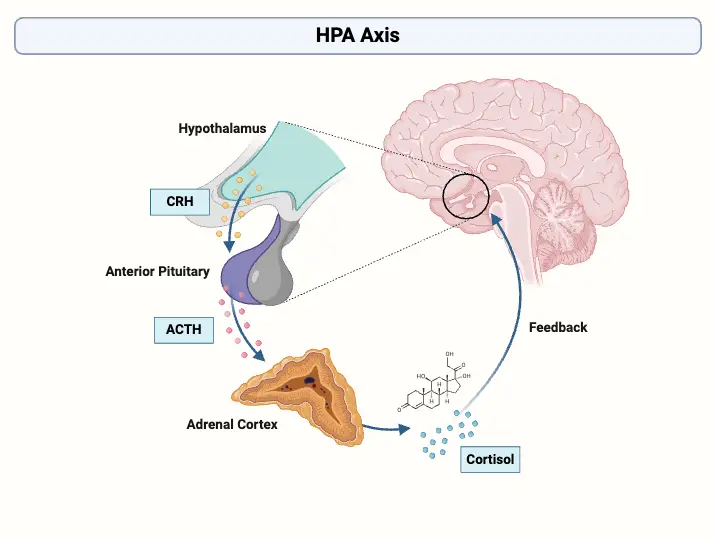
- The HPA axis is pivotal in regulating the production of glucocorticoids and adrenal androgens.
- The process begins when paraventricular neurons (PVN) in the hypothalamus release corticotropin-releasing hormone (CRH) in response to stressors or circadian rhythms.
- CRH binds to receptors on the anterior pituitary gland, leading to the synthesis and release of adrenocorticotropic hormone (ACTH) from pre-pro-opiomelanocortin (pre-POMC).
- This process also generates other hormones such as alpha-melanocyte-stimulating hormone (MSH) through the cleavage of POMC.
- ACTH circulates in the bloodstream and interacts predominantly with melanocortin type 2 receptors (MC2-R) in the zona fasciculata of the adrenal cortex, stimulating the synthesis of glucocorticoids.
- Glucocorticoids exert negative feedback on the hypothalamus and anterior pituitary to regulate their own levels, preventing excessive hormone production.
- The secretion of ACTH follows a pulsatile pattern, peaking in the morning and declining throughout the day, which is essential for maintaining circadian rhythms of cortisol levels.
- Renin-Angiotensin-Aldosterone System (RAAS):
- The RAAS is crucial for regulating the synthesis of mineralocorticoids, particularly aldosterone.
- Unlike glucocorticoids, mineralocorticoid production is primarily regulated by renin and potassium levels rather than ACTH.
- When renal perfusion decreases, the juxtaglomerular apparatus in the kidneys releases renin.
- Renin converts angiotensinogen, produced by the liver, into angiotensin I, which is then transformed into angiotensin II (AT-II) by angiotensin-converting enzyme (ACE) in the lungs.
- AT-II stimulates aldosterone synthesis in the zona glomerulosa of the adrenal cortex by activating aldosterone synthase, thereby promoting sodium reabsorption and potassium excretion, which are vital for fluid balance and blood pressure regulation.
- Adrenal Medulla and the Sympathetic Nervous System:
- The adrenal medulla, part of the adrenal gland, is closely associated with the sympathetic nervous system.
- This relationship facilitates the rapid secretion of catecholamines, specifically epinephrine and norepinephrine, during stressful situations.
- These hormones play essential roles in the “fight or flight” response, enhancing metabolic activity, increasing heart rate, and redirecting blood flow to vital organs.

Vasculature of Adrenal Gland
The vasculature of the adrenal glands is critical for their function, ensuring an adequate blood supply necessary for the synthesis and release of hormones. This extensive vascular network is comprised of three primary arteries and respective venous drainage systems. Understanding the anatomy and physiological significance of these blood vessels is essential for grasping how adrenal glands operate within the endocrine system.
- Arterial Supply:
- The adrenal glands receive their blood supply from three main arteries:
- Superior Adrenal Artery:
- Originates from the inferior phrenic artery.
- Supplies blood to the upper portion of the adrenal gland.
- Middle Adrenal Artery:
- Arises directly from the abdominal aorta.
- Provides a substantial blood flow to the central areas of the adrenal gland.
- Inferior Adrenal Artery:
- Branches off from the renal arteries.
- Primarily nourishes the lower parts of the adrenal gland.
- Superior Adrenal Artery:
- The adrenal glands receive their blood supply from three main arteries:
- Venous Drainage:
- Each adrenal gland is drained by distinct venous structures that facilitate the removal of blood from the gland:
- Right Adrenal Vein:
- Drains directly into the inferior vena cava.
- This direct connection allows for efficient drainage and transport of hormones into systemic circulation.
- Left Adrenal Vein:
- Drains into the left renal vein.
- Although slightly more complex, this pathway also efficiently channels blood away from the gland.
- Right Adrenal Vein:
- Each adrenal gland is drained by distinct venous structures that facilitate the removal of blood from the gland:
- Significance of Vascularization:
- The rich vascular network ensures a high rate of hormone delivery directly into the bloodstream.
- Adequate blood supply is vital for the synthesis of steroid hormones such as cortisol, aldosterone, and adrenal androgens.
- The rapid transport of these hormones influences numerous physiological processes, including metabolism, immune response, and stress management.
- Physiological Interactions:
- Blood flow through these arteries is regulated to meet the demands of the body, particularly in response to stress or hormonal signals.
- Increased blood flow may enhance hormone secretion during times of physiological need, such as during stress or low blood pressure.
Diseases and Disorders of Adrenal Gland
The adrenal glands are crucial components of the endocrine system, and various diseases and disorders can affect their functionality, leading to significant health consequences. Understanding these conditions is essential for recognizing their symptoms and underlying mechanisms, which can aid in diagnosis and treatment.
- Cushing’s Syndrome:
- Cushing’s syndrome arises from an overproduction of cortisol, primarily a glucocorticoid hormone secreted by the adrenal cortex.
- The hypersecretion can be attributed to adrenal tumors or excessive adrenocorticotropic hormone (ACTH) secretion from the pituitary gland.
- Common manifestations include:
- Adiposity: Characteristic fat distribution leads to rounded faces, increased fat around the neck, and abdominal obesity.
- Protein Breakdown: The condition is associated with significant tissue protein degradation, leading to muscle weakness.
- Bone Health: Increased gluconeogenesis can contribute to osteoporosis, making bones more susceptible to fractures.
- Metabolic Changes: Patients often experience hyperglycemia (high blood sugar) and glycosuria (presence of glucose in urine), indicating altered glucose metabolism.
- Adrenal Hypoplasia:
- Adrenal hypoplasia refers to the underdevelopment of the adrenal cortex, which may result from various clinical factors.
- This condition can be categorized as:
- Primary Hypoplasia: Directly affects the adrenal gland, resulting in a decrease in the secretion of essential adrenal hormones.
- Secondary Hypoplasia: Less common and generally milder, this form affects hormone secretions without significantly altering the gland’s structural integrity.
- Addison’s Disease:
- Addison’s disease is characterized by insufficient production of glucocorticoids and mineralocorticoids, typically due to autoimmune destruction of adrenal tissue.
- The immune system may generate autoantibodies targeting the adrenal cortex, leading to decreased hormone production.
- Key symptoms include:
- Muscle Weakness: General fatigue and muscle weakness can significantly impact daily activities.
- Hypoglycemia: Low blood sugar levels may occur, causing dizziness and confusion.
- Increased Pigmentation: Patients often exhibit hyperpigmentation, particularly in areas exposed to the sun, due to elevated ACTH levels stimulating melanocyte activity.
- Adrenocortical Adenomas:
- These are benign tumors of the adrenal cortex that can lead to excess cortisol production, resulting from disruptions in intracellular signaling pathways, specifically high levels of cyclic adenosine monophosphate (cAMP) or protein kinase A.
- While typically benign, these adenomas can cause hypersecretion of adrenal hormones if they produce other hormones.
- The growth of these adenomas is often stimulated by ACTH binding to its receptors, triggering pathways that promote cortical cell proliferation.
Functions of Adrenal Gland
The adrenal glands serve multiple critical functions in the body, primarily through the hormones they produce. These functions are integral to maintaining homeostasis and enabling the body to respond effectively to various physiological demands.
- Regulation of Electrolytes:
- The mineralocorticoids, particularly aldosterone, are crucial for regulating sodium (Na⁺) and potassium (K⁺) levels in blood and extracellular fluids.
- This regulation directly impacts blood volume and pressure, ensuring the body maintains fluid balance and proper cardiovascular function.
- Metabolic Regulation:
- Cortisol, a glucocorticoid produced by the adrenal cortex, plays a vital role in metabolic processes.
- It influences glucose metabolism by promoting gluconeogenesis, which is the synthesis of glucose from non-carbohydrate sources, thereby ensuring a stable supply of energy during stress or fasting.
- Additionally, cortisol modulates the metabolism of fats, proteins, and carbohydrates, ensuring that energy production meets the body’s demands.
- Immune System Modulation:
- Cortisol also exerts significant effects on the immune system. It has anti-inflammatory properties that help regulate immune responses, preventing excessive inflammation that could harm the body.
- Cardiovascular Function:
- The actions of cortisol and other adrenal hormones support cardiovascular health by regulating blood pressure and influencing vascular tone through the modulation of vasoconstrictors.
- Stress Response:
- The adrenal glands produce hormones that facilitate the body’s adaptation to stress.
- Catecholamines, specifically epinephrine and norepinephrine, are secreted by the adrenal medulla in response to acute stressors, activating the “fight or flight” response.
- This response prepares the body for immediate physical action by increasing heart rate, enhancing blood flow to muscles, and promoting energy availability.
- Reproductive System Development:
- The adrenal glands also synthesize androgens, which are weak sex hormones.
- These androgens are converted into more potent sex hormones in peripheral tissues, playing a role in the development and function of the reproductive system.
- They are particularly important for the development of secondary sexual characteristics during puberty and contribute to libido in both males and females.
- Megha R, Wehrle CJ, Kashyap S, et al. Anatomy, Abdomen and Pelvis: Adrenal Glands (Suprarenal Glands) [Updated 2022 Oct 17]. In: StatPearls [Internet]. Treasure Island (FL): StatPearls Publishing; 2024 Jan-. Available from: https://www.ncbi.nlm.nih.gov/books/NBK482264/
- Dutt M, Wehrle CJ, Jialal I. Physiology, Adrenal Gland. [Updated 2023 May 1]. In: StatPearls [Internet]. Treasure Island (FL): StatPearls Publishing; 2024 Jan-. Available from: https://www.ncbi.nlm.nih.gov/books/NBK537260/
- Hasan, Najat. (2018). Adrenal gland: Structure, Hormones and clinical disorders – part 1.
- https://www.geeksforgeeks.org/adrenal-gland/
- https://teachmeanatomy.info/abdomen/viscera/adrenal-glands/
- https://www.adrenal.com/adrenal-gland/anatomy
- https://en.wikipedia.org/wiki/Adrenal_gland