What is Bright Field Microscope?
- A bright-field microscope represents the most fundamental and commonly utilized form of optical microscopy. In this setup, a specimen is illuminated from beneath using white light and observed from above, resulting in a dark image against a bright background.
- This instrument, often referred to as a compound light microscope, employs a combination of lenses—namely the condenser, objective, and ocular—to attain magnifications generally ranging from 40× to 1000×, with a feasible upper limit of approximately 1300×.
- Contrast arises from variations in light absorption and refractive properties within different regions of a specimen; areas that are dense or stained tend to absorb more light, resulting in a darker appearance against the illuminated background.
- The iris or aperture diaphragm, along with the condenser, plays a crucial role in focusing the light beam. Köhler illumination is employed to ensure uniform lighting while minimizing glare.
- Staining is frequently necessary for enhancing contrast in colorless or transparent specimens, which often restricts the use of live cells in various situations.
- The benefits encompass straightforwardness, cost-effectiveness, user-friendliness, and versatility, allowing for the integration of digital imaging or camera systems.
- The drawbacks include inadequate contrast when dealing with unstained specimens, resolution constrained by the principles of optical physics (approximately 200 nm), and the risk of photodamage due to intense light exposure.
- Oil immersion Utilizing objective lenses along with colored or polarizing filters can enhance resolution and emphasize structures.
- Typical uses encompass histology, pathology, microbiology (such as Gram-stained bacteria), parasitology, cell biology, medical diagnostics, and mineralogy.
- This method is fundamental in educational settings and laboratories, establishing a baseline for more sophisticated techniques such as phase-contrast or dark-field microscopy.

What is Dark Field Microscope?
- A dark-field microscope represents a specific type of optical microscope that employs unique illumination techniques to present specimens as luminous images against a dark backdrop.
- The setup utilizes a dark-field condenser or patch stop, which obstructs the direct light beam, permitting only a hollow cone of oblique light to illuminate the specimen.
- The method exclusively captures light that is either scattered or diffracted by the specimen; any unscattered light does not reach the objective, resulting in images with high contrast.
- Perfect for examining unstained, transparent, or living specimens, including bacteria (such as spirochetes), algae, protozoa, blood cells, and delicate structures like flagella.
- To effectively collect scattered light, it is essential to utilize high numerical aperture condensers, typically oil-immersion, along with compatible objectives.
- Provides improved contrast and resolution relative to bright-field microscopy; however, the overall image brightness tends to be reduced because of light loss.
- Little sample preparation is required; staining is not needed, maintaining the integrity of live cells.
- The benefits encompass live imaging, the absence of staining, exceptional contrast, the capability to identify non-culturable organisms, and the exposure of surface details.
- The limitations encompass the necessity for robust illumination, which poses a risk of photodamage, heightened sensitivity to dust and debris, and reduced efficacy when dealing with thick or dense samples.
- Typical applications include microbiology (such as spirochete detection), parasitology, live-cell imaging, materials science, forensic analysis, and the study of nanoparticles or minerals.
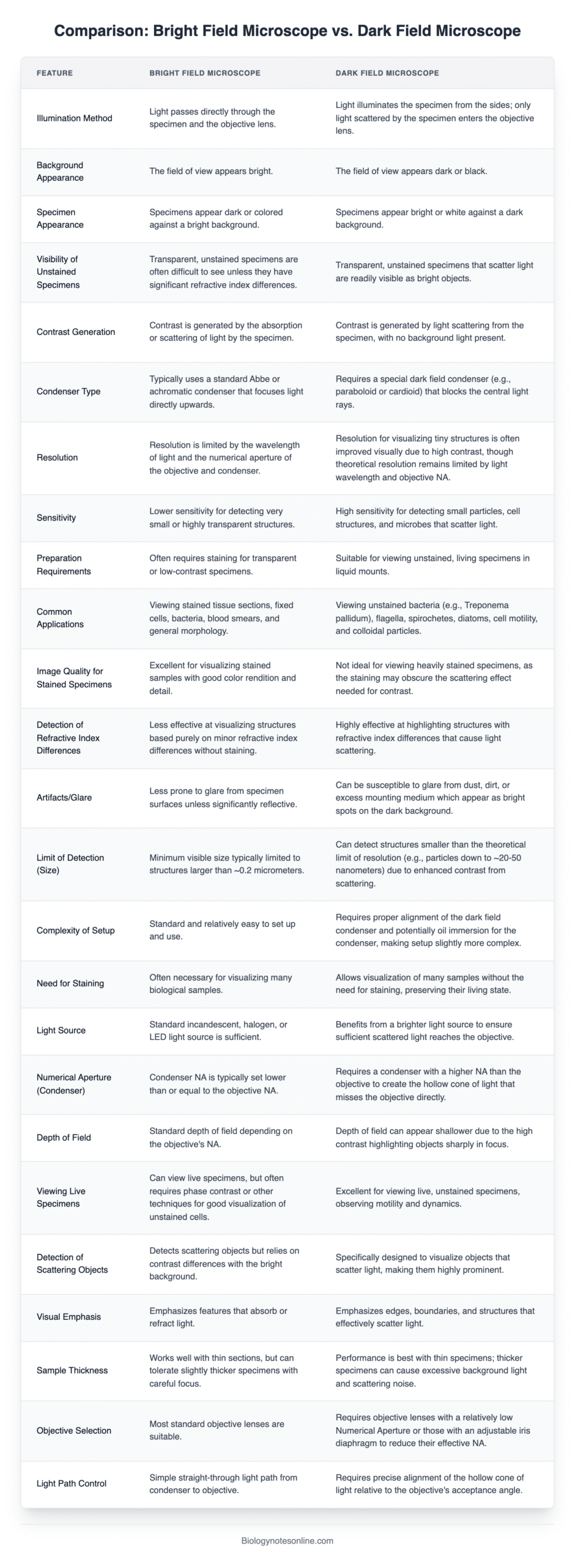
Difference Between Bright Field Microscope and Dark Field Microscope
| Feature | Bright‑Field Microscope | Dark‑Field Microscope |
|---|---|---|
| Image Background | Bright | Dark |
| Specimen Appearance | Dark/stained specimens appear visible | Unstained specimens appear bright against dark |
| Illumination | Transmitted white light through specimen | Oblique/scattered light; direct light blocked |
| Contrast Mechanism | Light absorption by specimen | Light scattering/reflection from specimen |
| Sample Preparation | Often requires staining, fixation | No staining/fixation needed |
| Internal Structures | Shows internal morphology well | Better for external surface details |
| Specimen Types | Fixed, stained, live | Primarily live, unstained, delicate specimens |
| Condenser Type | Abbe, achromatic, variable focal length | Paraboloid/cardioid condenser with patch stop |
| Opaque Stop/Filter | Not used | Required (central stop) |
| Resolution | Limited by light absorption; typical up to ~1300× | High contrast reveals fine structures, though lower resolution due to low light |
| Light Intensity | Higher light levels | Lower light level, requires strong illumination |
| Ease of Use | Simple setup, widely available | More complex setup and alignment |
| Cost | Affordable, commonly used | More expensive; specialized optics |
| Suitability for Thick Specimens | Works well with thick/opaque samples | Less effective with thick specimens |
| Opaque Materials | Not suitable | Can image minerals, metals, crystals |
| Image Artifacts | Minimal halos; low out-of-focus blur | Sensitive to dust; halos; needs clean optics |
| Live Specimens | Staining may damage live cells | Ideal for observing motility and live behavior |
| Applications | Histology, pathology, stained slides, medical diagnostics | Microbiology, spirochetes (e.g., Treponema), nanoparticles, blood cells |
| Quantitative Analysis | Easier due to even illumination | More difficult due to uneven light; requires care |
| Photodamage Potential | May cause photodamage in sensitive live samples | Potentially gentler on live samples due to less direct light |

Reference
- https://public.wsu.edu/~omoto/papers/darkfield.html
- https://elsevier.blog/dark-field-microscopy/
- https://www.bioscience.com.pk/en/topics/microbiology/dark-field-microscopy
- https://virtual-labs.github.io/exp-microscopy-iitk/theory.html
- https://evidentscientific.com/en/insights/what-is-darkfield-microscopy
- https://library.snls.org.sz/boundless/boundless/microbiology/textbooks/boundless-microbiology-textbook/microscopy-3/other-types-of-microscopy-30/dark-field-microscopy-244-343/index.html
- https://kiraoptical.com/what-are-dark-field-microscopes
- https://searchnsucceed.in/dark-field-microscope-principle-applications
- https://microbeonline.com/dark-field-microscopy
- https://en.wikipedia.org/wiki/Dark-field_microscopy
- Text Highlighting: Select any text in the post content to highlight it
- Text Annotation: Select text and add comments with annotations
- Comment Management: Edit or delete your own comments
- Highlight Management: Remove your own highlights
How to use: Simply select any text in the post content above, and you'll see annotation options. Login here or create an account to get started.