What is Urogenital System?
- The urogenital system is an essential component of human anatomy, encompassing both the urinary and reproductive systems. The term “urogenital” is derived from the integration of these two systems, which are interconnected both anatomically and functionally. This intricate relationship is evident in males, where the urinary and reproductive pathways converge, sharing common channels such as the male urethra. Therefore, understanding the urogenital system requires a comprehensive examination of both its urinary and reproductive components.
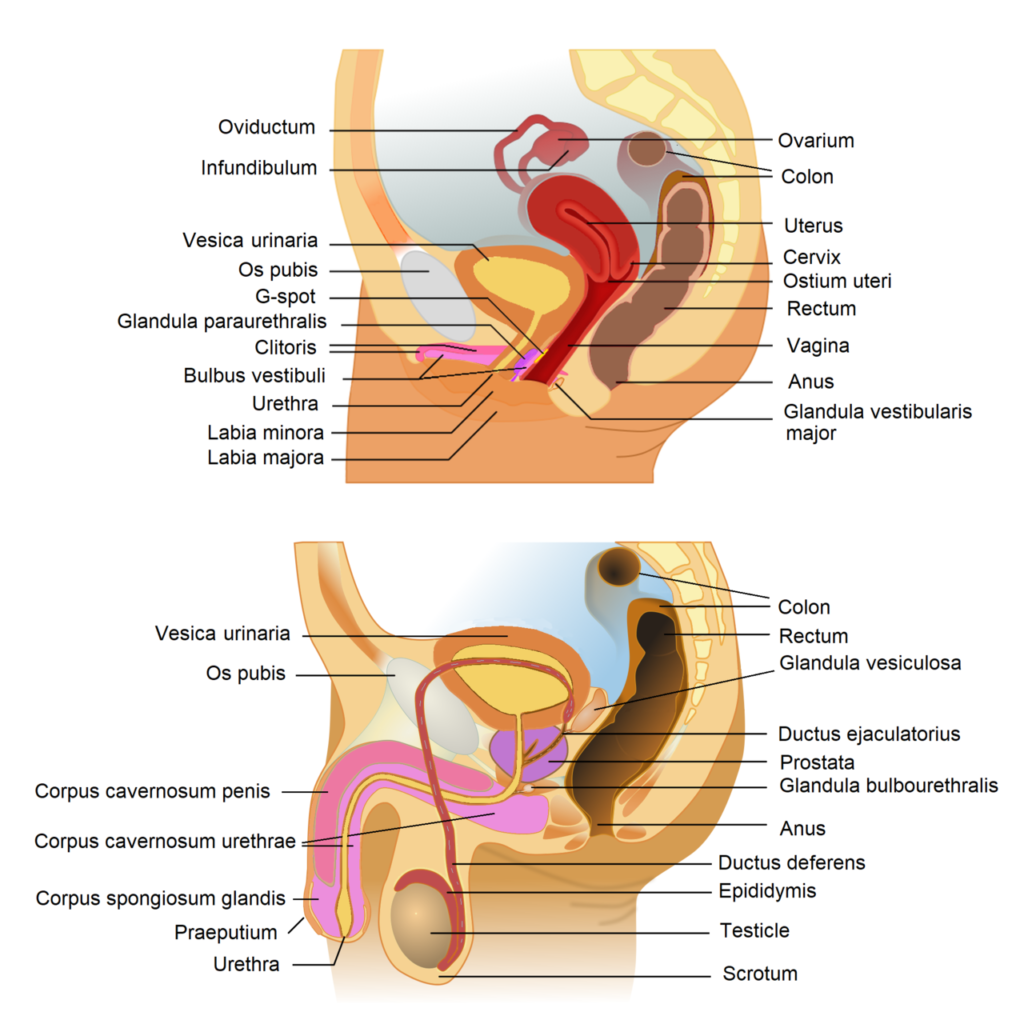
- In males, the urogenital system comprises several key structures, including the epididymis, testes, vas deferens, ejaculatory ducts, penis, urethra, prostate, and various accessory glands. The testes are responsible for producing sperm and hormones, while the epididymis serves as a storage and maturation site for sperm. The vas deferens transports sperm from the epididymis to the ejaculatory duct, where it merges with seminal fluid from the seminal vesicles, contributing to semen formation. The urethra, extending through the penis, plays a dual role by allowing the excretion of urine and the passage of semen during ejaculation.
- Conversely, the female urogenital system consists of the ovaries, uterus, clitoris, uterine tubes, and vagina. The ovaries are vital for producing ova and hormones, while the uterus serves as the site for fetal development during pregnancy. The uterine tubes, also known as fallopian tubes, facilitate the transport of ova and are the typical site for fertilization. The vagina functions as both a canal for childbirth and a conduit for sperm during intercourse.
- The term “urinogenital” is often used interchangeably with “urogenital,” emphasizing the interrelated nature of the urinary and reproductive systems. Specifically, the urethra in males is referred to as the urinogenital duct, highlighting its role in transporting both urine and reproductive fluids. This duct is situated along the length of the penis, where it serves as the exit point for urine during micturition and for sperm during sexual intercourse.
- The urogenital duct, as a functional unit, underscores the complexity of the urogenital system, wherein urine is expelled from the urinary system while sperm is delivered from the reproductive system. This dual functionality is crucial for maintaining homeostasis and reproductive health.
Male and Female Excretory System
The male and female excretory systems share significant similarities in structure, particularly concerning the kidneys, ureters, bladder, and urethra. Despite these commonalities, there are notable differences, particularly in the anatomy and function of the urethra.
- Kidneys:
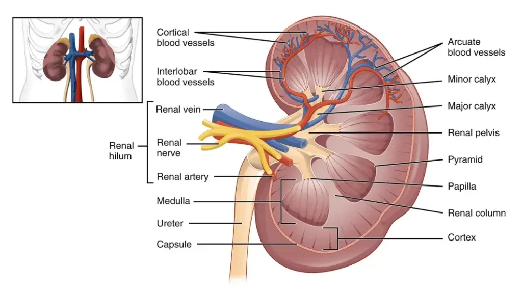
- The kidneys serve as the principal excretory organs in humans, responsible for filtering blood and producing urine.
- They develop from the lateral plate mesoderm, which forms nephric ridges that differentiate into nephrotomes.
- These nephrotomes further develop into nephrons, the basic structural and functional units of the kidneys.
- Ureters:
- Ureters are muscular tubes that transport urine from the kidneys to the bladder.
- The smooth muscle in the ureters contracts rhythmically, facilitating the movement of urine through peristaltic waves.
- Bladder:
- The urinary bladder acts as a temporary storage site for urine.
- In mammals, the bladder is typically large and lined with a specialized epithelium, which allows it to stretch as it fills.
- Unlike birds and reptiles, which excrete uric acid directly into the cloaca, mammals store liquid urine until it is expelled.
- Urethra:
- The urethra is the tubular structure responsible for transporting urine from the bladder to the exterior of the body.
- In males, the urethra is significantly longer, measuring approximately 17-20 cm, compared to the female urethra, which measures about 2.5-4 cm.
- The male urethra is divided into four distinct segments: the prostatic urethra, which passes through the prostate; the membranous urethra; the bulbar urethra; and the pendulous or penile urethra.
- In females, the urethra is shorter and opens directly above the vaginal opening, emphasizing the anatomical differences between the sexes.
- Sex Differences:
- Most anatomical differences in the urinary system between males and females occur at the bladder neck and continue distally to the urethra.
- The presence of the vas deferens, a male reproductive structure, also differentiates male urinary anatomy from that of females, but overall, the primary components remain structurally similar.
Development of Urogenital System
The development of the urogenital system is a complex process originating from the intermediate mesoderm, which plays a crucial role in the formation of both the urinary and reproductive structures. Understanding this developmental trajectory provides insights into the organization and functionality of these systems in adults.
- Urogenital Ridge Formation:
- The urogenital system begins to form when the intermediate mesoderm generates a urogenital ridge on both sides of the aorta.
- This ridge is pivotal in the subsequent development of the nephric structures, which will contribute to both renal and reproductive functions.
- Nephric Structures:
- Three distinct tubular nephric structures develop sequentially from the urogenital ridge: pronephros, mesonephros, and metanephros.
- These structures progress from head to tail along the developing embryo.
- Pronephros:
- The pronephros is the most cranial pair of nephric tubes and is generally transient, as it usually regresses during development.
- Although it may perform some initial renal functions, its role is limited and is largely replaced by subsequent structures.
- Mesonephros:
- The mesonephros forms in the midsection of the embryo and consists of mesonephric tubules and the mesonephric duct, also known as the Wolffian duct.
- Initially, the mesonephric tubules perform essential kidney functions, contributing to the excretory processes of the embryo.
- However, as development progresses, many of these tubules undergo regression, while the mesonephric duct persists.
- The mesonephric duct establishes a connection to the cloaca at the tail end of the embryo, facilitating the transport of excretory and reproductive products.
- Metanephros:
- The metanephros ultimately gives rise to the adult kidney, marking the final and most advanced stage of nephric development.
- Its formation involves the metanephric blastema, which develops from the condensation of adjacent neurogenic intermediate mesoderm.
- This process is complemented by the extension of the caudal mesonephric duct, leading to the emergence of the ureteric bud.
- The ureteric bud plays a vital role in establishing the connection between the developing kidney and the urinary tract, facilitating the maturation of renal structures.
Male Urogenital System
The male urogenital system comprises a series of structures integral to reproductive and excretory functions. This system includes the testes, epididymis, vas deferens, ejaculatory ducts, urethra, penis, prostate, and accessory glands. Understanding each component’s role provides a clearer picture of male reproductive physiology.
- Testes:
- The testes are the primary reproductive organs, located within the scrotum, with the left testis positioned slightly lower than the right.
- Their primary function is the production of sperm.
- Two significant cell types, Sertoli cells and Leydig cells, are present in the testes.
- Sertoli Cells: These cells support and nourish developing sperm cells.
- Leydig Cells: Responsible for producing testosterone, these cells are largely inactive before puberty, at which point testosterone levels significantly increase, influencing various reproductive functions.
- Epididymis:
- The epididymis is a coiled structure approximately 20 meters in length when fully stretched and consists of three regions: the head, body, and tail.
- Sperm produced in the seminiferous tubules of the testes enter the epididymis for maturation.
- After spending 18 to 24 hours in the epididymis, non-motile sperm gain the capacity for movement, although their actual motility is temporarily inhibited by substances secreted within the epididymis.
- Vas Deferens:
- The vas deferens is a muscular tube measuring about 45 cm that transports sperm from the epididymis to the ejaculatory ducts.
- This tube plays a crucial role in propelling sperm during ejaculation through muscular contractions.
- Ejaculatory Ducts:
- Formed by the union of the vas deferens and ducts from the seminal vesicles, the two ejaculatory ducts are approximately 2 cm long.
- They enter the prostate gland and connect to the urethra, facilitating the passage of sperm and seminal fluid into the urethra.
- Urethra:
- The male urethra measures 18 to 20 cm in length, extending from the internal opening of the urinary bladder to the external urethral orifice at the tip of the penis.
- It serves a dual function, allowing for the expulsion of urine and the delivery of sperm during ejaculation.
- Penis:
- The penis serves as the external genitalia of the male and consists of two main parts: the root and the body.
- Nerve responses regulate the penis’s function during ejaculation, triggering contractions in the vas deferens and stimulating the prostate gland and seminal vesicles to release fluids that mix with sperm.
- Prostate Gland:
- The prostate gland is a fibromuscular organ encircling the prostatic section of the urethra.
- It produces a thin, milky fluid containing citrate ions, phosphate ions, and enzymes, which are crucial for sperm vitality.
- This prostatic fluid is alkaline, counteracting the acidic environment that can be detrimental to sperm fertility.
- Accessory Glands:
- The accessory glands of the male urogenital system include the seminal vesicles and bulbourethral glands.
- Seminal Vesicles: These glands produce a significant portion of seminal fluid, contributing nutrients to sperm.
- Bulbourethral Glands: These glands secrete a lubricant that aids in the lubrication of the penis during erection and ejaculation, enhancing the reproductive process.
Female Urogenital System
The female urogenital system encompasses a variety of organs essential for reproduction and the excretion of urine. This system comprises the kidneys, bladder, ureters, urethra, and reproductive organs, including the ovaries, uterus, fallopian tubes, and vagina. Each component plays a vital role in maintaining both reproductive health and urinary function.
- Kidneys:
- The kidneys are bean-shaped organs situated in the lower back.
- Their primary function is the production of urine, which serves to eliminate waste products from the body.
- Urine is formed in the kidneys and subsequently transported via the ureters to the urinary bladder for storage.
- Ureters:
- Ureters are slender tubes that carry urine from the kidneys to the urinary bladder.
- These tubes facilitate the flow of urine through rhythmic contractions known as peristalsis, ensuring efficient transport to the bladder.
- Urinary Bladder:
- The bladder serves as a reservoir for urine.
- It can expand to hold varying volumes of urine and contracts to expel urine through the urethra during urination.
- Urethra:
- The female urethra is a short tubular structure that extends from the bladder to the external opening.
- It is responsible for the excretion of urine from the body, playing a key role in the urinary system.
- Uterus:
- The uterus is a hollow, muscular organ where fetal development occurs during pregnancy.
- The cervix, which is the lower part of the uterus, opens into the vaginal canal, connecting the reproductive and urinary systems.
- Fallopian Tubes:
- The fallopian tubes are delicate structures that connect the ovaries to the uterus.
- These tubes are essential for the transport of eggs from the ovaries to the uterus, making them critical for fertilization and early embryonic development.
- Ovaries:
- Females possess two ovaries, which are responsible for the production of eggs (ova) and the secretion of key hormones, including estrogen and progesterone.
- These organs are linked to the uterus by supportive ligaments and receive blood supply from ovarian arteries.
- Vagina:
- The vagina is a muscular tube measuring approximately 6 to 7.5 cm in length, connecting the uterus to the external environment.
- It serves multiple functions: facilitating copulation, allowing menstrual blood to exit the body, and serving as the birth canal during childbirth.

Types of uteri in mammals
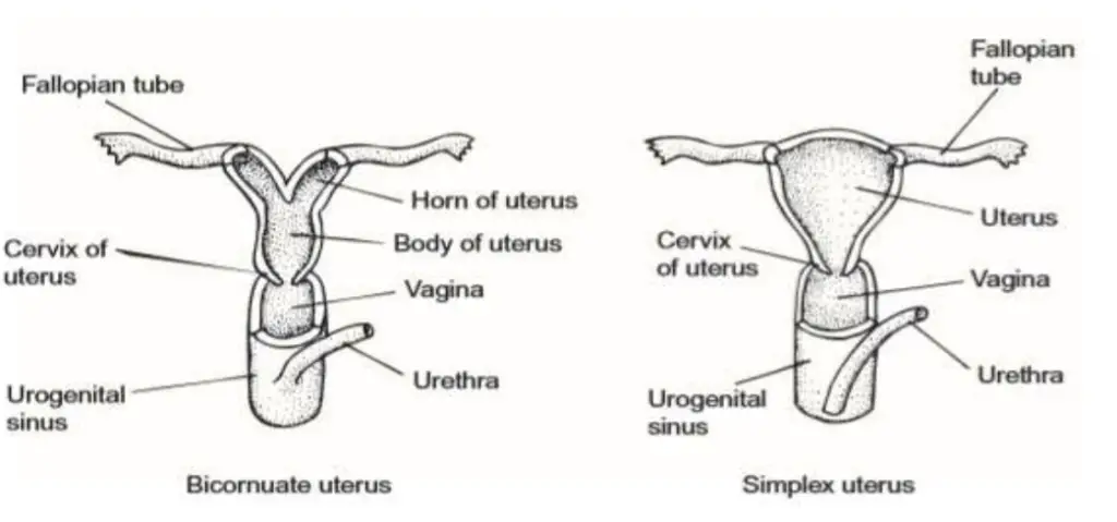
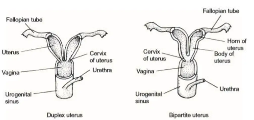
Eutherian mammals exhibit four primary types of uteri based on the degree of fusion at the distal end, each with distinct anatomical and functional characteristics.

- Duplex Uterus:
- In species with a duplex uterus, there are two distinct uteri, each of which opens separately into the vagina.
- This configuration allows for independent reproductive capabilities in each uterus.
- Notable examples include elephants, many rodents, and certain bats. In some cases, such as in certain rodents, there may even be two separate vaginas corresponding to each uterus, which further emphasizes reproductive independence.
- Bipartite Uterus:
- The bipartite uterus features two uteri that are partly fused, allowing them to open through a single vaginal aperture.
- This structure is common in various mammalian species, including most carnivores, pigs, cattle, and some rodents and bats.
- The partial fusion facilitates a more integrated reproductive system while still maintaining some degree of separation between the two uteri.
- Bicornuate Uterus:
- In the bicornuate uterus, the two uteri are more than half fused, forming two distinct horns or cornua where the developing young reside.
- This type of uterus is observed in species such as rabbits, whales, sheep, insectivores, most bats, and some carnivores and hoofed mammals.
- Each horn may have separate passageways, though this is not always externally visible.
- Typically, one horn may be larger and longer than the other, and while both ovaries can produce viable eggs, the blastocyst may preferentially implant in the larger horn.
- Simplex Uterus:
- The simplex uterus is characterized by a complete fusion of the two uteri into a single structure without uterine horns.
- This configuration is found in species such as armadillos, apes, and humans.
- The fusion begins at the ends of short oviducts, resulting in a singular uterine body where the blastocyst implants.
- Typically, only one fetus develops per pregnancy, although armadillos are unique in their ability to give birth to identical quadruplets, showcasing an exceptional reproductive adaptation.

Succession of Vertebrate Kidney
The succession of vertebrate kidney development reveals a fascinating journey through which the kidney evolves from simple structures to more complex forms suited for efficient waste filtration and water regulation. Understanding this progression involves examining the stages of kidney formation, which can be categorized into three distinct types: pronephros, mesonephros, and metanephros. Each stage reflects the anatomical and functional adaptations of vertebrates as they evolve.
- Embryonic Development of the Kidneys:
- The kidneys arise from the postero-dorsal mesoderm of the embryo, where it expands to form a nephric ridge.
- The nephric ridge subsequently gives rise to the paired nephrotome, a segmental structure containing nephrocoel, a coelomic chamber that may connect to the coelom via a ciliated peritoneal funnel.
- The glomerulus develops from the median part of the nephrotome, while ducts grow from its lateral ends, merging to form a common nephric duct. This structure becomes the nephric tubule, a fundamental component of the urinary system.
- Tripartite Concept of Kidney Organization:
- The nephric tubules differentiate structurally along the nephric ridge, leading to the tripartite concept of kidney organization.
- This concept categorizes the nephric tubules into three regions, each contributing to the eventual formation of the adult kidneys:
- Pronephros: Arising from the anterior region of the nephric ridge, the pronephros develops briefly during embryonic stages. It consists of pronephric tubules that form a common duct, opening into the cloaca. Pronephric tubules and glomeruli function in larval cyclostomes and some adult fishes.
- Mesonephros: The middle region of the nephric ridge gives rise to the mesonephros. Mesonephric tubules do not create a new duct but connect to the existing pronephric duct, now referred to as the mesonephric duct. While the mesonephros remains functional during embryonic stages, it is also seen in some adult fishes and amphibians.
- Metanephros: This kidney type appears later, originating from the ureteric diverticulum at the base of the pre-existing mesonephric duct. It promotes the growth of metanephric tubules, ultimately forming the definitive kidney in adult amniotes, where the metanephric duct is termed the ureter.
- Kidney in Lower Vertebrates:
- In hagfish, pronephric tubules fuse to create a pronephric duct. Anterior tubules lack glomeruli, while posterior tubules are associated with glomeruli but do not connect to the coelom. The mesonephros becomes the functional kidney in adult hagfish.
- Lamprey larvae (ammocoetes) possess a pronephric kidney composed of coiled tubules serviced by a glomus, a structure different from a glomerulus, as it supplies several tubules.
- Kidney in Amphibians:
- Amphibians transition from a pronephros to a mesonephros during larval stages, which is later replaced by an opisthonephros in adults. This dual functionality enables the anterior kidney tubules to transport sperm, illustrating the integration of the reproductive and urinary systems.
- Kidney in Other Vertebrates:
- In amniotes, the mesonephros is replaced by the metanephros in adults, which discharges waste through a new urinary duct, the ureter. The metanephric tubules are well-differentiated into proximal, intermediate, and distal segments, with the intermediate portion being notably elongated, constituting the major part of the loop of Henle.
- Loop of Henle:
- The loop of Henle is a critical structure found in metanephric tubules, specifically in animals capable of producing concentrated urine, such as mammals and birds. The loop consists of:
- A straight portion of the proximal tubule.
- A thin-walled intermediate region.
- A straight portion of the distal tubule.
- The loop serves as both a positional and structural feature of the nephron. The descending limb carries fluid from the cortex to the medulla, while the ascending limb returns to the cortex. The distinction between thick and thin segments is based on the height of the epithelial cells, with cuboidal cells being thick and squamous cells being thin.
- The loop of Henle is a critical structure found in metanephric tubules, specifically in animals capable of producing concentrated urine, such as mammals and birds. The loop consists of:

Evolution of the vertebrate kidney
The evolution of the vertebrate kidney is a complex narrative that reflects how various vertebrate classes adapted to their distinct external osmotic environments. These adaptations are crucial for maintaining internal water balance and effectively excreting nitrogenous wastes. The progression of kidney types—pronephric, mesonephric, and metanephric—demonstrates successful evolutionary responses to environmental pressures. The variations in kidney structure among vertebrates correlate significantly with the habitats they occupy, leading to adaptations in the number, complexity, arrangement, and location of kidney tubules.
- Embryological Origin:
- The kidney in all vertebrates originates from the intermediate mesoderm, specifically the nephrogenic mesoderm.
- The kidney is composed of two main elements: the kidney duct and the kidney tubules.
- The functional units of the kidney, known as nephrons, are evolutionary modifications of the nephridia.
- Kidney development is complex, involving the formation of two or three distinct kidney types in a temporal and spatial sequence:
- The pronephric kidney is the first and largest to develop, located anteriorly.
- The mesonephric kidney forms second.
- In birds, reptiles, and mammals, a third kidney type, the metanephric kidney, develops posterior to the mesonephros.
- Effects of Environment on Nephron Structure and Function:
- Higher vertebrates, including humans, possess nephrons made up of several components:
- Glomerulus: Filters blood.
- Bowman’s Capsule: Also filters blood and contains podocytes that prevent large molecules, such as blood proteins and cells, from passing into Bowman’s space.
- Proximal Convoluted Tubule: Reabsorbs water, salts, glucose, and amino acids.
- Loop of Henle: Reabsorbs water and small molecules.
- Distal Convoluted Tubule: Secretes H2_22 ions, potassium, and certain drugs.
- Higher vertebrates, including humans, possess nephrons made up of several components:
- Types of Nephrons Across Species:
- There are three types of nephrons, each adapted to different environments:
- Primitive Nephrons:
- Found in amphibians, freshwater bony fishes, and elasmobranchs.
- Characterized by a large renal corpuscle, allowing for high water output to combat potential overdilution.
- Marine Teleost Nephrons:
- Common in many marine teleosts and reptiles.
- Feature small or absent corpuscles with shortened renal tubules to increase salt excretion and conserve water, resulting in low water output.
- Aglomerular kidneys, seen in seahorses and pipefishes, eliminate water output through renal, gill, and rectal gland mechanisms.
- Mammalian and Avian Nephrons:
- Present in birds and mammals.
- Feature a large glomerulus and a complex tubule system with a long loop of Henle, facilitating significant water reabsorption.
- Produce relatively concentrated urine despite high water output at the glomerulus.
- Primitive Nephrons:
- There are three types of nephrons, each adapted to different environments:
- Adaptations Correlated with Environmental Factors:
- Freshwater Vertebrates:
- Inhabiting a medium more dilute than their bodily fluids, these vertebrates face the risk of overdilution. To counter this, they eliminate large amounts of water through the presence of a large corpuscle.
- Marine Vertebrates:
- Living in high salinity conditions, these vertebrates need to conserve water while excreting excess salt. The reduction or absence of glomeruli in some species reflects this need.
- Terrestrial vertebrates experience similar challenges in dry environments, leading to reduced renal corpuscle size and decreased water output.
- Birds and Mammals:
- These animals have developed efficient mechanisms for water conservation despite high glomerular output, resulting in the production of concentrated urine through specialized nephron structures.
- Freshwater Vertebrates:
Evolution of Gonads and Urinogenital ducts in vertebrates
The evolution of gonads and urinogenital ducts in vertebrates reflects significant adaptations to reproductive strategies and environmental challenges. Understanding this evolution involves examining the anatomical and functional developments across different vertebrate classes, highlighting the diverse mechanisms that have arisen to facilitate reproduction and waste management.
- Gonads Overview:
- Gonads are the primary sex glands in vertebrates, typically existing as paired structures: testes in males and ovaries in females. Most vertebrates are dioecious, meaning they have distinct male and female individuals.
- Exceptions include hermaphroditic cyclostomes and certain bony fishes, where individuals may exhibit both male and female reproductive traits.
- In hagfish, the gonad is bipartite: the anterior portion produces ova, while the posterior section produces sperm. Despite this dual structure, each gonad functions primarily as either an ovary or testis at maturity.
- Gonadal Development:
- The mesodermal coelomic epithelium gives rise to elongated genital ridges, which lie along the dorsal surface of the coelom near developing kidneys.
- These genital ridges are longer than the mature gonads. The middle part of the mesoderm becomes the gonads, while the anterior (progonal) and posterior (epigonal) sections remain sterile.
- The germinal epithelium, which forms from the peritoneum covering the gonads, is responsible for producing ova in the ovaries and spermatozoa in the testes.
- The testes consist of highly coiled seminiferous tubules lined with germinal epithelium, while the ovaries contain ova at various developmental stages.
- Male Gonads and Urinogenital Ducts:
- In cartilaginous and bony fishes, testes are paired, and the sexes are typically distinct. Conversely, in cyclostomes, gonads are unpaired, and ducts are absent.
- The anterior portion of the mesonephric kidney in anamniotes evolves to serve as part of the male genital system. This anterior section loses its renal functions, converting into the vasa efferentia that connect to the seminiferous tubules of the testes.
- The mesonephric duct serves as both a urinary and genital duct in males. In some elasmobranchs, such as dogfish, special accessory urinary ducts are formed to separate urinary function from the genital duct, which becomes solely a vas deferens.
- In amphibians, testes connect directly or via mesonephric tubules to the archinephric duct, leading to the cloaca. Male amniotes possess metanephric kidneys with separate ureters, while the persistent mesonephric duct becomes the vas deferens, forming an epididymis.
- Various reptiles possess specialized copulatory organs for sperm transfer, such as hemipenes in lizards and snakes, while crocodiles and turtles possess a structure homologous to the mammalian penis.
- Female Gonads and Urinogenital Ducts:
- Female gonads, or ovaries, generally consist of connective tissue enveloped by a germinal epithelium, producing ova that are released into the coelom.
- In female anamniotes, the nephrotome forms coelomic funnels that develop into oviducts, or Müllerian ducts, which open into the cloaca. The Müllerian duct also appears in males but degenerates under the influence of androgens.
- In elasmobranchs, the original pronephric duct bifurcates, with one duct becoming the mesonephric duct for urine and the other forming the oviduct for ova, showcasing the evolutionary adaptation of these systems.
- In cartilaginous and bony fishes, females typically possess two oviducts. In elasmobranchs, these oviducts are fused at the upper ends, leading to a single ostium for egg passage. The presence of shell glands indicates adaptations for internal fertilization and egg encasement.
- In amphibians, ovaries are paired, often containing lymph-filled cavities, and oviducts may be paired or fused, with specialized structures for temporary egg storage. Reptile ovaries and oviducts follow similar patterns, with added modifications for egg development and protection.
- In birds, typically, only the left ovary and oviduct remain functional, while in female mammals, oviducts are differentiated into distinct regions: the Fallopian tubes, uterus, and vagina. Monotremes exhibit unique reproductive structures, such as separate uteri and lack of a vagina.
- Variations in Uterine Structures in Eutherians:
- Female eutherian mammals exhibit four types of uteri based on the degree of fusion of the two uteri:
- Duplex Uterus: Found in rodents and some bats, where the two uteri remain separate.
- Bipartite Uterus: Seen in carnivores, pigs, and cattle, where the lower ends of the two uteri fuse to open through a single vaginal aperture.
- Bicornuate Uterus: Present in ungulates and whales, where the uteri fuse with a single cavity but lack a partition wall.
- Simplex Uterus: Characteristic of primates, where the two uteri completely fuse into a single body with one cavity.
- Notably, the variations in uterine structure do not correlate with phylogenetic relationships among mammalian orders, indicating evolutionary adaptation over time.
- Female eutherian mammals exhibit four types of uteri based on the degree of fusion of the two uteri:
Significance of Urogenital System
- Urine Production and Elimination:
- The urogenital system is responsible for the production, storage, and excretion of urine.
- This process begins in the kidneys, which filter blood to produce urine, and continues through the ureters, bladder, and urethra, ensuring that waste products are efficiently removed from the body.
- Regulation of Urogenital Functions:
- Urogenital activities are regulated by both autonomic and somatic efferent pathways originating in the lumbosacral spinal cord.
- The autonomic nervous system (ANS) is responsible for involuntary actions, while the somatic nervous system controls voluntary actions, illustrating the complexity of urogenital function regulation.
- Reflex Circuits:
- Certain functions, such as penile erection, are managed by reflex circuits in the spinal cord and brain stem, operating automatically without conscious control.
- In contrast, micturition (urination) involves more complex processes, necessitating voluntary control from the cerebral cortex, thereby integrating higher-level brain functions with basic physiological actions.
- Reproductive Functionality:
- The urogenital system is integral to reproductive activities, enabling sexual intercourse and the potential for conception.
- It ensures the proper functioning of reproductive organs and processes, which are vital for the continuation of human life.
- Consequences of Urogenital Defects:
- Disorders in the urogenital system can have serious implications. Conditions such as bilateral renal agenesis (failure of both kidneys to develop), bladder agenesis (absence of the bladder), and complete vulvar opening aplasia (underdeveloped external genitalia) can be fatal if not detected and managed early.
- In males, issues like uncertain sexual delineation and hypospadias (a condition where the opening of the urethra is not located at the tip of the penis) can also occur, underscoring the need for early diagnosis and intervention.
- https://egyankosh.ac.in/bitstream/123456789/16528/1/Unit-9.pdf
- https://dducollegedu.ac.in/Datafiles/cms/ecourse%20content/URINOGENITAL%20SYSTEM%20part-1%2025.03.2020.pdf
- https://dducollegedu.ac.in/Datafiles/cms/ecourse%20content/EVOLUTION%20OF%20UROGENITAL%20DUCTS%20part-2%2027.03.2020.pdf
- https://www.upcollege.ac.in/Upload/econtent/1338.pdf
- https://sajaipuriacollege.ac.in/pdf/zoology/Zoology_Sem-IV_CC8_unit-5_Urogenital_System_Prof.-Mowmita-Saha.pdf
- https://gcwgandhinagar.com/econtent/document/15880661673.2.1%20SUCCESSION%20OF%20VERTEBRATE%20KIDNEY%20%20UNIT%203.pdf
- https://gcwgandhinagar.com/econtent/document/15880662033.2.2%20Evolution%20of%20%20Gonads%20and%20Urinogenital%20ducts%20in%20vertebrates.pdf
- https://juniperpublishers.com/apbij/pdf/APBIJ.MS.ID.555554.pdf