- In 1875, Reoch theorised that microorganisms were responsible for the alkaline fermentation of urine (urea) and the subsequent formation of ammonia.
- Subsequently, numerous types of urea-degrading bacteria were isolated and their urease activity was examined. Urease activity is a crucial criterion for the identification of Proteus species and permits Proteus to be separated from non-lactose-fermenting Enterobacteriaceae members.
- Stuart devised a chemically defined medium that was demonstrated to be differential for Proteus species.
- In this medium, Proteus are able to use urea as their only nitrogen source and create enough ammonia to overcome the medium’s high buffering capacity, resulting in a yellow to bright pink colour shift (fuschia).
- Christensen later developed a medium that would support the growth of Enterobacteriaceae members that cannot use ammonia as their main nitrogen source.
- This medium also has a reduced buffering capacity, enabling the detection of smaller amounts of alkali created by the breakdown of urea.
- Consequently, Christensen’s urea agar can detect microorganisms with a weak or delayed urease activity.
What is Urease Test?
- The purpose of the urease test is to detect microorganisms capable of hydrolyzing urea with the enzyme urease.
- Reoch proposed in 1875 to evaluate the alkaline fermentation of urea and the formation of ammonia by bacteria in order to determine an organism’s ability to manufacture urease. Alkalinization causes a purplish-red hue in the presence of phenol red.
- It is frequently employed to differentiate the genus Proteus from other intestinal bacteria. Ammonia is produced as one of the byproducts of the hydrolysis of urea.
- This weak base raises the pH of the medium above 8.4, causing phenol red, a pH indicator, to change from yellow to pink.
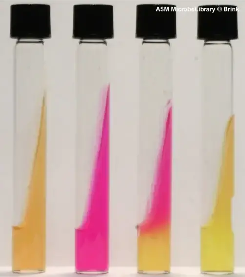
- Proteus mirabilis is an efficient urea hydrolyzer (center tube pictured here).
- The tube on the far right was inoculated with an organism lacking urease, whereas the tube on the far left was not infected.
- Urease is an enzyme produced by plaque bacteria in order to maintain the pH of the dental biofilm by hydrolyzing urea from saliva and gingival exudate into ammonia.
- The purpose of this work was to evaluate the urease activity of oral bacterial species using the rapid urease test (RUT) in a microplate format and to see if this test could be used to measure urease activity in site-specific supragingival dental plaque samples ex vivo.
Purpose of Urease Test
- The urease test finds organisms with the ability to hydrolyze urea into ammonia and carbon dioxide.
- To differentiate Proteeae with urease from other Enterobacteriaceae.
- It can be used to detect Helicobacter pylori infection.
Principle of Urease Test
- Urease is an endogenously produced enzyme that breaks down urea into carbon dioxide and ammonia.
- Urease test media contain 2% urea and pH indicator phenol red.
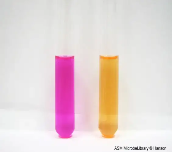
- Due to the creation of ammonia, an increase in pH causes a colour change from yellow (pH 6.8) to bright pink (pH 8.2).
- Stuart’s urea broth is a highly buffered medium that requires considerable doses of ammonia to increase the pH over 8.0, causing a change in hue.
- This medium contains all of the required nutrients for Proteus, which makes it unique.
- Christensen’s urea agar contains peptones and glucose and has a lower buffer content.
- This medium enables the growth of many enterobacteria, making it possible to observe urease activity.
(NH2)2CO + H2O → CO2 + 2NH3
Media Used for Urease Test
Two types of media are typically employed to detect urease activity. Christensen’s urea agar is employed to identify urease activity in numerous bacteria. Stuart’s urea broth is largely used to distinguish between Proteus species. Both forms of media are commercially accessible as tubes or as a powder.
Christensen’s Urea Agar
Composition of Christensen’s Urea Agar
| Ingredient | Amount |
| Peptone | 1 g |
| Dextrose | 1 g |
| Sodium chloride | 5 g |
| Potassium phosphate, monobasic | 2 g |
| Urea | 20 g |
| Phenol red | 0.012 g |
| Agar | 15 to 20 g |
Preparation of Christensen’s Urea Agar
- Dissolve the first six ingredients in 100 cc of distilled water and filter sterilise to prepare the urea base (0.45-mm pore size).
- Suspend the agar in 900 ml of distilled water, heat until completely dissolved, and autoclave for 15 minutes at 121oC and 15 psi.
- Cool the agar to 50 to 55 degrees Celsius.
- Aseptically combine 100 ml of urea base that has been filter-sterilized with the cooled agar solution.
- Distribute 4 to 5 ml each sterile tube (13 x 100 mm) and slant the tubes until firm during cooling.
- Long slant and short butt are excellent qualities.
- The prepared media will be yellow-orange in hue.
- Refrigerate the prepared medium at 4 to 8 degrees Celsius until use.
- Do not reheat the medium once it has been made, or the urea will disintegrate.
Stuart’s Urea Broth
Composition of Stuart’s Urea Broth
| Ingredient | Amount |
| Yeast extract | 0.1 g |
| Potassium phosphate, monobasic | 9.1 g |
| Potassium phosphate, dibasic | 9.5 g |
| Urea | 20 g |
| Phenol red | 0.01 g |
Preparation of Stuart’s Urea Broth
- Dissolve all ingredients in 1 litre of distillated water, then filter-sterilize the solution (0.45-mm pore size).
- Distribute 3 cc of prepared broth per sterile tube (13 x 100 mm) (13 x 100 mm).
- The prepared media will be yellow-orange in hue.
- Refrigerate the prepared broth between 4 and 8 degrees Celsius until use.
- Do not heat the medium, as urea will breakdown if heated.
Procedure of Urease Test
Urease Test using Christensen’s Urea Agar – Solid Media
- Apply a strong inoculum derived from an 18- to 24-hour pure culture to the entire slant surface.
- Do not stab the butt, as it will be used as a colour control.
- Incubate tubes with loose caps at 35oC.
- Observe the slope for a change in hue every six hours, twenty-four hours, and every day for up to six days.
Urease Test using Stuart’s Urea Broth – Liquid Media
- Use a heavy inoculum from a pure culture that has been going for 18 to 24 hours to start the broth.
- Gently shake the tube to move the bacteria around.
- At 35oC, put tubes with loose caps in an incubator.
- At 8, 12, 24, and 48 hours, watch the broth for a change in colour.
Result of Urease Test
Christensen’s Urea Agar
- Positive Test: A bright pink (fuchsia) colour on the slant that may go into the butt shows that urea is being made. Keep in mind that any amount of pink is a good sign.
- Negative Test: If the organism doesn’t have urease, the culture medium will stay yellow.
Note
- If the test is left out for too long, proteins in the medium may break down and give a false positive result.
- A control medium without urea should be used to rule out protein hydrolysis as the cause of a positive test.
- Rapidly urease-positive Proteeae (Proteus spp., Morganella morganii, and some Providencia stuartiistrains) will cause a strong positive reaction within 1 to 6 hours of incubation.
- Delayed-positive organisms, like Klebsiella or Enterobacter, will usually show a weak positive reaction on the slant after 6 hours, but the reaction will get stronger and spread to the butt with more time (up to 6 days).

Stuart’s Urea Broth
- Positive Test: The presence of urea is shown by a bright pink (fuchsia) colour in the broth.
- Negative Test: No color change to pink.
Note:
- Rapidly urease-positive Proteeae (Proteus spp., Morganella morganii, and some Providencia stuarti strains), for which this medium is different, will produce a strong positive reaction as early as 8 hours, but always within 48 hours of incubation.
- Due to the high buffering capacity of this medium, organisms that show a delayed reaction, like Enterobacter, will not show a positive reaction.

Urease positive and negative test Organisms
| BacteriaUrease positiveVariable ureaseNegative ureaseEnterobacteriaceaeProteus , Klebsiella (lent)/Salmonella, Citrobacter, Escherichia coli, Edwardsiella, Shigella, Providencia, Enterobacter, Hafnia alvei, Serratia marcescens et liquefaciensYersinia spp. | Y.enterocolitica, Y.pseudotuberculosis | / | Y.pestis |
|---|---|---|---|
| Vibrionaceae | / | / | Aeromonas, Plesiomonas, Vibrio |
| Haemophilus influenzae | biotypes : I II III IV | / | biotypes : V VI VII VIII |
| Bordetella spp. | B. parapertussis(24 h), B. bronchiseptica(24 h) | / | B. pertussis, B. trematum, B. holmesii |
| Brucella spp. | B. suis , B. canis (fast) | B. melitensis B. abortus (plus long ou peut être négatif) | / |
| Bartonella spp. | / | / | Bartonella spp. |
| Helicobacter species | H. pylori, H. heilmannii | / | H. cinaedi, H. fennelliae |
| Ureaplasma urealyticum | Ureaplasma urealyticum | / | / |
| Streptococcus spp | S. vestibularis (Group Salivarius) | / | Group Mitis, Group Anginosus, Group Mutans |
Comments And Tips
- Rapid urine test kits can be bought in stores.
- By adding a loopful of culture to 0.5 ml of Stuart’s urea broth, you can also get good results.
- On urea agar slants, Proteus spp. can grow quickly (in about 1 or 2 hours) if a heavy inoculum is put in one spot halfway up the slant.
- Helicobacter pylori is also urease positive, and this is a clinically important bacteria.
- In the clinical lab, there are many different tests and media that can be used to find H. pylori.
Helicobacter pylori case
Helicobacter pylori breaks down urea quickly, usually in 30 seconds. One thing that sets H. pylori apart from other Helicobacter species is how fast it moves. In the clinical laboratory, many different tests and media have been made to find H. pylori.
1. Rapid urease test
- The Urease Rapid Test, which is also called the Campylobacter-like organism (CLO) test, is a quick way to find out if you have Helicobacter pylori.
- The test is based on how well H. pylori can release the enzyme urease. The sample, such as a gastric biopsy, is put in a gel that contains urea.
- H. pylori’s urease quickly breaks down urea into ammonia and water, which changes the colour of the indicator.
Results
- Results can be read in 1 minute to 3 hours, but in some cases, the CLO test can be put on hold for up to 24 hours.
2. Urea breath test
- Most people think that the urea breath test is a simple, non-invasive, and accurate way to show that you have Helicobacter pylori.
- The idea behind the test is easy to understand. Isotopically labelled urea taken by mouth, either with 14C or 13C (13C is better), is broken down by the enzyme urease of H. pylori, and the labelled CO2 is breathed out.
- Either a scintillation counter (Carbon-14) and isotope ratio mass spectrometry or mass correlation spectrometry can find the labelled CO2 (Carbon-13).
- With a sensitivity and specificity between 95 and 100%, the main use for UBT is to confirm that eradication was successful. For testing to be accurate, it should be done 4-6 weeks after treatment ends and 5 days after antacids.
- The test is also a great way to check for infection when an ulcer is found during an endoscopy but a biopsy cannot be done because the patient is on anticoagulant medication.
Uses
- It is used to find out if a tissue biopsy sample has H. pylori in it.
- It is used to tell the difference between Candida albicans and Cryptococcus neoformans. The first one is urease negative, and the second one is urease positive.
- Some Enterobacteriaceae species, like Klesiella and Proteus, can be found with this test.
- The brucella and Cryptococcus species can be found with a urease test.
Limitations
- Microorganisms like H. pylori and Brucella split urea quickly, which could change the test result.
- A biochemical test and/or a serological test should be done for a full identification.
- Don’t use inoculum from a suspension of broth to help urea grow and break down.
- If the culture is kept in the incubator for a long time, a false-positive alkaline reaction will be seen.
- Urea is sensitive to heat, so don’t put the urea agar slant in direct heat. When urea is exposed to heat, it breaks down.
- If you are testing Proteus species, make sure to check the inoculation within six hours to see if anything happened.
- Make sure the cap is loose when you incubate the medium. If you don’t, it can change the result.
- Store the test tube somewhere dark because urea breaks down on its own when exposed to light.
- Urea agar shouldn’t be the only way to figure out how much urease is working because different organisms have different abilities and hydrolysis rates.
Quality Control
- Positive control (PC): Proteus mirabilis (Urease positive bacteria)
- Negative Control (NC): Escherichia coli (Urease negative bacteria)
- Uninoculated (UN): Along with the test medium, an uninoculated medium is cultured to compare the colour change.
References
- https://asm.org/getattachment/ac4fe214-106d-407c-b6c6-e3bb49ac6ffb/urease-test-protocol-3223.pdf
- https://microbiologie-clinique.com/urease-test-principle-protocol-results.html
- https://microbenotes.com/urease-test-principle-procedure-and-result/
- https://microbeonline.com/urease-test-principle-procedure-interpretation-and-urease-positive-organsims/
- http://www.uwyo.edu/molb2021/additional_info/summ_biochem/urease.html
- https://bmcoralhealth.biomedcentral.com/articles/10.1186/s12903-018-0541-3
- https://www.austincc.edu/microbugz/urease_test.php
- https://bmcgastroenterol.biomedcentral.com/articles/10.1186/1471-230X-5-38
- https://vlab.amrita.edu/?sub=3&brch=76&sim=214&cnt=1
- https://himedialabs.com/TD/M1828.pdf
- https://www.labtestsguide.com/urease-test
- https://universe84a.com/urease-test/