What are Tissues?
- Tissues are groups of cells that share a common structure and function. They represent a level of organization in biological systems that lies between individual cells and organ systems. Tissues play a crucial role in the formation of organs, which are composed of functional groups of tissues working collaboratively to perform specific tasks.
- The study of tissues is referred to as histology, which involves examining the structure and function of tissues at both the microscopic and macroscopic levels. Histologists utilize various techniques to prepare tissue samples for examination, including the embedding of tissues in paraffin and sectioning them into thin slices for observation under a microscope. This method allows for the detailed analysis of cellular architecture and the identification of specific tissue types.
- Furthermore, the examination of tissues in relation to disease is called histopathology. This field focuses on understanding the alterations in tissue structure and function caused by various pathological conditions. Through histopathological analysis, medical professionals can diagnose diseases, assess the severity of conditions, and guide treatment options.
- Tissues are categorized into four primary types: epithelial, connective, muscle, and nervous tissues. Each tissue type serves distinct functions within the body. Epithelial tissue, for example, covers body surfaces and lines cavities, providing protection and facilitating absorption and secretion. Connective tissue provides structural support and connects different tissues and organs. Muscle tissue is responsible for movement, while nervous tissue is integral to the transmission of signals throughout the body.
Definition of Tissues
Tissues are groups of similar cells that work together to perform specific functions in an organism. They serve as the structural and functional units that form organs, playing critical roles in various biological processes.
Characteristics of Tissues
- Supported by an extracellular matrix that gives structural integrity and promotes cell communication, tissues are collections of similar cells arranged to serve particular purposes.
- The cells within a tissue exhibit specialized shapes, sizes, and biochemical properties that reflect their functional roles, ensuring uniformity and efficiency in tissue performance
- Tissues show a clear structural arrangement, with cells stacked in layers or patterns allowing coordinated activities including protection, absorption, and secretion.
- In connective tissues, the extracellular matrix—made of proteins like collagen and elastin—is essential for mechanical strength, elasticity, and signaling cues that control cell behavior.
- Clear differences between meristematic tissues—which are actively dividing and in charge of growth—and permanent tissues with specialized purposes including support, photosynthesis, and nutrient movement define plant tissues.
- Tissues have different regenerative capacity; for example, epithelial tissues often have high turnover rates while nerve tissues show limited regenerative potential, reflecting variations in underlying cellular mechanisms.
- Histologically, tissues are defined by their staining properties and microscopic architecture, which are used to identify normal versus pathological states in clinical diagnostics
- The integration of cells with their extracellular matrix in tissues is fundamental for organ formation, ensuring that each tissue type contributes effectively to the overall function and homeostasis of an organism
Location of Tissues
- The structural and functional units of organs and organ systems are found in the tissues scattered across the bodies of multicellular life.
- Whereas connective tissue supports organs like bones, tendons, and blood vessels, epithelium tissue lines surfaces and cavities including the skin, the digestive tract, and the respiratory system in animals.
- Muscle tissues found in areas needing movement—the heart, limbs, the digestive tract—allow contraction and coordinated motion.
- Found mostly in the brain, spinal cord, and peripheral nerves, nervous tissue forms the communication system guiding bodily activities.
- In plants, meristematic tissues at the growth points—such as the terminals of roots and shoots—as well as permanent tissues (dermal, vascular, and ground tissues) found in structures like leaves, stems, and roots.
- The general arrangement of an organism depends on the placement of tissues as every kind of tissue is positioned deliberately to support certain purposes and preserve homeostasis.
Formation of Tissues
The formation of tissues, known as histogenesis, involves the differentiation of cells into specialized types that collaborate to perform specific functions. In animals, this process originates from the three primary germ layers established during embryonic development:
- Ectoderm: Gives rise to tissues such as the epidermis and nervous system.
- Mesoderm: Forms connective tissues, muscles, and the circulatory system.
- Endoderm: Develops into the lining of the digestive and respiratory tracts.
These germ layers differentiate into the four fundamental tissue types in animals:
- Epithelial Tissue: Covers body surfaces and lines cavities.
- Connective Tissue: Provides support and structure.
- Muscle Tissue: Facilitates movement.
- Nervous Tissue: Conducts nerve impulses.
In plants, tissue formation begins with meristematic tissues, which are regions of actively dividing cells located at growth points like root and shoot tips. These meristematic cells differentiate into various permanent tissues:
- Dermal Tissue: Forms the outer protective covering.
- Vascular Tissue: Comprises xylem and phloem for water and nutrient transport.
- Ground Tissue: Performs functions such as photosynthesis, storage, and support.
Types of Tissues
Tissues are classified into two main categories: plant tissues and animal tissues. Each category is characterized by distinct structures and functions that are vital to the overall functioning of the respective organisms.
- Plant Tissue
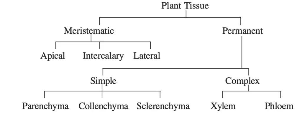
Plant tissues can be divided into two main types: meristematic and permanent tissues.- Meristematic Tissue
- This tissue consists of actively dividing cells, which are undifferentiated and capable of continuous growth.
- Located at the tips of roots and shoots, meristematic tissue contributes to the elongation and development of the plant.
- Cells in this tissue are small, have thin cell walls, and contain large nuclei.
- Permanent Tissue
- Permanent tissues arise from meristematic tissues after they differentiate.
- They are classified into two subcategories: simple and complex tissues.
- Simple Tissues
- Composed of one type of cell.
- Examples include parenchyma (storage and photosynthesis), collenchyma (flexible support), and sclerenchyma (rigid support).
- Complex Tissues
- Made up of more than one type of cell that work together.
- Examples include xylem (water transport) and phloem (nutrient transport).
- Meristematic Tissue
- Animal Tissue
Animal tissues are categorized into four primary types: epithelial, connective, muscle, and nervous tissues.- Epithelial Tissue
- This tissue covers body surfaces, lines cavities, and forms glands.
- Epithelial cells are tightly packed, providing protection and facilitating absorption and secretion.
- They can be classified based on the number of layers (simple or stratified) and the shape of the cells (squamous, cuboidal, or columnar).
- Connective Tissue
- Connective tissue provides structural support and binds other tissues together.
- It consists of cells dispersed within an extracellular matrix, which can be liquid, gel-like, or solid.
- Types of connective tissue include loose connective tissue (provides flexibility), dense connective tissue (provides strength), adipose tissue (stores fat), blood (transports nutrients and waste), and bone (provides structure).
- Muscle Tissue
- Muscle tissue is responsible for movement and is classified into three types: skeletal, cardiac, and smooth muscle.
- Skeletal muscle is voluntary and striated, enabling body movements.
- Cardiac muscle, found only in the heart, is involuntary and striated, facilitating heartbeat.
- Smooth muscle is involuntary and non-striated, controlling movements in hollow organs like the intestines and blood vessels.
- Nervous Tissue
- Nervous tissue is specialized for communication and control within the body.
- It consists of neurons, which transmit signals, and glial cells, which support and protect neurons.
- This tissue is essential for processing information and coordinating responses to stimuli.
- Epithelial Tissue
Types of Animal Tissues
Animal tissues are classified into four primary types: connective, muscle, nervous, and epithelial tissues. Each type has distinct characteristics and functions that contribute to the overall physiology of the organism. Understanding these tissues is crucial for comprehending how organs are structured and how they perform their designated functions.
- Connective Tissue
Connective tissues consist of cells that are separated by non-living material known as the extracellular matrix. This matrix plays a vital role in providing shape and maintaining the position of various organs. Connective tissue types include:- Fluid Connective Tissue: Includes blood, which transports nutrients and wastes throughout the body.
- Fibrous Connective Tissue: Comprises tendons and ligaments, providing support and stability.
- Skeletal Connective Tissue: Includes bone and cartilage, offering structural integrity and support.
- Providing shape and maintaining the position of organs.
- Acting as the primary supporting tissue in the body.
- Insulating the body and conserving heat.
- Binding organs together and offering support.
- Protecting against pathogens through phagocytic activity.
- Transporting water, nutrients, minerals, hormones, gases, and waste products within the body.
- Muscle Tissue
Muscle tissues are responsible for producing force and generating motion, facilitating both locomotion and movements within internal organs. The three types of muscle tissue include:- Skeletal Muscle: Typically attached to bones, allowing voluntary movement.
- Cardiac Muscle: Found exclusively in the heart, responsible for involuntary contractions that pump blood.
- Visceral or Smooth Muscle: Located in the inner walls of organs, aiding in involuntary movements.
- Facilitating movements such as walking, running, lifting, and chewing.
- Maintaining posture and an erect position.
- Constricting organs and blood vessels to regulate flow.
- Pumping blood and controlling blood flow in arteries.
- Regulating respiration through involuntary movements that drive air in and out of the body.
- Nervous Tissue
Nervous tissue is a critical component of the central nervous system, which includes the brain and spinal cord, as well as the peripheral nervous system, which forms cranial and spinal nerves.The primary functions of nervous tissue include:- Serving as a communication network for the nervous system, crucial for information processing.
- Responding to stimuli and transmitting information within the body.
- Playing a significant role in emotions, memory, and reasoning.
- Maintaining homeostasis and creating awareness of the surrounding environment.
- Coordinating various metabolic activities throughout the body.
- Epithelial Tissue
Epithelial tissues are formed by cells that cover the external surfaces of body organs and line the surfaces of internal cavities. They are found in areas such as the skin, reproductive tract, airways, and the inner lining of the digestive tract.The functions of epithelial tissue are varied and include:- Participating in sensory reception, excretion, filtration, and metabolic activities.
- Providing mechanical strength and resistance to underlying cells and tissues.
- Facilitating the movement of materials through filtration, diffusion, and secretion.
- Protecting internal organs against pathogens, toxins, physical trauma, and radiation.
- Secreting hormones, enzymes, mucus, and other products, transporting them to the circulatory system.
Types of Plant Tissues
Plant tissues can be classified based on two key criteria: the different parts of the plants they are associated with and the various types of cells they contain. This classification leads to a deeper understanding of how plant tissues function and contribute to the overall growth and health of the plant.
- Classification Based on Plant Parts
Plant tissues are broadly categorized into three tissue systems based on their location and function:- Epidermal Tissue:
- Comprises cells that form the outermost surface of leaves and stems.
- Functions primarily in protection against mechanical injury, water loss, and pathogen invasion.
- Vascular Tissue:
- Responsible for the internal transport of fluids and nutrients.
- Consists of xylem, which transports water and minerals from roots to other parts of the plant, and phloem, which distributes sugars and nutrients produced during photosynthesis.
- Ground Tissue:
- Involved in the production and storage of nutrients through photosynthesis.
- Serves as a site for metabolic processes and provides structural support to the plant.
- Epidermal Tissue:
- Classification Based on Cell Types
Plant tissues can also be divided into two types based on the kinds of cells they contain:- Meristematic Tissue:
- Comprises young cells that are continuously dividing, facilitating the increase in both length and width of the plant.
- Different types of meristematic tissues can be classified according to their position, function, plane of division, origin, and development. The three main types are:
- Apical Meristem: Located at the tips of roots and shoots, promoting primary growth.
- Lateral Meristem: Responsible for secondary growth, increasing the girth of the plant.
- Intercalary Meristem: Found at the base of leaves or internodes, contributing to the growth of new leaves or stems.
- The functions of meristematic tissue include:
- Responsible for the growth of new organs.
- Involved in the movement of water and nutrients within the plant.
- Facilitates both primary and secondary growth.
- Provides protection from mechanical injury as the outermost tissue.
- Gives rise to other tissues, including the epidermis, cortex, endodermis, ground tissue, and vascular tissue.
- Permanent Tissue:
- Composed of a group of cells that are similar in origin, structure, and function, which have undergone complete growth and differentiation after the cessation of meristematic activity.
- Permanent tissues can be classified into three types:
- Simple Permanent Tissues: Consist of one type of cell and include parenchyma, collenchyma, and sclerenchyma.
- Complex Permanent Tissues: Composed of more than one type of cell, such as xylem and phloem.
- Special or Secretory Tissues: Involved in producing specific substances like resins or essential oils.
- The functions of permanent tissues encompass:
- Assisting in flotation in aquatic plants.
- Storing food in the form of starch, proteins, oils, and fats.
- Providing hardness and protection to fruits such as nuts and coconuts.
- Containing chloroplasts for conducting photosynthesis.
- Involvement in secretion, transportation, and providing mechanical support to the plant.
- Meristematic Tissue:
The Plant Tissues
The plant tissues are mainly of two catagories:
- Meristematic (Gk. meristos : dividing)
- Permanent (non-dividing)
A. Meristematic tissues
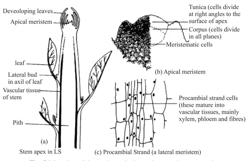
Meristematic tissues are essential components of plant growth, consisting of undifferentiated cells that are actively dividing. These tissues are primarily located in regions of the plant where growth occurs, such as at the tips of roots and shoots, as well as in the cambium layer. The presence of meristematic tissues enables plants to increase in both length and girth, which is crucial for their development and overall health.
- Characteristics of Meristematic Tissue:
- Active Cell Division: Cells in meristematic tissue are continuously dividing, which is vital for plant growth.
- Dense Cytoplasm and Large Nucleus: Each meristematic cell contains a significant amount of cytoplasm and typically features a single, prominent nucleus.
- Compact Arrangement: The cells are closely packed together, exhibiting little to no intercellular space.
- Thin Cell Walls: The cell walls of meristematic cells are made of a thin layer of cellular material, allowing for flexibility and growth.
- Varied Cell Shapes: These cells may be spherical, oval, polygonal, or rectangular in shape, adapting to their functional requirements.
- Vacuole Presence: Depending on the specific type of meristematic tissue, the cells may or may not contain vacuoles.
- Functions of Meristematic Tissue:
- Continuous Cell Division: Meristematic tissues possess the ability to divide, which results in the continuous production of new cells. These new cells subsequently differentiate into specialized cell types that perform various functions within the plant.
- Increase in Length: The cells located at the root tip and shoot tip contribute to the elongation of the plant, enabling it to grow taller and deeper into the soil.
- Increase in Girth: The cambium, found in the lateral regions of the plant, is responsible for increasing the thickness or girth of the plant, allowing it to support additional weight and resist external pressures.
Types of Meristematic Tissue
Meristematic tissues are classified based on their location and functional roles within the plant. Each type serves distinct purposes in facilitating growth and development. The three primary types of meristematic tissue include apical meristem, lateral meristem, and intercalary meristem. Understanding these types is crucial for comprehending how plants grow in length and girth.
- Apical Meristem:
- Location: Apical meristems are situated at the tips of roots and stems.
- Function: This type of meristem is primarily responsible for the increase in length of the plant axis. By contributing to the elongation of roots and shoots, apical meristems enable plants to explore new areas for water and nutrients, enhancing their overall growth potential.
- Lateral Meristem (Cambium):
- Location: Lateral meristems are found on the lateral sides of stems and roots.
- Function: These tissues play a critical role in increasing the thickness or girth of the stem and root. This thickening process, known as secondary growth, is essential for providing structural support to mature plants, allowing them to withstand external pressures and maintain stability.
- Intercalary Meristem:
- Location: Intercalary meristems are located at the bases of leaves or internodes in twigs.
- Function: This type of meristem is responsible for the longitudinal growth of plants, particularly contributing to the increase in length of the internodal regions. Intercalary meristems enable quick growth responses, particularly in monocots like grasses, allowing them to recover rapidly from damage or herbivory.
B. Permanent tissues
Permanent tissues are specialized structures that arise from meristematic tissues as a result of cell differentiation. This process involves cells taking on a permanent shape, size, and function, marking a transition from the undifferentiated state of meristematic tissues. Understanding permanent tissues is crucial for comprehending how plants maintain their structure and perform various functions.
- Origin: Permanent tissues originate from meristematic tissues, which are characterized by actively dividing cells. As these cells differentiate, they develop into permanent tissues, taking on specific roles in the plant.
- Characteristics:
- Loss of Division: Cells in permanent tissues have lost their ability to divide, marking a significant transition from the meristematic phase.
- Definite Form and Size: The cells exhibit a definite shape and size, which allows them to perform specialized functions effectively.
- Specific Functions: Each cell within permanent tissues is differentiated, meaning it carries out specific functions essential for the plant’s growth and survival.
- Cell Composition: The cells can be either living or dead, contributing to various functions in the plant. For instance, thin-walled permanent tissues are typically living, while thick-walled tissues can be either living or dead.
- Cell Wall Structure: The cell walls of permanent tissues can be thin or thick, depending on the tissue type. This variation in wall thickness affects the tissue’s overall function and durability.
- Cytoplasm: Permanent tissue cells are generally large and often contain vacuolated cytoplasm, which aids in storage and other cellular processes.
- Functionality:
- Permanent tissues serve various functions within the plant, reflecting their specific adaptations. For example, living permanent tissues contribute to metabolic processes, while dead permanent tissues may provide structural support or aid in water retention.
- The functionality of permanent tissues can be seen in their role in storage, support, and transport within the plant.
Types of Permanent Tissues
Permanent tissues in plants are crucial components that have undergone differentiation from meristematic tissues, taking on specific structures and functions to support various plant activities. They are categorized into two main types: simple permanent tissues and complex permanent tissues, each serving distinct roles within the plant.
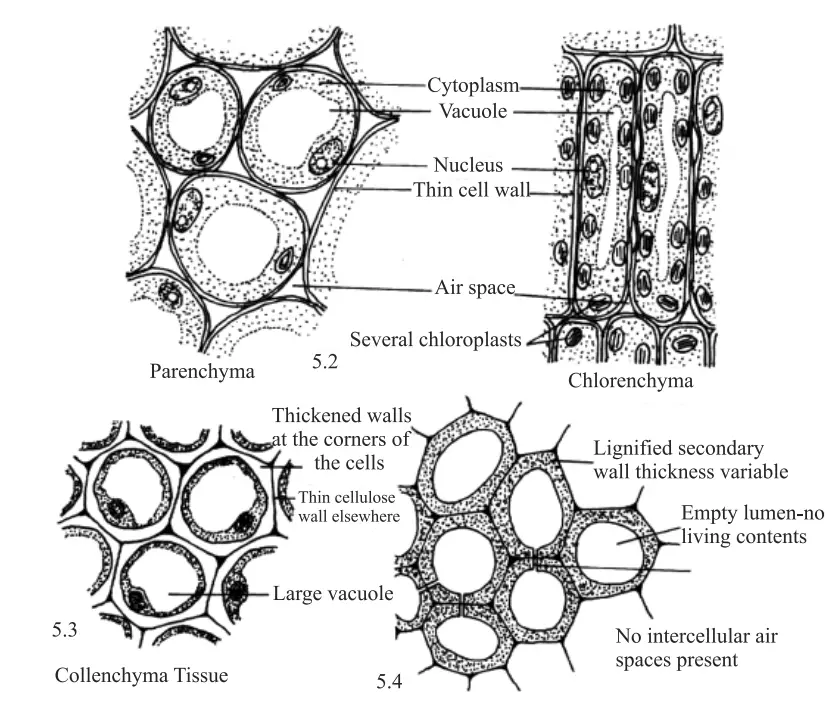
- Simple Permanent Tissues: These tissues are composed of structurally similar cells, meaning they consist of only one type of cell. Simple permanent tissues are further divided into three categories based on the nature of their cells:
- Parenchyma:
- This tissue is regarded as the most primitive and fundamental among plant tissues, being the most common type found in plants.
- Parenchyma cells are living and can take on various shapes, including spherical, oval, or polygonal.
- The cell walls are thin, primarily composed of cellulose, which contributes to their flexibility.
- These cells are loosely packed with substantial intercellular spaces, allowing for efficient gas exchange and storage.
- A large central vacuole typically occupies much of the cell’s interior, surrounded by dense peripheral cytoplasm containing a distinct nucleus.
- Parenchyma is commonly found in soft parts of the plant, such as the cortex of roots, ground tissues in stems, and the mesophyll of leaves.
- Function: The primary role of parenchyma is the storage of food and other substances.
- Specialized Parenchyma:
- Chlorenchyma: Parenchyma containing chloroplasts, involved in photosynthesis.
- Aerenchyma: Parenchyma with air cavities, facilitating buoyancy in aquatic plants.
- Stellate/Steel Parenchyma: Star-shaped parenchyma, contributing to structural integrity.
- Idioblast: Parenchyma that contains ergastic substances, which are metabolic waste products.
- Collenchyma:
- Cells of collenchyma are living and characterized by their elongated shapes.
- The cell walls are irregularly thickened at the corners, providing support and flexibility.
- Intercellular spaces are minimal, enhancing structural integrity.
- Function: Collenchyma provides mechanical strength and flexibility to the plant, allowing for easy bending without breaking, particularly in leaves and stems.
- Sclerenchyma:
- Unlike the previous types, the cells of sclerenchyma are dead at maturity.
- The cell walls are long, narrow, and significantly thickened due to lignin deposition, which acts as a cementing agent that hardens the cell walls.
- These cells lack intercellular spaces, providing robust structural support.
- Sclerenchyma is typically found in stems, around vascular bundles, within leaf veins, and in the hard coverings of seeds and nuts.
- In older plants, the epidermis undergoes changes, forming several layers of thick cork or bark, composed of dead and compactly arranged cells.
- Cork cells contain suberin, a substance that renders them impervious to gases and water.
- Parenchyma:
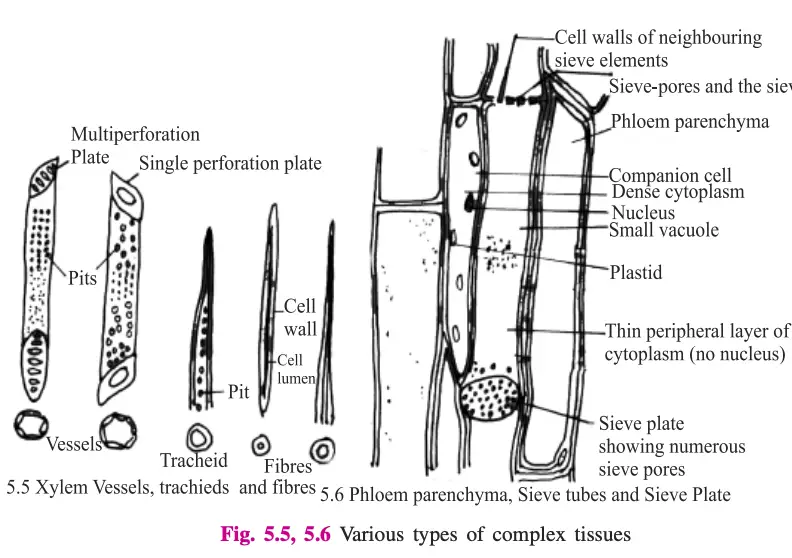
- Complex Permanent Tissues: In contrast to simple permanent tissues, complex permanent tissues consist of structurally different cells that coordinate to perform a common function. The primary examples of complex permanent tissues are xylem and phloem, both of which are conducting tissues forming vascular bundles.
- Xylem:
- Xylem is composed of four types of cells:
- Tracheids: Dead, thick-walled, lignified tubular cells that conduct water.
- Vessels/Tracheae: Also dead and thick-walled, vessels are found exclusively in the xylem of angiosperms and are absent in gymnosperms and pteridophytes.
- Xylem Parenchyma: Living cells with thin cell walls, involved in storage and transport.
- Xylem Fibers: Dead cells with thick walls that provide support to the xylem tissue.
- Xylem is composed of four types of cells:
- Phloem:
- Phloem, also known as bast, consists of four types of cells:
- Sieve Tubes: Tubular cells with perforated end walls, allowing for the flow of nutrients; these cells lack a nucleus but contain a thin layer of cytoplasm.
- Companion Cells: Small, elongated cells with dense cytoplasm and a prominent nucleus, supporting sieve tube function.
- Phloem Parenchyma: Thin-walled cells that play a significant role in the storage and transportation of food.
- Phloem Fibers: Thick-walled, elongated dead sclerenchymatous cells that provide mechanical strength to the phloem tissue.
- Phloem, also known as bast, consists of four types of cells:
- Xylem:
- Functionality of Complex Tissues: The primary function of both xylem and phloem is to facilitate the transport of water, minerals, salts, and food materials throughout the plant. Xylem transports water and nutrients from the roots to the rest of the plant, while phloem is responsible for moving food from the leaves to storage organs and growing regions.
Structure, Function and Distribution of simple tissues
| Tissue | Living or Dead | Structure | Function | Distribution |
|---|---|---|---|---|
| 1. Parenchyma | Living | (i) Oval or round, thin-walled with sufficient cytoplasm. (ii) Has a prominent nucleus and intercellular spaces. (iii) Wall made up of cellulose. | (a) Forms large parts of various organs in most plants. (b) Acts as storage cells. (c) Chlorenchyma carries out photosynthesis. (d) Turgid parenchyma gives rigidity to the plant body. | (1) Pith and cortex of stem and root. (2) Mesophyll of leaves. (3) Endosperm of seed. (4) Xylem and phloem parenchyma in vascular tissue. (5) Occur in leaves and stems of aquatic plants. |
| (a) Chlorenchyma | Living | Parenchyma containing chloroplasts. | Carries out photosynthesis. | Present in the mesophyll of leaves. |
| (b) Aerenchyma | Living | Parenchyma with large air spaces or intercellular spaces. | Facilitates buoyancy in aquatic plants. | Found in aquatic plants. |
| 2. Collenchyma | Living | (i) Elongated cells with thick primary walls, thickenings more in the corners of the cells. (ii) Wall material is cellulose and pectin. (iii) Intercellular spaces present. | Provides mechanical support and flexibility to the plant body. | Occurs in the peripheral regions of stems and leaves. |
| 3. Sclerenchyma | Dead | (i) Consists of thick-walled cells, walls uniformly thick with lignin. (ii) Elongated cells with pointed ends. (iii) Walls are thick with lignin and irregular in shape. | (a) Provides mechanical support to the plant body. (b) Protects inner thin-walled cells from damage. | (1) Found in the stems, around vascular bundles, in leaves, fruits, and seeds. (2) Fibers occur in patches or continuous bands in various parts of stems in many plants. (3) Sclereids occur commonly in fruits and seeds. |
Structure and function of the components of xylem and phloem
| Tissues | Living or Dead | Structure | Function |
|---|---|---|---|
| Xylem | Dead | 1. Tracheids (i) Long cells with pointed ends. (ii) Walls thick with lignin. (iii) Have pores on the walls. | Conducts water and minerals upward from roots to leaves. |
| 2. Vessels (i) Shorter and broader than tracheids. (ii) Walls thick with lignin and have pores. (iii) End walls open, forming long tubes. | Conducts water and minerals upward from roots to leaves. | ||
| 3. Xylem Fibers (i) Long cells with very thick lignin deposition on the walls. (ii) No pores on the walls. | Provides mechanical support to the plant. | ||
| Living | 4. Xylem Parenchyma (i) Small thin-walled cells with cellulose walls. | Storage and transport of nutrients. | |
| Phloem | Living | 1. Sieve Tube (i) Elongated sieve elements join to form sieve tubes. (ii) Cell wall made of cellulose. (iii) End walls have perforations. | Translocates food assimilated in the leaves by photosynthesis to different parts of the plant. |
| Living | 2. Companion Cell (i) Long, rectangular cells associated with sieve cells. (ii) Cell wall made of cellulose. | Assists in the transport of nutrients through the sieve tubes. | |
| Dead | 3. Phloem Fiber (i) Very long cells with thick lignified walls. | Provides mechanical support to the phloem tissue. | |
| Living | 4. Phloem Parenchyma (i) Elongated cells. (ii) Cell walls thin and made of cellulose. | Storage and transportation of nutrients. |
Theories explaining growth of the plant at its shoot apex and root tip
The growth of plants at their shoot apex and root tip is a fundamental aspect of their development and is explained through two primary theories: the Tunica Corpus Theory and the Histogen Theory. Both theories elucidate the processes and mechanisms governing the division and differentiation of cells in these crucial growth regions.
- Tunica Corpus Theory:
- Developed specifically for the vegetative shoot apex, this theory posits that the apical meristem is organized into two distinct zones of tissue: the tunica and the corpus.
- The tunica (derived from the Latin word for “cover”) consists of one or more layers of peripheral cells that primarily undergo anticlinal divisions. This means that the cells divide perpendicularly to the surface, contributing to surface growth.
- The corpus (from the Latin word for “body”) is a mass of cells enclosed by the tunica, where cell division occurs irregularly and in various planes. This results in an increase in the volume of the mass.
- The tunica ultimately gives rise to the epidermis and cortex, while the corpus develops into the endodermis, pericycle, pith, and vascular tissue.
- Histogen Theory:
- This theory describes the apical meristem of both stems and roots as a compact mass of meristematic cells, which are similar and exhibit rapid division. This collective of cells is referred to as the promeristem.
- The promeristem differentiates into three specific zones, known as dermatogen, periblem, and plerome. Each zone consists of a group of initials termed a histogen (tissue builder), which plays a critical role in tissue formation.
- The dermatogen is responsible for forming the epidermis of stems and the epiblema of roots, serving as the protective outer layer.
- The periblem, located in the middle layer, develops into the cortex of both stems and roots, playing a vital role in storage and transport.
- The plerome forms the central meristematic region, which contributes to the development of the pericycle, pith, and vascular tissues, essential for the plant’s structural integrity and nutrient transport.
Both theories provide a comprehensive understanding of how plants grow at their shoot and root tips, highlighting the intricacies of cellular division and differentiation in apical meristems.
Animal tissues
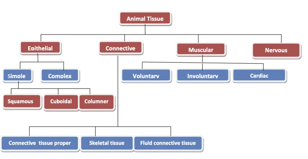
A. Epithelial Tissue
Epithelial tissue plays a crucial role in the structure and function of animal bodies, serving as a protective barrier that covers surfaces and lines tubes and cavities. This type of tissue, often referred to as covering and lining tissue, is integral to maintaining the integrity of organs and facilitating various physiological processes.
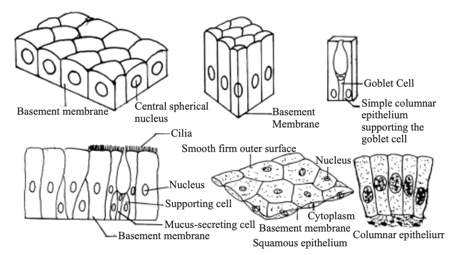
- Characteristics of Epithelial Tissue:
- Cells within epithelial tissue are tightly packed together, forming continuous sheets that provide a cohesive layer.
- There is minimal intercellular space, with only a small amount of connecting material between the cells.
- Epithelial tissue is typically separated from underlying connective tissues by an extracellular fibrous basement membrane, which provides support and anchorage.
- This tissue is avascular, meaning it does not contain blood vessels, relying instead on diffusion from adjacent tissues for nutrient and waste exchange.
- Classification of Epithelial Tissue: Depending on the cell shape and function, epithelial tissue can be classified into two main categories: simple epithelial tissue and complex epithelial tissue.
- Simple Epithelial Tissue:
- Squamous Epithelium (Pavement Epithelium):
- Composed of extremely thin and flat cells, this tissue forms delicate linings.
- Locations include the lining of the esophagus, mouth, blood vessels, alveoli of lungs, and Bowman’s capsule of the nephron.
- Cuboidal Epithelium:
- Cells are cube-shaped and provide a more robust lining.
- Found in the lining of kidney tubules and the ducts of salivary glands, this type plays a role in secretion and absorption.
- Columnar Epithelium:
- Characterized by tall, pillar-like cells, this tissue is essential for absorption and secretion, especially in the inner lining of the intestines.
- The cells can be modified into three specialized types:
- Ciliated Epithelium: Features cilia on their free ends, facilitating the movement of small particles. This type lines the fallopian tubes and respiratory tract.
- Glandular Epithelium: Cells that have adapted to form glands, responsible for the secretion of various substances.
- Sensory Epithelium: Modified to form sensory cells, which receive stimuli; examples include rod and cone cells in the eyes and taste buds in the tongue.
- Squamous Epithelium (Pavement Epithelium):
- Complex Epithelial Tissue:
- Stratified Squamous Epithelium:
- Composed of multiple layers of epithelial cells, this tissue is designed to withstand wear and tear.
- Commonly found in the skin, it provides a protective barrier against mechanical injury, pathogens, and water loss.
- Stratified Squamous Epithelium:
- Simple Epithelial Tissue:
B. Connecitve tissue
Connective tissue plays a vital role in the animal body, serving to connect, support, and bind various organs and tissues together. This diverse category of tissue is essential for maintaining the structural integrity of the body and facilitating numerous physiological processes.
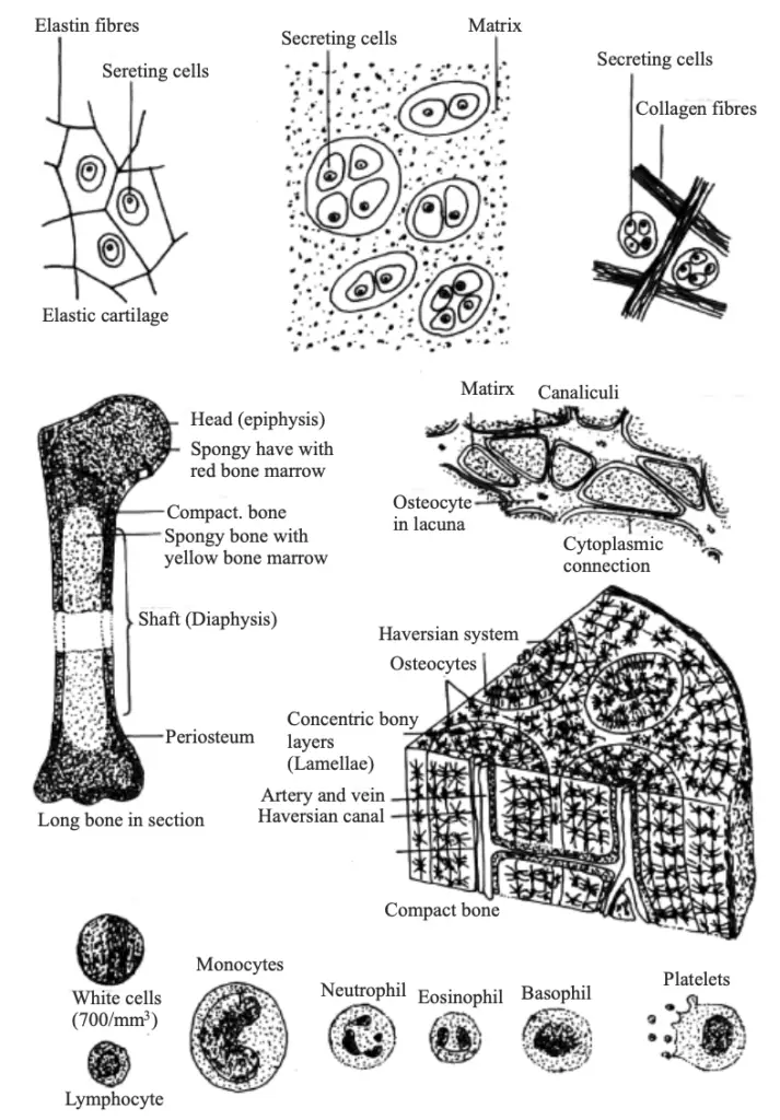
- Characteristics of Connective Tissue:
- The cells of connective tissue are living and loosely spaced, allowing for flexibility and adaptability within the tissue structure.
- These cells are embedded in an intercellular matrix, which can vary in consistency. The matrix may be jelly-like, fluid, dense, or rigid, depending on the specific type and function of the connective tissue. This matrix is a secretion from the cells of the tissue and serves to support and bind cells together.
- Types of Connective Tissue: Connective tissue can be classified into three main categories: connective tissue proper, skeletal tissue, and fluid connective tissue.
- Connective Tissue Proper (Packing Tissue):
- Areolar Tissue (Loose Connective Tissue):
- This type of tissue provides support and elasticity to various organs and is involved in the cushioning of structures.
- Fibrous Tissue:
- Fibrous connective tissue is further categorized into two structures:
- Tendons: These connect muscles to skeletal tissue, such as cartilage and bone. Tendons are strong and non-flexible, allowing for the efficient transfer of force from muscle to bone.
- Ligaments: Ligaments connect skeletal tissues to each other, providing stability to joints. They are elastic and flexible, allowing for movement while maintaining structural integrity.
- Fibrous connective tissue is further categorized into two structures:
- Adipose Tissue:
- This tissue is distributed abundantly beneath the skin and is primarily responsible for the storage of fat. Adipose tissue also plays a role in insulation and energy storage.
- Areolar Tissue (Loose Connective Tissue):
- Skeletal Tissue:
- Skeletal tissue comprises both cartilage and bone, contributing to the framework of the body.
- Cartilage: This connective tissue provides support and flexibility to various body parts and smoothens bone surfaces at joints. It is found in locations such as the nose, ears, trachea, and larynx, serving critical functions in cushioning and facilitating movement.
- Bone: Bone is a strong and non-flexible connective tissue due to the presence of calcium and phosphorus, which provide hardness. Bone serves as the primary structural component of the skeleton, supporting the body and protecting vital organs.
- Fluid Connective Tissue:
- This category includes blood and lymph, which are essential for transportation and immune responses.
- Blood:
- Blood consists of a liquid matrix called plasma, which accounts for approximately 55% of its volume. Plasma contains water, proteins, nutrients, electrolytes, metabolic wastes, dissolved gases, hormones, and anticoagulants such as heparin.
- Blood corpuscles include three main types:
- Red Blood Cells (RBCs or Erythrocytes): Responsible for transporting respiratory gases, primarily oxygen and carbon dioxide.
- White Blood Cells (WBCs or Leukocytes): These cells play a crucial role in the immune response, helping to fight infections by producing antibodies.
- Blood Platelets: These components are essential for blood clotting, preventing excessive bleeding after injury.
- Lymph:
- Lymph is a colorless fluid that contains plasma and white blood cells. It escapes from blood capillaries into body tissues and flows through lymph vessels.
- The primary functions of lymph include aiding in the exchange of materials between tissues and blood, as well as protecting the body against infections through the immune response.
- Connective Tissue Proper (Packing Tissue):
C. Muscular tissue
Muscular tissue is essential for movement within the animal body, consisting primarily of elongated cells known as muscle fibers. This specialized tissue facilitates various types of movement in body parts and plays a crucial role in locomotion. Muscular tissue contains specific proteins, referred to as contractile proteins (including myosin, actin, troponin, and tropomyosin), which enable the contraction and relaxation of muscle fibers, thus facilitating movement.
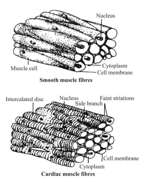
- Types of Muscular Tissue: Muscular tissue is categorized into three distinct types based on the structure of muscle fibers:
- Striated / Stripe / Voluntary / Skeletal Muscular Tissue:
- This type is predominantly associated with the skeletal system, as these muscles are primarily attached to bones.
- The term “striated” refers to the presence of alternating light and dark bands or striations, which are visible under a microscope. This striation pattern results from the organized arrangement of contractile proteins.
- The cells within skeletal muscular tissue are characterized as long, cylindrical, unbranched, and multinucleated, meaning they contain multiple nuclei. This structure is adapted for the rapid and forceful contractions required for voluntary movements.
- Involuntary Muscular Tissue / Smooth Muscular Tissue / Unstriated Muscular Tissue:
- In contrast to striated muscles, involuntary muscular tissue operates without conscious control, meaning movements are not under voluntary control.
- It is referred to as “smooth” muscular tissue because it is primarily found in the walls of smooth, visceral organs.
- The absence of striations distinguishes this tissue, which leads to the term “unstriated.”
- The cells in smooth muscular tissue are typically long with pointed ends, giving them a spindle shape, and are uninucleated, possessing a single nucleus. This tissue type is located in various structures, including the alimentary canal, blood vessels, the iris of the eye, ureters, and bronchi of the lungs.
- Cardiac Muscular Tissue / Striated Involuntary Muscular Tissue:
- Cardiac muscular tissue is specific to the heart, forming the heart wall, which is crucial for its pumping action.
- This type of muscle is both striated and involuntary, as it features striations similar to skeletal muscle while operating independently of conscious control.
- The cells of cardiac muscular tissue are cylindrical, branched, and uninucleated, reflecting a unique structure that allows for coordinated contractions necessary for effective heart function.
- Striated / Stripe / Voluntary / Skeletal Muscular Tissue:
D. Nervous Tissue
Nervous tissue is a specialized type of tissue integral to the functioning of the nervous system, which governs all body activities. This tissue is primarily composed of nerve cells, or neurons, which are uniquely adapted to receive stimuli and transmit signals rapidly throughout the body. The brain and spinal cord are the central components of this tissue, playing pivotal roles in processing and coordinating information.
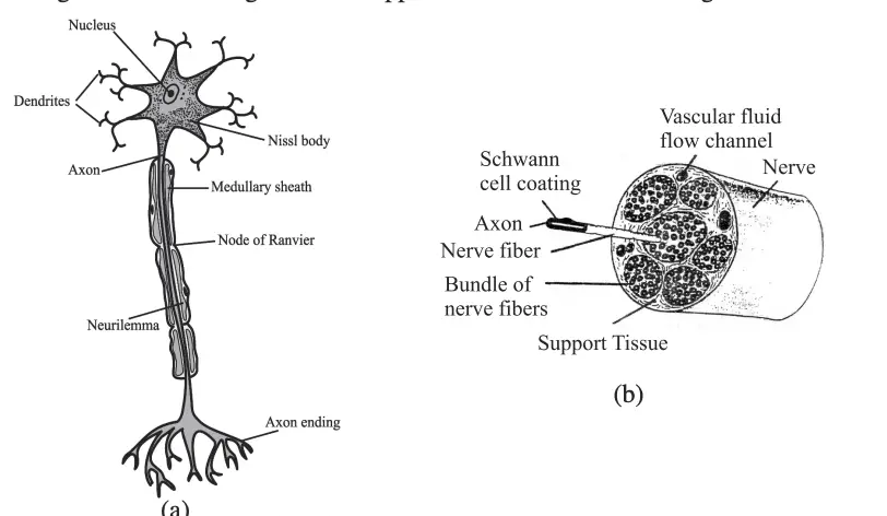
- Components of Neurons: Each neuron consists of three main parts:
- Cyton (Cell Body): The cyton houses the large central nucleus and cytoplasm, from which long, thin, hair-like extensions emerge. This structure supports the overall function of the neuron by providing metabolic support and housing the nucleus, which contains genetic material.
- Dendrites: These are short, branched fibers that receive nerve impulses. Dendrites play a critical role in collecting signals from other neurons or sensory receptors, thereby facilitating communication within the nervous system.
- Axon: In contrast to dendrites, the axon is a single, long conducting fiber that transmits impulses away from the cell body. The axon may be myelinated, which enhances the speed of impulse conduction.
- Functions of Nerve Cells: Neurons serve several essential functions within the body:
- Control of Body Activities: Nerve cells are responsible for regulating and controlling all bodily functions, from voluntary movements to involuntary processes.
- Coordination Among Body Parts: They facilitate communication and coordination between different body systems, ensuring that responses to stimuli are timely and appropriate.
- Transmission of Nerve Impulses: Dendrites carry nerve impulses towards the cyton, whereas the axon conducts these impulses away from the cyton, thereby maintaining the flow of information throughout the nervous system.
- Synapse: The synapse is the junction or region where the axon of one neuron connects with the dendrite of another neuron. This specialized area is crucial for the transfer of nerve impulses, as it allows chemical and electrical signals to pass between neurons, enabling communication within the nervous system.
- Characteristics of Axon and Dendrite:
- The axon is typically single in number and can be long, with possible branching, facilitating long-distance signal transmission.
- In contrast, dendrites may be one or more in number and are always branched, allowing for the reception of multiple incoming signals.
- Nerve Impulse: A nerve impulse is the information transmitted through neurons in the form of chemical and electrical signals. This transmission is vital for the functioning of the nervous system, allowing for rapid communication between various body parts.
Functions of Tissues
- Groups of like cells and extracellular materials work together in tissues to carry out particular physiological activities.
- On internal cavities and body surfaces, epithelial tissues serve as protective barriers mediating activities like absorption, secretion, and sensory perception.
- Connective tissues support organs structurally, bond, and cushion; they also help in nutrition movement, immunological reactions, and energy storage.
- Through contraction of specialized cells, muscle tissues generate force and movement, therefore supporting both intentional and involuntary motions.
- By sending electrical and chemical impulses between several areas, nervous tissues enable quick coordination and communication inside the body.
- Meristematic tissues—which drive development—as well as permanent tissues including vascular tissues for water and nutrient transport, cutaneous tissues for protection, and root tissues for photosynthesis and storage split tissue activities in plants.
- Maintaining homeostasis and general organismal health depends on the coordinated activities of these many tissue types; their organization and performance are therefore crucial indicators in both normal physiology and disease diagnosis.
- Chen, Ting-Hsuan. (2014). Tissue Regeneration: From Synthetic Scaffolds to Self-Organizing Morphogenesis. Current stem cell research & therapy. 9. 10.2174/1574888X09666140507123401.
- https://www.longdom.org/open-access-pdfs/formation-of-tissues-of-the-body.pdf
- https://ilovepathology.com/tissue-repair-by-connective-tissue-deposition-angiogenesis-tissue-remodeling/
- https://byjus.com/biology/tissues/
- https://organismalbio.biosci.gatech.edu/growth-and-reproduction/plant-development-i-tissue-differentiation-and-function/
- https://en.wikipedia.org/wiki/Tissue_%28biology%29
- https://www.eolss.net/Sample-Chapters/C03/E6-71-03-06.pdf
- https://organismalbio.biosci.gatech.edu/growth-and-reproduction/plant-development-i-tissue-differentiation-and-function/