The nervous system represents one of the most intricate and essential systems in multicellular organisms, functioning as the primary coordinator of actions, sensory information, and responses to environmental stimuli. This complex network is responsible for detecting changes in the environment, processing that information, and enabling organisms to react accordingly, working closely with the endocrine system to regulate physiological responses.
Historically, nervous tissue is believed to have originated in wormlike organisms approximately 550 to 600 million years ago. In vertebrates, the nervous system is categorized into two principal components: the central nervous system (CNS) and the peripheral nervous system (PNS). The CNS encompasses the brain and spinal cord, serving as the main processing center for sensory input and motor commands. Conversely, the PNS comprises nerves, which are bundles of long axonal fibers that extend from the CNS to various parts of the body, facilitating communication between the CNS and peripheral organs.
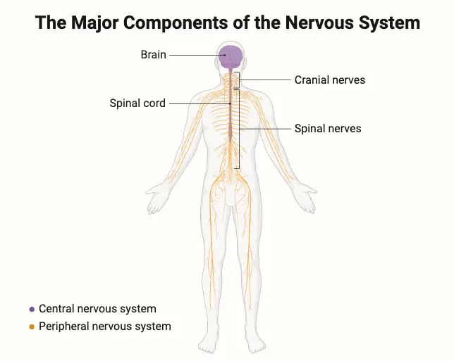
Within the PNS, nerves can be divided into two distinct categories: motor nerves (efferent), which convey signals from the brain to execute movements, and sensory nerves (afferent), which transmit sensory information from the body back to the CNS. The PNS is further subdivided into the somatic and autonomic nervous systems. The somatic nervous system oversees voluntary movements and sensory information transmission, while the autonomic nervous system regulates involuntary functions such as heart rate and digestion, subdividing further into the sympathetic, parasympathetic, and enteric nervous systems. The sympathetic nervous system activates during emergencies, preparing the body for ‘fight or flight’ responses, while the parasympathetic system promotes a ‘rest and digest’ state.
At a cellular level, the nervous system is characterized by specialized cells known as neurons. Neurons possess unique structures that enable the rapid transmission of electrochemical impulses. Signals travel along axons, reaching neighboring cells through electrical synapses or triggering the release of neurotransmitters at chemical synapses. The activity at these synapses can lead to excitation, inhibition, or modulation of the receiving cell’s behavior. The intricate connections formed between neurons create neural pathways, circuits, and networks that underlie perception and behavioral responses. Supporting these neurons are glial cells, which provide essential structural and metabolic support, and together with the vascular components, they constitute the neurovascular unit, which regulates blood flow to meet the high energy demands of active neurons.
Nervous systems are prevalent in most multicellular animals, although they exhibit a wide range of complexities. Notably, sponges, placozoans, and mesozoans lack any form of nervous system, while organisms such as ctenophores and cnidarians feature a diffuse nerve net. Most other animal species possess a more complex organization that includes a centralized brain and nerve cords, with sizes ranging from several hundred neurons in simpler organisms to an astounding 300 billion neurons in larger mammals, such as African elephants.
The central nervous system is pivotal for transmitting signals between different cells and regions, while also receiving feedback to inform ongoing processes. Dysfunction within this system may arise from genetic mutations, trauma, toxic exposure, infections, or age-related degeneration. Neurology is the branch of medicine dedicated to understanding and treating disorders affecting the nervous system. In the peripheral nervous system, common issues include nerve conduction failures, often resulting from conditions like diabetic neuropathy or demyelinating diseases such as multiple sclerosis.
The nervous system’s functionality is fundamentally reliant on the specialized communication between neurons, which transmit signals across synapses via neurotransmitters. This intricate communication enables organisms to perceive their environment, maintain homeostasis, and engage in complex behaviors. An understanding of neuroanatomy, the study of the nervous system’s structure, is crucial for comprehending its functions and the localization of specific tasks within different brain regions.
Gross Organization of the Mammalian Nervous System
Central Nervous System (CNS)
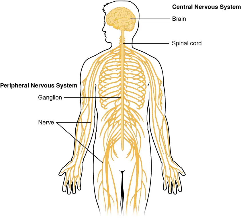
The central nervous system (CNS) is a vital component of the nervous system, composed of the brain and spinal cord, both of which are encased in bony structures for protection. This system serves as the primary control center for processing sensory information and coordinating bodily functions.
- The CNS comprises two major structures:
- The Brain
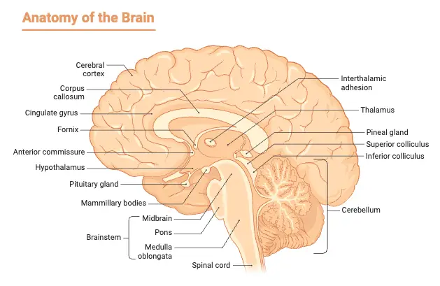
- Located entirely within the skull, the brain is the most complex organ in the body, responsible for higher cognitive functions, emotional regulation, memory, and sensory processing.
- Cerebrum:
- The cerebrum is the largest part of the brain, divided into two hemispheres.
- Each hemisphere is responsible for sensations and motor functions on the opposite side of the body; the left hemisphere governs the right side, while the right hemisphere controls the left.
- The cerebrum facilitates higher-level functions such as reasoning, problem-solving, and voluntary muscle movement.
- Cerebellum:
- Positioned posterior to the cerebrum, the cerebellum is significantly smaller yet contains a vast number of neurons, comparable to the total in both cerebral hemispheres.
- It primarily functions as a movement control center, ensuring coordination, balance, and fine motor skills.
- Each side of the cerebellum is dedicated to managing movements on the corresponding side of the body.
- Brain Stem:
- The brain stem acts as a conduit between the cerebrum, cerebellum, and spinal cord.
- It is integral for regulating essential life functions, including breathing, heart rate, and temperature control.
- The brain stem is the most primitive part of the brain; damage to it can be fatal, while injuries to other brain regions may be survivable.
- The Brain
- The Spinal Cord:
- The spinal cord extends from the brain stem down the vertebral column, acting as the primary pathway for information exchange between the brain and the rest of the body.
- It is responsible for transmitting sensory information from the body to the brain and motor commands from the brain to the muscles.
- Any injury resulting in a transection of the spinal cord can lead to anesthesia or paralysis in the areas of the body below the site of injury, as the brain loses its ability to control those regions.
- The spinal cord communicates with the peripheral nervous system through spinal nerves that exit the cord between each vertebra.
- Each spinal nerve has two branches:
- Dorsal Root:
- Contains sensory axons that transmit information to the spinal cord, such as sensations from the skin.
- Ventral Root:
- Contains motor axons that convey signals away from the spinal cord, enabling muscle movement in response to stimuli.
- Dorsal Root:
Peripheral Nervous System (PNS)
The peripheral nervous system (PNS) encompasses all components of the nervous system outside the brain and spinal cord, functioning as a critical link between the central nervous system (CNS) and the rest of the body. The PNS can be broadly categorized into two distinct systems: the somatic nervous system and the visceral nervous system.
- Somatic Peripheral Nervous System (sPNS)
- This system is responsible for voluntary movements and the sensory perception of external stimuli.
- Components:
- Comprised of spinal nerves that innervate the skin, joints, and muscles under voluntary control.
- Motor axons, originating from motor neurons in the ventral spinal cord, facilitate muscle contractions.
- The cell bodies of these motor neurons reside within the CNS, but their axons extend into the PNS.
- Sensory Function:
- Somatic sensory axons collect information from the body’s periphery and transmit it to the CNS.
- These axons enter the spinal cord via the dorsal roots, and their cell bodies are located in dorsal root ganglia, with one ganglion for each spinal nerve.
- Components:
- This system is responsible for voluntary movements and the sensory perception of external stimuli.
- Visceral Peripheral Nervous System (vPNS)
- Also referred to as the autonomic nervous system (ANS), this system regulates involuntary bodily functions.
- Components:
- Composed of neurons that innervate internal organs, blood vessels, and glands.
- Visceral sensory axons provide feedback regarding the internal environment, such as blood pressure and oxygen levels.
- Motor Function:
- Visceral motor fibers control the contraction and relaxation of smooth muscles lining the organs, regulate cardiac muscle contraction, and modulate glandular secretions.
- For instance, the visceral PNS plays a role in blood pressure regulation by adjusting heart rate and blood vessel diameter.
- Components:
- Also referred to as the autonomic nervous system (ANS), this system regulates involuntary bodily functions.
- Afferent and Efferent Axons
- Understanding the terms afferent and efferent is essential in discussing the PNS.
- Afferent Axons:
- These axons, which transport sensory information toward the CNS, are responsible for relaying signals about external and internal stimuli.
- Efferent Axons:
- These axons emerge from the CNS to innervate muscles and glands, executing motor commands and autonomic functions.
- Afferent Axons:
- Understanding the terms afferent and efferent is essential in discussing the PNS.
- Cranial Nerves
- The PNS includes 12 pairs of cranial nerves that originate from the brain stem, primarily innervating the head and neck.
- Diversity of Function:
- Each cranial nerve is associated with specific functions and can be categorized as part of the CNS, somatic PNS, or visceral PNS.
- Many cranial nerves contain a mix of afferent and efferent axons, allowing them to perform various roles, such as sensory perception and motor control.
- Diversity of Function:
- The PNS includes 12 pairs of cranial nerves that originate from the brain stem, primarily innervating the head and neck.
Development of the Nervous System
The development of the nervous system is a complex and intricate process that begins early in embryonic development and continues throughout fetal growth. Understanding this process provides insight into the organization and functionality of the central nervous system (CNS). The CNS originates from a structure called the neural tube, which forms during the initial stages of development. This overview will highlight key stages of neural development, emphasizing the structural organization and functional implications of various components.
- Formation of the Neural Tube
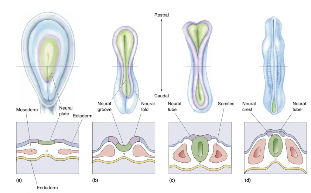
- The early embryo is structured as a flat disk composed of three primary layers: the endoderm, mesoderm, and ectoderm. The endoderm eventually develops into internal organ linings, while the mesoderm gives rise to the skeletal system and musculature. The ectoderm is responsible for forming the nervous system and skin.
- Approximately 17 days post-conception in humans, the ectoderm thickens to form the neural plate. As development progresses, the neural plate forms a groove, termed the neural groove, flanked by neural folds. These folds subsequently fuse to create the neural tube, the precursor to the CNS.
- The neural crest, which emerges as some ectodermal tissue is pinched off during the fusion process, gives rise to neurons with cell bodies in the peripheral nervous system.
- Neurulation
- The transformation of the neural plate into the neural tube is called neurulation, occurring around 22 days after conception. Improper closure of the neural tube can lead to birth defects, underscoring the importance of maternal nutrition during this critical developmental window.
- Primary Brain Vesicles
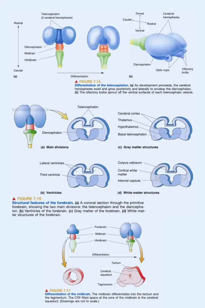
- Differentiation marks the next critical phase, where the rostral end of the neural tube expands to form three primary vesicles: the prosencephalon (forebrain), mesencephalon (midbrain), and rhombencephalon (hindbrain). The rostral-most vesicle is the prosencephalon, followed by the mesencephalon, and finally the rhombencephalon, which connects to the developing spinal cord.
- Differentiation of the Forebrain
- In the forebrain, secondary vesicles sprout from the prosencephalon, leading to the formation of the optic and telencephalic vesicles. The diencephalon remains central, flanked by these newly formed structures.
- The optic vesicles evolve into the optic nerves and retinas, establishing a vital connection between the sensory systems and the brain. The telencephalon, or “endbrain,” develops into the cerebral hemispheres and engages in several differentiation processes: growth over the diencephalon, formation of olfactory bulbs, division of cell walls into various structures, and development of white matter systems, including the corpus callosum and internal capsule.
- Forebrain Structure-Function Relationships
- The cerebral cortex, the most evolved area of the forebrain, plays a critical role in sensory processing, perception, and voluntary movement. Information from sensory neurons, including those from the olfactory bulbs, eyes, ears, and skin, is relayed to the cortex via the thalamus, often referred to as the “gateway to the cerebral cortex.”
- The hypothalamus, located beneath the thalamus, regulates autonomic functions and visceral responses, orchestrating activities like the fight-or-flight response and homeostatic processes.
- Differentiation of the Midbrain
- The midbrain undergoes minimal differentiation compared to other brain regions. Its dorsal surface forms the tectum, while the tegmentum develops on its ventral side. The cerebral aqueduct, a narrow channel for cerebrospinal fluid (CSF), connects the midbrain to the third ventricle.
- The midbrain is involved in sensory processing and motor control. Structures like the superior colliculus and inferior colliculus are pivotal in visual and auditory processing, respectively, while the tegmentum contains neurons that influence movement and modulate consciousness.
- Differentiation of the Hindbrain
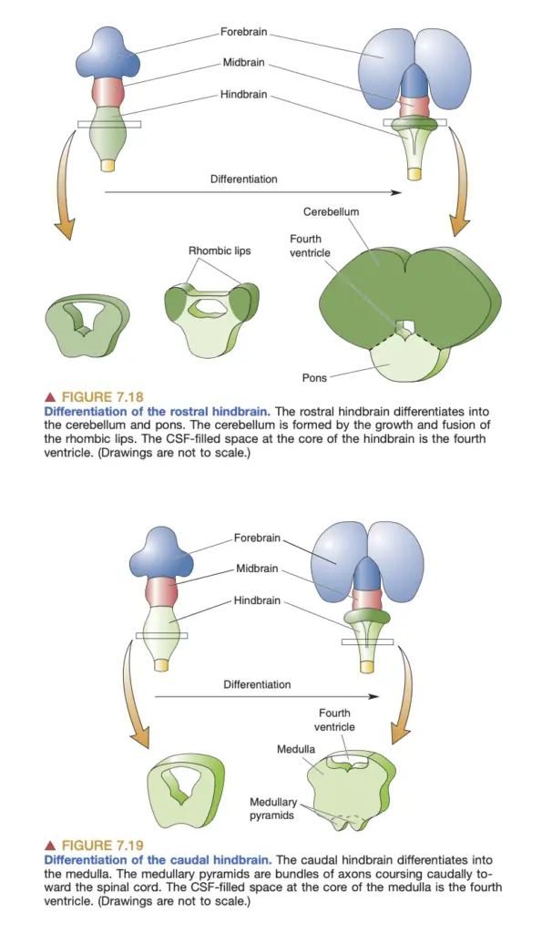
- The hindbrain differentiates into three major structures: the cerebellum, pons, and medulla oblongata. The cerebellum, which develops from the rostral hindbrain, is crucial for coordinating movement and balance. The pons serves as a relay station between the cerebral cortex and cerebellum, integrating sensory and motor information.
- The medulla, forming from the caudal hindbrain, regulates essential autonomic functions and houses neural pathways critical for sensory processing, particularly auditory and somatic sensation.
- Differentiation of the Spinal Cord
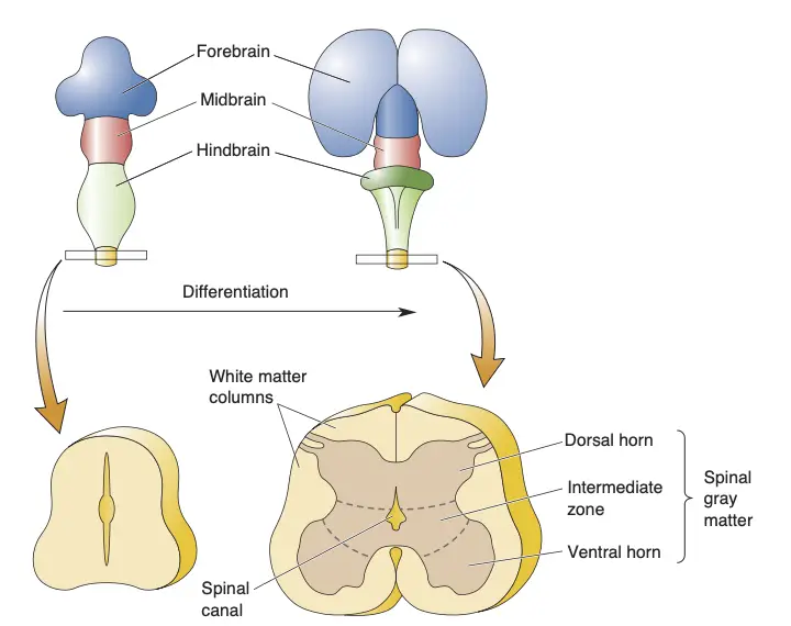
- The spinal cord develops from the caudal neural tube and features a characteristic butterfly shape in cross-section, composed of gray matter (neurons) and white matter (axons). The dorsal horn receives sensory input, while the ventral horn contains motor neurons projecting to muscles.
- The dorsal columns transmit somatic sensory information, facilitating communication between the body and brain. The organization of these columns reflects the pathway by which sensory signals ascend to higher brain centers.
Special Features of the Human CNS
The human central nervous system (CNS) exhibits unique structural and functional features that distinguish it from other mammals while adhering to a fundamental mammalian blueprint. Understanding these features is vital for comprehending the complexities of human cognition, behavior, and overall neurological function.
- Basic Structure of the Human CNS
- The CNS comprises the brain and spinal cord, which are interconnected and work collaboratively to facilitate bodily functions and responses.
- Telencephalon: The largest part of the brain, responsible for higher cognitive functions.
- Diencephalon: Located centrally, surrounding the third ventricle, it includes structures such as the thalamus and hypothalamus.
- Brain Stem: Comprises the midbrain, pons, and medulla, facilitating basic life functions such as respiration and heart rate.
- Cerebellum: Positioned posteriorly, it is essential for coordination, balance, and motor control.
- The CNS comprises the brain and spinal cord, which are interconnected and work collaboratively to facilitate bodily functions and responses.
- Cerebral Cortex Characteristics
- The cerebral cortex is a highly convoluted structure, displaying a complex arrangement of grooves (sulci) and bumps (gyri).
- Surface Area Expansion: The extensive folding of the cortex increases its surface area, which is crucial for higher-order processing. In adults, the cortical surface area measures approximately 1,100 cm².
- Functional Significance: The cortex is integral to reasoning, cognition, and voluntary movement. Damage to this region can lead to profound impairments in sensory and motor functions.
- The cerebral cortex is a highly convoluted structure, displaying a complex arrangement of grooves (sulci) and bumps (gyri).
- Lobes of the Cerebrum
- The human cerebrum is divided into four distinct lobes, each associated with different functions:
- Frontal Lobe: Located under the frontal bone, involved in executive functions, decision-making, and voluntary motor control.
- Parietal Lobe: Positioned posterior to the frontal lobe, it processes sensory information and spatial awareness.
- Temporal Lobe: Arcing around the side, under the temporal bone, this lobe is critical for auditory processing and memory functions.
- Occipital Lobe: Found at the posterior of the cerebrum, it is primarily responsible for visual processing.
- The human cerebrum is divided into four distinct lobes, each associated with different functions:
- Ventricular System
- The ventricular system consists of interconnected cavities filled with cerebrospinal fluid (CSF), which cushions the brain and helps maintain homeostasis.
- Third Ventricle: Located in the diencephalon, it is surrounded by key brain structures.
- Cerebral Aqueduct: Connecting the third and fourth ventricles, this channel facilitates the flow of CSF.
- Fourth Ventricle: Positioned between the cerebellum and the brain stem, it further aids in CSF circulation.
- The ventricular system consists of interconnected cavities filled with cerebrospinal fluid (CSF), which cushions the brain and helps maintain homeostasis.
- Comparative Anatomy with Other Mammals
- While the human brain shares common structures with other mammals, notable differences exist, particularly in size and complexity.
- Olfactory Bulb Size: Humans possess a smaller olfactory bulb compared to many other mammals, reflecting a shift in sensory reliance.
- Cerebral Hemispheres: The human cerebral hemispheres are significantly more developed, enabling advanced cognitive functions that are not as pronounced in other species.
- While the human brain shares common structures with other mammals, notable differences exist, particularly in size and complexity.
- Functional Implications of Structural Features
- The unique structural characteristics of the human CNS facilitate advanced capabilities, including:
- Enhanced Cognition: The complexity of the cerebral cortex underlies sophisticated reasoning, problem-solving, and language.
- Motor Control: The intricately folded cortex allows for refined motor skills and coordination.
- Sensory Integration: The structural layout of the lobes enables the integration of sensory inputs, enhancing perception and response to the environment.
- The unique structural characteristics of the human CNS facilitate advanced capabilities, including:
The Cerebral Cortex
The cerebral cortex, a crucial component of the brain, exhibits distinct structural characteristics that facilitate its diverse functions across various species, particularly in mammals. Understanding the types of cerebral cortex is essential for appreciating the complexities of neural processing and cognitive functions.
- General Features of the Cerebral Cortex
- The cerebral cortex is organized into layers, with neuron cell bodies arranged in parallel sheets.
- Layering: These layers provide a structural framework that supports various neural functions.
- Molecular Layer: The most superficial layer, known as layer I, lacks neurons and lies between the pia mater and the neuron layers.
- Pyramidal Cells: At least one of the cortical layers contains pyramidal neurons characterized by prominent apical dendrites that extend into the molecular layer.
- The cerebral cortex is organized into layers, with neuron cell bodies arranged in parallel sheets.
- Types of Cerebral Cortex
- The cerebral cortex can be classified based on its cytoarchitecture, which varies across different regions:
- Neocortex:
- Found exclusively in mammals, the neocortex comprises multiple layers and is involved in higher cognitive functions.
- It features a complex arrangement of six distinct layers, each with unique types of neurons and connections.
- The expansion of the neocortex during human evolution correlates with advanced cognitive capabilities.
- Hippocampus:
- Despite its convoluted structure, the hippocampus is characterized by a single cell layer.
- It plays a significant role in memory formation and spatial navigation.
- The hippocampus is connected to the neocortex and participates in processing contextual information.
- Olfactory Cortex:
- This cortex has only two layers and is continuous with the olfactory bulb, emphasizing its role in the sense of smell.
- It is situated ventrally and laterally to the hippocampus, separated by the rhinal fissure.
- The olfactory cortex is essential for processing olfactory information and has direct connections to other areas involved in emotion and memory.
- Neocortex:
- The cerebral cortex can be classified based on its cytoarchitecture, which varies across different regions:
- Structural and Functional Relationships
- The structural characteristics of each cortical type reflect their functional specializations:
- Neocortex:
- The layered structure facilitates complex processing tasks, such as sensory perception, decision-making, and voluntary motor control.
- Connections with the thalamus position the neocortex as the primary site for sensory information integration.
- Hippocampus:
- Its unique single-layer architecture is adapted for the processing of memory and learning tasks, supporting long-term potentiation and synaptic plasticity.
- It is integral to the limbic system, contributing to emotional regulation.
- Olfactory Cortex:
- The simpler structure allows for rapid processing of olfactory stimuli, crucial for survival and behavioral responses.
- The olfactory cortex is closely linked to emotional centers of the brain, highlighting its role in the emotional aspects of smell.
- Neocortex:
- The structural characteristics of each cortical type reflect their functional specializations:
- Implications for Neuroscience
- Understanding the different types of cerebral cortex aids in exploring neurological disorders and cognitive impairments.
- The distinct functions and structures suggest specific vulnerabilities in various brain regions, which can inform therapeutic approaches.
- Insights into the organization and function of the neocortex, hippocampus, and olfactory cortex are critical for developing interventions for conditions such as Alzheimer’s disease, epilepsy, and other cognitive disorders.
- Understanding the different types of cerebral cortex aids in exploring neurological disorders and cognitive impairments.
Areas of Neocortex
The neocortex, a critical structure in the mammalian brain, is distinguished by its layered organization and diverse functional areas. This region plays a vital role in sensory perception, motor control, and higher cognitive functions. The classification of neocortical areas has significantly advanced through the work of various neuroscientists, particularly the German neuroanatomist Korbinian Brodmann, who developed a cytoarchitectural map of the neocortex in the early 20th century.
- Brodmann’s Areas
- Brodmann’s work laid the foundation for identifying distinct cortical areas based on differences in cellular organization.
- Each region is assigned a number that corresponds to its unique cytoarchitecture, for example, area 17 in the occipital lobe is designated as the primary visual cortex, while area 4 in the frontal lobe is recognized as the primary motor cortex.
- Brodmann hypothesized that these areas not only differ in structure but also in function, a notion supported by contemporary research demonstrating specific functional roles.
- Brodmann’s work laid the foundation for identifying distinct cortical areas based on differences in cellular organization.
- Functional Areas of the Neocortex
- The neocortex can be broadly categorized into primary sensory areas, secondary sensory areas, and motor areas.
- Primary Sensory Areas:
- These areas are the first to receive and process sensory input from the respective sensory modalities.
- For example, area 17 (primary visual cortex) receives direct input from the retina via the thalamus, making it essential for visual processing.
- Other examples include the primary auditory cortex and the primary somatosensory cortex, each corresponding to specific sensory pathways.
- These areas are the first to receive and process sensory input from the respective sensory modalities.
- Secondary Sensory Areas:
- These areas are characterized by extensive interconnections with primary sensory areas, facilitating the integration and interpretation of sensory information.
- Secondary areas enhance perceptual processing by refining and associating sensory inputs.
- For instance, the secondary visual areas further process information received from the primary visual cortex to extract complex visual features.
- These areas are characterized by extensive interconnections with primary sensory areas, facilitating the integration and interpretation of sensory information.
- Motor Areas:
- The motor areas are primarily involved in planning and executing voluntary movements.
- The primary motor cortex (area 4) directly sends projections to motor neurons in the spinal cord, allowing for precise control of muscle contractions.
- Other motor-related areas, such as the supplementary motor area, contribute to the planning and coordination of movement.
- The motor areas are primarily involved in planning and executing voluntary movements.
- Primary Sensory Areas:
- The neocortex can be broadly categorized into primary sensory areas, secondary sensory areas, and motor areas.
- Evolutionary Perspective
- The evolution of the neocortex across different mammalian species has revealed insights into its functional expansion.
- Brodmann suggested that the neocortex has expanded through the addition of new areas, particularly between existing primary and secondary sensory areas.
- While the basic structure of the neocortex has remained relatively constant, the surface area has increased dramatically in some species, particularly in humans.
- This expansion is particularly evident in regions related to complex sensory processing and higher cognitive functions, with primates exhibiting a significant increase in secondary sensory areas.
- The evolution of the neocortex across different mammalian species has revealed insights into its functional expansion.
- Association Areas
- A notable feature of the neocortex, especially in primates, is the presence of association areas, which are implicated in integrative and higher-order functions.
- These areas do not correspond directly to specific sensory or motor functions but are involved in processing information from multiple modalities.
- The frontal and temporal lobes contain substantial portions of association cortex, which are critical for complex cognitive tasks such as reasoning, planning, and social cognition.
- The expansion of the frontal cortex is particularly associated with the development of higher cognitive functions and the capacity for understanding unobservable mental states, such as intentions and beliefs.
- A notable feature of the neocortex, especially in primates, is the presence of association areas, which are implicated in integrative and higher-order functions.
- Implications for Neuroscience
- Understanding the organization and function of the neocortex is essential for exploring neurological and psychological disorders.
- Disruptions in specific neocortical areas can lead to distinct cognitive deficits, influencing areas such as perception, movement, and personality.
- Research continues to focus on mapping the intricate connections within the neocortex, aiming to uncover the underlying circuitry that supports its complex functions.
- Understanding the organization and function of the neocortex is essential for exploring neurological and psychological disorders.
New Methods in Neuroanatomy
Neuroanatomy, which focuses on the structural organization of the nervous system, has undergone significant transformations due to the introduction of innovative imaging techniques and methodologies. These advancements have provided researchers and clinicians with unprecedented capabilities to visualize and analyze brain structures and their functions in living organisms. Below is a detailed overview of key methods that are reshaping the field of neuroanatomy.
- Imaging Techniques
- Computed Tomography (CT):
- Utilizes X-ray technology to create detailed cross-sectional images of the brain.
- Rotates an X-ray source around the head, producing a digital reconstruction that reveals both gray and white matter and the position of the ventricles.
- Particularly effective in identifying structural abnormalities, such as tumors, hemorrhages, or strokes.
- Magnetic Resonance Imaging (MRI):
- Employs strong magnetic fields and radio waves to generate high-resolution images.
- Excels in visualizing soft tissue structures within the brain, providing extensive anatomical detail.
- Functional MRI (fMRI):
- An extension of MRI that measures brain activity by detecting changes in blood flow.
- Allows for the examination of brain function in real time, facilitating studies on cognitive processes and neural responses.
- Diffusion Tensor Imaging (DTI):
- A specialized MRI technique that maps the diffusion of water molecules in brain tissue.
- Provides insights into the integrity and orientation of white matter tracts, essential for understanding connectivity between different brain regions.
- Particularly valuable in studying brain networks and their alterations in various neurological conditions.
- Computed Tomography (CT):
- Histological Techniques
- While imaging techniques provide non-invasive insights, histological methods are crucial for examining the cellular and molecular architecture of the brain.
- Immunohistochemistry:
- Utilizes antibodies to identify specific proteins within brain tissue sections.
- Fluorescent markers help visualize the distribution and density of various cell types, enhancing the understanding of neuronal and glial cell connections.
- In Situ Hybridization:
- Allows for the localization of specific RNA molecules within tissue sections.
- Provides critical information about gene expression patterns across different brain regions, contributing to knowledge about neural development and function.
- Electrophysiological Techniques
- These methods are essential for investigating the functional characteristics of neurons and their networks.
- Patch-Clamp Recording:
- Enables researchers to measure the electrical activity of individual neurons by isolating a small patch of membrane.
- Provides in-depth information about ion channel behavior and synaptic transmission, advancing the understanding of neuronal excitability.
- Multi-Electrode Arrays:
- Allow for simultaneous recordings from multiple neurons.
- Facilitate the study of neural network dynamics and responses to various stimuli, enhancing insights into collective neural behavior.
- Optogenetics
- This pioneering technique integrates genetics and optics to control the activity of specific neurons with light.
- By introducing light-sensitive proteins into targeted neurons, researchers can activate or inhibit these cells using precise light pulses.
- This method has yielded significant insights into the functional roles of specific neural circuits in behavior, cognition, and various neurological disorders.
- Advancements in Computational Neuroscience
- The incorporation of computational models alongside neuroanatomical data has improved the understanding of brain function.
- Advanced algorithms and machine learning techniques are being applied to analyze complex neuroimaging datasets, enabling the identification of patterns and the prediction of outcomes based on structural and functional connectivity.
- These computational approaches facilitate a more comprehensive understanding of how neural circuits operate and how alterations in these circuits may relate to various brain disorders.
- https://www.nichd.nih.gov/health/topics/neuro/conditioninfo/parts#:~:text=The%20nervous%20system%20has%20two,all%20parts%20of%20the%20body.
- https://www.ncbi.nlm.nih.gov/books/NBK542179/
- https://openstax.org/books/anatomy-and-physiology-2e/pages/12-1-basic-structure-and-function-of-the-nervous-system
- Text Highlighting: Select any text in the post content to highlight it
- Text Annotation: Select text and add comments with annotations
- Comment Management: Edit or delete your own comments
- Highlight Management: Remove your own highlights
How to use: Simply select any text in the post content above, and you'll see annotation options. Login here or create an account to get started.