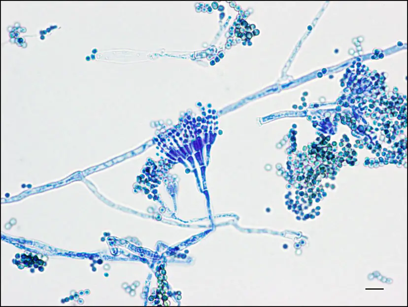
- Penicillium ascomycetous fungi are minute organisms that are of primary significance in the natural habitat, in the making of foods, and in the pharmaceutical trade.
- In medicine, a well-known antibiotic penicillin is used to fight against infection from certain kinds of bacteria.
- There are other species of penicillium which are used in the food industries for the production of cheese.
- Penicillium can be found in a freezer that has not been cleaned for a duration of 2 months or longer.
Characteristics
- It is a saprophytic fungi and mainly found in the soil, in the air, and in decaying organic matter.
- It is also known as a green or blue mold.
- They contain numerous and closely packed brush-like structures. These structures help in spore formation which is known as penicillin (sing.: penicillus).
- These brush-like structures are the main identification significance of penicillium.
- They contain slightly elongated simple or branching structures and the end portion of flask-shaped clusters is known as phialides and are called conidiophores.
- The spores of Penicillium are known as conidia. Spores are constructed in dry chains and they arise from the tops of the phialides. The apex of the phialides is occupied by the oldest spores while the most newborn spores are discovered at the bottom of the phialides.
- The branching structure is the characteristic feature of penicillium species. The P. glabrum contains an unbranched structure and they merely carry one cluster of phialides holding the tip of the stipe.
- It has a vegetative body called mycelial and is to a vast degree branched with septate hyphae, which is made of thin-walled cells and possess one or more nuclei.
- To maintenance the cytoplasmic continuity each septum contains a central pore.
- In some vegetative bodies, the mycelia develop much deeper within the substratum to receive food nutrients while others insist on the substrate to produce a mycelial felt.
- They stored their foods in the form of oil globules.
Reproduction
Penicillium reproduced by vegetative, asexual, and sexual reproductive methods. These are discussed below;
Vegetative reproduction
In this method, the vegetative mycelium is divided into two or more parts then these parts individually grow into a new mycelium just like the parent mycelium.
Asexual Reproduction
This method is accomplished with the formation of asexual spores which are also known as conidiophores.
Conidiophores: The vegetative body develops simple and long conidiophores which branch at approximately two-thirds of the distance to the apex, in a manner that’s expected of a broom. The conidiophore branches stop in a bunch of conidiogenous cells called the phialides that give off chains of conidia at their apex.
Conidia: These are tiny, spore-like structures and contain a single nucleus. They contain pigmented walls. The walls is consist of two layers such as the inner wall and the outer wall. The outer wall is a thick ornamented layer, called exine; whereas the inner wall is thin and smooth known as the intine. The conidia are discharged from the conidiophores and formed airborne to settle on a convenient substratum for germination by the development of a germ tube which prolongs and becomes separates, developed into new hyphae.
Sexual Reproduction
The sexual reproduction of penicillium is accomplished by the Fertilization. The penicillium contains male sex organs known as antheridia and female sex organs are called ascogonia.
During Fertilization, the apex of the antheridium reaches the walls of the ascogonium, and after the areas of touch between the two organs diffuse and develop a pore.
The gametangia’s protoplast affects each organ throughout this pore. The nuclei of the antheridial and ascogonial develops a number of pairs with each pair called dikaryon.
Study of Penicillium using Temporary Mount
In this practical, we will prepare a temporary mount of Penicillium to examine their structure, shape, and arrangement.
Aim
To prepare a temporary mount of Penicillium in order to examine the shape, structure, and arrangement.
Requirements
For preparation of temporary mount the following instruments and stains are required;
- Source of penicillium.
- Microscope slide.
- Compound microscope with power supply and illuminants.
- Hematoxylin stain.
- Clean coverslip.
- Forceps.
- Immersion oil.
Procedure
- Transfer the mold from the food sample to a clean microscopic slide by using a sterile forceps.
- Then place 2-3 drops of freshly prepared hematoxylin stain and wait until the stain is soaked by the mold.
- Now place a clean coverslip over the mold and make sure no air bubbles are formed between the coverslip and glass slide.
- Observe the slide under a microscope. At first use the low-power objective lens then use 40x objective lens for more detailed observation.
- After that placed a drop of immerse oil at the top of the coverslip and then put the immerse lense for further detailed image.
Observation
- Under 40X total magnification, the hematoxylin stained fungal cells appear as tortuous masses of small stalks, called hyphae. Sometimes these sections end in complex arrangements that assisted closer and more accurate inspection.
- Under 100X total magnification, a much clear image of these complex structures is observed, it appears as a squashed flower-like structure. This squashed flower-like appearance is known as conidiophore.
- Under 400X total magnification, a spherical structure is seen, known as the conidia.
Result
Base on the observation the stained fungal cell is penicillium sp.

- Text Highlighting: Select any text in the post content to highlight it
- Text Annotation: Select text and add comments with annotations
- Comment Management: Edit or delete your own comments
- Highlight Management: Remove your own highlights
How to use: Simply select any text in the post content above, and you'll see annotation options. Login here or create an account to get started.