Stratified squamous epithelium Definition
- The stratified squamous epithelium consists of squamous (flattened) epithelial cells arranged in layers upon a basal membrane.
- A stratified squamous epithelium contains many sheets of cells, where the cells in the apical layer and several layers present deep to it are squamous, but the cells in deeper layers vary from cuboidal to columnar.
- The number of the layer of cells is considered as the important difference between the simple epithelium and the stratified epithelium and also the functions of these layers are different, whereas the structure is more or less similar.
- Due to the continual cell division in the lower (basal) layers, the cells in the superficial layers are pushed towards the surface, where they are shed.
Stratified squamous epithelium diagram and Structure
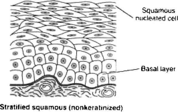
- It has two or more layers of cells. The cells in the apical layer and several layers deep to it are squamous while the cells in deeper layers vary from cuboidal to columnar.
- Only one layer in the stratified squamous epithelium is directly attached to the basement membrane while the rest of the layers are attached to one another to keep the structural integrity.
- During the basal cells division, the arising daughter cells push older cells upward toward the apical surface.
- The cells within the epithelium are tightly attached with very few intracellular spaces.
- The outermost layer of the tissue in the stratified epithelium is exposed towards the lumen at the apical surface. All the other surfaces of the cells are connected to other cells by cell junctions and adhesions.
- These cells contain many desmosomes and other adhesins and become more irregular in shape and then flatten as they move towards the surface.
- They become dehydrated and less metabolically active. while they move toward the surface and away from blood supply in underlying connective tissue.
- Tough proteins predominate as cytoplasm is reduced, and cells become sturdy, hard structures that eventually die.
- At the apical layer, after dead cells lose cell junctions, they are sloughed off, but they are continuously replaced as new cells emerge from basal cells.
- Just like all other epithelial tissue, the stratified squamous epithelium also doesn’t have a direct blood supply but has a distinct nerve supply.

Classification of stratified squamous epithelium
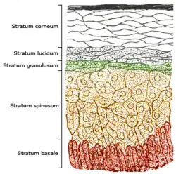
The stratified squamous epithelium is divided into two classes based on the accumulation of keratin by the cells towards the surface;
- Keratinized stratified squamous epithelium
- Non-keratinized stratified squamous epithelium
Keratinized stratified squamous epithelium
- It is a type of stratified squamous epithelium, in which the cells contain a tough layer of keratin within the apical segment of cells and many layers deep into it.
- This fibrous intracellular protein or Keratin protects the skin and underlying tissues from heat, microbes, and chemicals.
- As the cells move upwards, they accumulate keratin in the process of keratinization, where they become thin, metabolically inactive pockets (squames) of keratin lacking nuclei.
- When they move away from the nutritive blood supply the relative amount of keratin started to increases within the cell and the organelles eventually die.
- The cells on the top layer lose all their function due to keratinization and they are only involved in providing protection against dehydration and other mechanical stress.
Non-keratinized stratified squamous epithelium
- It is a type of epithelium in which the cells do not contain a huge amount of keratin, but rather are moisturized by mucus from the salivary or the mucus glands.
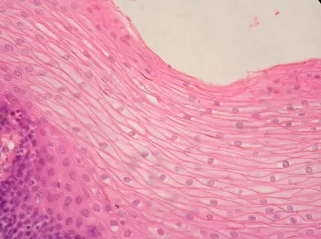
- The flattened cells of the surface layer maintain their nuclei and most metabolic functions.
- This epithelium doesn’t contain as many keratin deposits, that’s why it is normally found in areas that need to be kept hydrated or are affected by dryness.
- It does have some amount of keratin which, in turn, depends on the generation of the layer of the tissue and the mechanical wear and tear it has been subjected to.
- Some cells of this epithelium keep the surface from drying out by producing trace amounts of mucus and other lubricating agents.

Function of stratified squamous epithelium
Stratified squamous epithelium play an important and crucial function is protection.
Protection
- It protects against mechanical stress, chemical abrasions, and even radiation.
- The keratinized epithelium whcih are present on the skin surface they blocks out the harmful radiation and prevents the exposure of internal tissues and organs to the radiation.
- Stratified squamous epithelium prevents water loss due to heat and damage to the internal organs by any physical distress.
- When damaged, this epithelium replaces the outermost layer, which also aids the effectiveness against any stress or invasion.
- The entry of foreign invaders is prevented by the non-keratinized stratified squamous epithelium as the surface is kept consistently moist.
- Both Keratinized stratified squamous epithelium and Non-keratinized stratified squamous epithelium functions as the first line of defense against microbial invasion or any physical harm.
Secretion
- The cells of non keratinized stratified squamous epithelium produces some amount of mucus to keep the surface moist.
- The mucus also helps to balance the pH of the surrounding, as in the case of the vaginal epithelium.
- It also functions as a lubricating agent in a region frequently subjected to friction like the buccal cavity.
Stratified Squamous Epithelium Location
- The keratinized epithelium lines the region exposed to the external environment and continuously subjected to stress like the superficial layer of the skin (keratinized stratified squamous epithelium location).
- It helps in formation of epidermis of the sole of the foot and the palm of the hand.
- The non keratinized epithelium forms the lining of the internal surfaces and cavities, which commonly endure friction and physical wear and tear.
- In the digestive system, the stratified squamous epithelium lines the surface of the tongue, the hard upper palate of the mouth, the esophagus, and the anus.
- It is also found in the female reproductive parts such as the vagina, cervix, and the labia majora.
- The upper respiratory tract that might come in contact with food is also lined with stratified epithelium.
- The stratified epithelium covers the cornea of the eye in order to protect the delicate tissues present in the eye.
- The epidermis of the urethra in the urinary system is also made up of stratified squamous epithelium.
Stratified squamous epithelium keratinized vs non keratinized
| Topic | Keratinized Epithelium | Nonkeratinized Epithelium |
| Definition | Keratinized Epithelium is a stratified squamous epithelium that forms the epidermis of land vertebrates. | Nonkeratinized Epithelium is a stratified squamous epithelium which lines the buccal cavity. pharynx and esophagus. |
| As An Effective Barrier | An effective barrier | A less effective barrier |
| Outermost Cell Layer | Contains dead cells | Contains living cells |
| Pervious OR Impervious To Water | It is Impervious | It is Pervious |
| KERATIN PROTEIN DEPOSITED ON THE SURFACE | Yes | No |
| Lamellate Granules | Lamellate Granules are Present | Lamellate Granules are Absent |
| Protection Against Abrasions | Provides better protection against abrasions | Provides moderate protection against abrasions |
| Increase In Cell Size | Lower | Greater in nonkeratinized epithelium |
| Tonofilaments | Tonofilaments are aggregated into bundles to form tonofibrils | Tonofilaments remain dispersed and so appear less conspicuous |
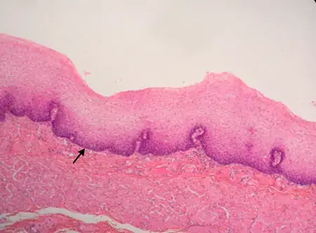
Stratified Squamous Epithelium Microscope


- Text Highlighting: Select any text in the post content to highlight it
- Text Annotation: Select text and add comments with annotations
- Comment Management: Edit or delete your own comments
- Highlight Management: Remove your own highlights
How to use: Simply select any text in the post content above, and you'll see annotation options. Login here or create an account to get started.