| Class | Lycopodiopsida |
| Order | Selaginellales |
| Family | Selaginellaceae |
| Genus | Selaginella |
Habitat of Selaginella
Selaginella species exhibit remarkable diversity in their habitats, being widely distributed across both tropical and temperate regions. The genus, comprising around 700 species, thrives in a variety of environmental conditions, adapting to both humid and arid ecosystems. Below is a detailed exploration of their habitat characteristics:
- Distribution:
Approximately 700 species of Selaginella are found across tropical and temperate regions, showcasing their adaptability to a range of climates. - Preferred Environments:
Most species of Selaginella flourish in damp and shady environments, where the conditions support their growth. These habitats offer the moisture and low-light conditions that are ideal for their survival and development. - Xerophytic Species:
Some species of Selaginella have adapted to xerophytic (dry) conditions. This adaptation allows them to survive in environments with limited water availability. Notable examples of xerophytic species include:- Selaginella lepidophylla
- Selaginella rupestris
- Epiphytic Species:
Certain species of Selaginella are epiphytic, meaning they grow on the surface of other plants, typically trees, rather than in soil. These species absorb moisture and nutrients from the air and the organic matter that accumulates on their host plants. An example of an epiphytic species is:- Selaginella oregana
- Common Species:
There are several common species of Selaginella that are frequently identified across their habitats. These species serve as representative examples of the genus’s adaptability to different environments:- Selaginella rupestris
- Selaginella megaphylla
- Selaginella kraussiana
- Selaginella bryopteris
Morphology of Selaginella
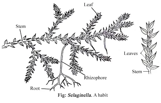
The plant body of Selaginella is a sporophyte that exhibits clear differentiation into roots, stems, leaves, ligules, and rhizophores. Each of these structures plays a vital role in the plant’s development and functionality. Below is a detailed breakdown of the morphology of Selaginella:
- Root:
- The roots of Selaginella are adventitious in nature, meaning they grow from unusual points such as the tip of the rhizophore or directly from the stem.
- These roots arise endogenously and branch through a process called dichotomous branching, where the root divides into two equal parts.
- In epiphytic species, the aerial roots possess a root cap that helps protect the root tip as it grows.
- The presence of root hairs allows for better absorption of water and nutrients from the surrounding environment.
- Stem:
- The stem of Selaginella is typically green and herbaceous, indicating that it performs photosynthesis. It is dichotomously branched, meaning it repeatedly splits into two branches.
- The stem can either be erect or prostrate, with erect branches growing from a horizontally spreading main stem.
- It is covered with leaves and exhibits pseudomonopodia, a form of growth where branches appear to arise from one point, but this growth pattern is not true monopodial branching.
- In most species, the shoot apex is characterized by a single apical cell, which governs the growth of the stem.
- Leaves:
- The leaves of Selaginella are microphyllous, which means they are small in size with a single unbranched midrib.
- Each leaf has a small, tongue-like membranous outgrowth called a ligule located on its upper (adaxial) surface.
- The leaves are dorsiventral, meaning they have a distinct upper and lower surface.
- Types of Leaf Conditions:
- Isophylly (Homoeophyllum):
In some species of Selaginella, all the leaves are of the same kind. This condition is known as isophylly, and the leaves are referred to as isophyllous. - Anisophylly (Heterophyllum):
In other species, two distinct types of leaves are present—small leaves and large leaves. This condition is called anisophylly, and the leaves are termed anisophyllous.
- Isophylly (Homoeophyllum):
- Ligule:
- The ligule is a distinctive feature of Selaginella leaves, found on the adaxial surface of the leaf. It is small, membranous, and tongue- or leaf-like in shape.
- A mature ligule has a prominent basal portion called the glossopodium, which anchors it to the leaf.
- Although the ligule is a consistent morphological feature, its exact function remains unclear.
- Rhizophore:
- The rhizophore is a unique, colorless, leafless, unbranched, and cylindrical structure in Selaginella.
- It arises from the prostrate stem at points where the stem dichotomizes, and it grows downward toward the soil.
- Once the free end of the rhizophore reaches the soil, it develops a tuft of adventitious roots, supporting the plant’s growth in new areas.
- Unlike typical roots, the rhizophore lacks a root cap but still develops adventitious roots at its tip.
Anatomy of Selaginella
The anatomy of Selaginella is complex, with a variety of tissues and structures that allow the plant to survive and adapt to different environments. Each part of the plant—stem, leaf, ligule, root, and rhizophore—has specific anatomical features that support its functions. Below is a detailed examination of the anatomy of Selaginella:
- Stem Anatomy:
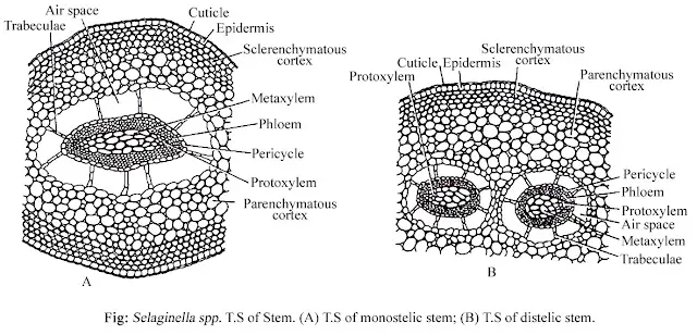
- The outermost layer of the stem is the epidermis, which lacks both hair and stomata. This epidermal layer is protected by a cuticle to prevent water loss.
- Beneath the epidermis lies a well-defined cortex, which can either be entirely parenchymatous or divided into an outer sclerenchymatous region and an inner parenchymatous region.
- The stele, which forms the central part of the stem, is typically protostelic, meaning the xylem is centrally located and surrounded by phloem cells. The number of steles may vary across different species of Selaginella.
- A central cavity, containing air spaces, separates the stele from the cortex.
- The endodermis connects the cortex to the central tissue through radially elongated cells known as trabeculae.
- Surrounding the stele is a single-layered pericycle. Notably, a pith is generally absent in the stem anatomy of Selaginella.
- Leaf Anatomy:
- The leaf’s epidermis is single-layered on both the upper and lower surfaces. Stomata are present on the upper surface, facilitating gas exchange.
- The epidermis also contains chloroplasts, which are essential for photosynthesis.
- The mesophyll tissue is uniformly composed of either spongy or palisade-like elongated cells with air spaces between them, facilitating gas exchange and photosynthesis.
- A simple vascular bundle is located in the center of the leaf, providing support and transporting water, nutrients, and sugars.
- Ligule Anatomy:
- The ligule, a unique feature of Selaginella, originates from several short rows of superficial cells.
- When fully developed, the ligule consists of a distinct, hemispherical basal region known as the glossopodium. In this region, the cells are large, thin-walled, and contain vacuolated cytoplasm.
- The glossopodium is surrounded by a sheath known as the glossopodium sheath, which helps in maintaining its structure.
- Root Anatomy:
- The root’s outermost layer is the epidermis, a large single layer covered by a protective cuticle. Root hairs arise from certain epidermal cells, aiding in water and nutrient absorption.
- Below the epidermis is a wide zone of cortex, divided into two distinct regions:
- The outer hypodermis, composed of sclerenchymatous cells that provide structural support.
- The inner parenchymatous region, which stores nutrients and aids in the transport of water and solutes.
- Although the endodermis is present, it is inconspicuous and not as prominent as in other plants.
- Just beneath the endodermis, there is a single-layered pericycle that surrounds the vascular tissue.
- The xylem, responsible for water transport, is centrally located and surrounded by phloem cells, which transport nutrients.
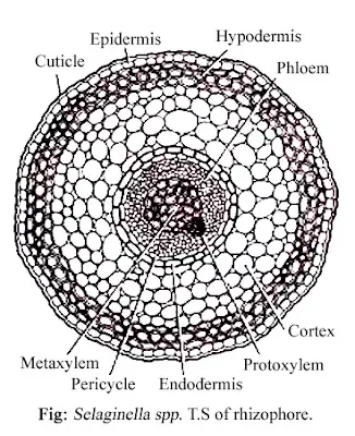
- Rhizophore Anatomy:
- The rhizophore’s outermost layer is a thick-walled epidermis, consisting of a single layer of cells. Unlike the roots, the rhizophore lacks root hairs.
- Beneath the epidermis lies the cortex, which is divided into two regions:
- The hypodermis, which is thick-walled and multi-layered, providing structural integrity.
- A thin-walled parenchymatous region that facilitates storage and nutrient transfer.
- The endodermis, located around the pericycle, separates the cortex from the vascular tissue.
- The pericycle, which is thin-walled, surrounds the vascular tissues.
- The stele within the rhizophore is protostelic, with xylem positioned centrally and phloem surrounding the xylem. This arrangement supports efficient transport of water and nutrients.
Reproduction of Selaginella
The reproduction of Selaginella occurs through both vegetative and sexual methods. Each method involves distinct structures and processes that contribute to the propagation of the species. Below is a detailed explanation of the reproductive strategies employed by Selaginella:
- Vegetative Reproduction:
- Fragmentation:
- Under humid conditions, in species like Selaginella rupestris, certain branches of the stem develop roots. These rooted branches can detach from the parent plant, ultimately growing into new individual plants.
- Tubers:
- Tubers are formed towards the end of the growing season and can be either aerial or underground. In species such as Selaginella chrysocaulos and Selaginella chrysorrhizos, tubers develop at the tips of underground branches. When conditions become favorable, these tubers germinate into new plants.
- Resting Buds:
- Resting buds develop at the tips of certain aerial branches. These buds can endure unfavorable conditions by becoming dormant. Once conditions improve, they give rise to rhizophores, which contribute to the development of new plants.
- Fragmentation:
- Sexual Reproduction by Spores:
- Sporophytic Phase:
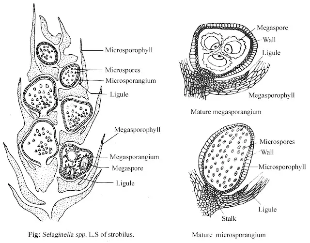
- Selaginella is a sporophytic plant that reproduces sexually. It is heterosporous, meaning it produces two distinct types of spores: megaspores and microspores. These spores are formed in specialized structures called megasporangia and microsporangia, respectively.
- Both types of sporangia are produced on fertile leaves known as megasporophylls (for megaspores) and microsporophylls (for microspores). These structures are typically grouped together to form a compact, terminal structure known as the strobilus.
- Sporophytic Phase:
- Strobilus:
- The strobilus is a reproductive structure formed by the aggregation of ligulate sporophylls at the apex of the branches of the stem. The length of the strobilus varies between species, ranging from ¼ inch to 2–3 inches. In some species, such as Selaginella cuspidata and Selaginella patula, the stem continues to grow beyond the strobilus in what is referred to as the “selago condition.”
- Sporangia:
- Mature sporangia are stalked structures with a short stalk and a capsule. The stalk is multicellular and multiseriate, while the capsule has a two-layered wall, referred to as the jacket.
- There are two types of sporangia:
- Megasporangia (larger and pale)
- Microsporangia (smaller and slightly elongated)
- Development of Sporangia:
- Sporangium development in Selaginella is of the eusporangiate type. The developmental process of megasporangia and microsporangia is similar up to the formation of the spore mother cells (SMCs).
- The sporangial initial divides to form outer jacket initials and inner archesporial initials. The archesporial initial gives rise to sporogenous tissue, while the outermost layer of sporogenous tissue forms the tapetum, which nourishes the developing spores.
- In microsporangia, all sporogenous cells become spore mother cells, which undergo meiosis to form microspores. However, in megasporangia, only one spore mother cell is functional, while the rest disintegrate. The functional spore mother cell undergoes meiosis to produce four megaspores.
- The development of megaspores begins before they are shed from the sporangium.
- Gametophytic Generation:
- Spore and Gametophyte Development:
- Spores represent the first cell of the gametophytic generation and are haploid. In Selaginella, two types of spores are produced:
- Microspores: These are tetrahedral in shape, with a two-layered wall composed of an outer exine (thick) and an inner intine (thin).
- Megaspores: These are also tetrahedral and feature a triradiate ridge. Their spore coat can either be two-layered (with exine and intine) or three-layered (with an additional mesospore between the exine and intine).
- Germination:
- Spore germination begins through segmentation. Initially, the spores remain within the sporangia, and they are shed as multicellular gametophytes.
- Microspores germinate to form male microgametophytes, while megaspores germinate to form female megagametophytes.
- Spores represent the first cell of the gametophytic generation and are haploid. In Selaginella, two types of spores are produced:
- Development of Microgametophyte:
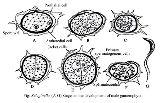
- The microspore undergoes an unequal division, producing a small prothallial cell and a larger antheridial initial. The prothallial cell remains vegetative, while the antheridial cell divides to form 12 cells—four of which form a central core (primary androgonial cells), with the others becoming jacket cells. At this stage, the microgametophyte consists of 13 cells: one prothallial cell, four androgonial cells, and eight jacket cells.
- Development of Megagametophyte:
- The nucleus of the megaspore divides repeatedly, forming a coenocytic mass of free nuclei. Cell walls then begin to form between these nuclei in the apical part of the spore, creating a cellular cushion-like structure. The gametophyte remains coenocytic below this tissue. Most of the superficial cells in the apical cushion develop into archegonial initials, which later give rise to archegonia.
- Spore and Gametophyte Development:
- Fertilization:
- Fertilization in Selaginella requires water. The swimming antherozoids (sperm) reach the egg through the neck of the archegonium, and the fusion of the antherozoid nucleus with the egg nucleus forms a zygote. This marks the end of the gametophytic generation and the beginning of the sporophytic generation.
- Embryo Development (Young Sporophyte):
- The zygote (or oospore) is the initial stage of the sporophytic generation. It undergoes a transverse division, forming an upper suspensor initial and a lower embryo initial. The suspensor initial further divides to form a multicellular suspensor, while the embryo initial divides vertically to form four cells. One of these cells will give rise to the shoot, and further divisions create the young sporophyte.
- https://www.slideshare.net/slideshow/selaginella-features-morphology-anatomy-and-reproduction/267938194
- https://premabotany.blogspot.com/2018/12/selaginella-classification-structure-of.html