Pteris is a widely distributed genus of ferns that can be found in various geographical locations around the world. Although this genus is cosmopolitan in nature, it thrives primarily in tropical and subtropical climates. Pteris species are typically located in well-drained environments or in the crevices of rocks, where they exhibit a remarkable adaptability to diverse habitats. Their prevalence along hilly slopes allows them to reach elevations of up to 1200 meters above sea level.
The genus Pteris encompasses approximately 250 to 280 recognized species, making it a significant group within the fern family. Notable species include P. quadriaurita, P. critica, P. vittata, P. pellucida, P. wallichiana, and P. stenophylla. Each of these species exhibits unique morphological and physiological traits that contribute to their survival in varying environmental conditions.
Morphologically, Pteris ferns are characterized by their distinctive fronds, which can vary significantly in shape, size, and color. The fronds often display a pinnate or pinnatifid structure, allowing for effective photosynthesis and nutrient absorption. This adaptation is essential for their growth, especially in shaded or under-canopy environments where light availability may be limited.
Pteris species possess various ecological functions. They play a vital role in soil stabilization due to their extensive root systems, which help prevent erosion. Additionally, these ferns contribute to the biodiversity of their habitats, providing shelter and habitat for a range of organisms. Therefore, their presence is crucial for maintaining ecological balance in tropical and subtropical ecosystems.
In terms of reproduction, Pteris ferns reproduce via spores rather than seeds. The sporangia, located on the underside of the fronds, release spores into the environment, which can then germinate into gametophytes. This unique reproductive strategy allows for colonization in new areas, enhancing their dispersal capabilities.

Occurrence and Distribution of Pteris
Pteris, a cosmopolitan fern, displays a remarkable distribution across nearly all geographical regions. This extensive range highlights its adaptability and resilience in diverse environments, although it shows a strong preference for tropical and sub-tropical climates.
- Habitat Preferences:
- Pteris typically thrives in well-drained areas, often found growing in the crevices of rocks, which provides stability and moisture retention.
- The species are commonly encountered along the slopes of hills, demonstrating their ability to colonize various altitudinal ranges. They have been observed at elevations reaching up to 1200 meters above sea level, indicating their adaptability to different climatic and environmental conditions.
- Species Diversity:
- The genus Pteris encompasses a substantial diversity, with approximately 250 to 280 species documented. This diversity underscores the genus’s evolutionary success and ecological versatility.
- Among the notable species found in India are P. quadriaurita, P. critica, P. vittata, P. pellucida, P. wallichiana, P. stenophylla, and P. biaurita. Each species exhibits unique morphological and ecological characteristics, contributing to the overall biodiversity of the region.
- Chemical Compounds:
- Research by Tanaka and Chin-ming (1982) identified specific compounds, such as Pterosin and Astragalin, from Pteridium aquilinum subsp. Wightianum. These findings illustrate the potential chemical diversity within the broader Pteris genus, suggesting various ecological roles and possible applications in medicine and industry.
Morphology of the Plant
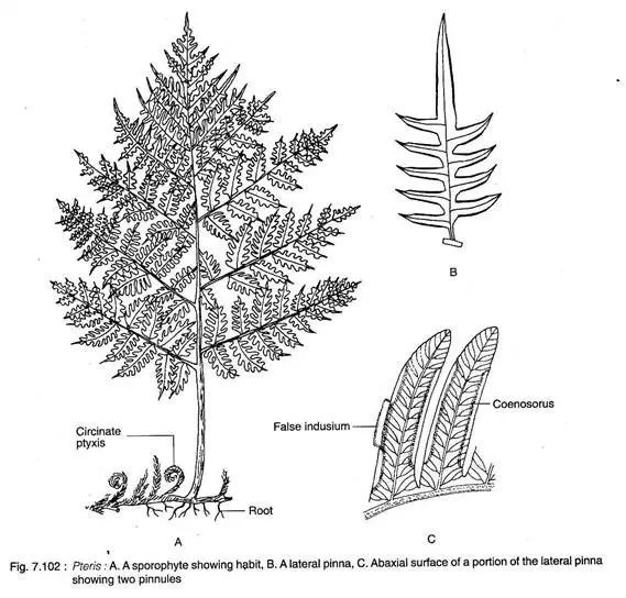
The morphology of Pteris is characterized by its unique structural features that enable it to thrive in diverse environments. This fern exhibits a rhizomatous stem, which plays a critical role in its growth and development, facilitating the production of roots and leaves.
- Rhizome Structure:
- The rhizome can display a range of growth forms, either creeping, as observed in P. grandiflora, or compact and erect, as seen in P. cretica and P. longifolia.
- The rhizome may or may not exhibit branching and is enveloped in protective scales.
- Roots typically emerge at the base of the leaf or from various points along the rhizome, contributing to the plant’s stability and nutrient uptake.
- The growing point of the rhizome is covered with ramenta, which are modified leaves that provide additional protection.
- Leaf Characteristics:
- Leaves originate from the upper surface of the rhizome, characterized by a long rachis, which serves as the central axis of the leaf.
- The leaf structure can be described as unipinnately compound, decompound, or multi-pinnately compound. This variety allows for adaptability in different ecological niches.
- The dissections of the pinnae are shallower compared to those in other ferns, such as Pteridium, which may influence the leaf’s overall surface area and light capture efficiency.
- Venation and Shape:
- The venation pattern of the leaves is of an open dichotomous type, which aids in the efficient transport of water and nutrients throughout the leaf structure.
- The pinnae exhibit a size variation; they are smaller near the base, larger towards the middle, and then again smaller towards the apex, as exemplified in P. vittata. This gradation can help the plant maximize light interception and reduce shading of lower leaves.
- Leaves display circinate vernation, a characteristic feature of true ferns, where the young leaves are coiled and unfurl as they mature.
- Texture and Reproductive Structures:
- Pinnae are often coriaceous, which refers to a leathery texture that can help reduce water loss and provide structural integrity.
- Reproductive structures, or sori, are located along the ventral margin of the pinnae. These sori are continuous along the margin, deliberately avoiding the apices of the segments, and are typically found in the sinuses between them. This arrangement allows for effective spore dispersal while minimizing the risk of damage to the reproductive structures.
Internal Structure of Pteris
The internal structure of Pteris reflects a remarkable organization and specialization that support its physiological functions. The plant’s anatomy varies across its different parts, including the rhizome, leaves, and roots, each contributing to its overall adaptability and efficiency.

- Rhizome Structure:
- The rhizome exhibits notable diversity in its stelar organization, which may be classified as either solenostelic, as seen in P. grandiflora and P. vittata, or dictyostelic.
- In P. biaurita, the rhizome displays a mixed protostele in its lower region, transitioning to a siphonostelic organization slightly above, and near the apex, it presents a polycyclic dictyostelic structure.
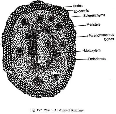
- The epidermis of the rhizome consists of a single layer that encases a broad cortex, which includes a hypodermal sclerenchyma layer.
- The primary bulk of the cortex is composed of parenchyma cells, providing storage and support.
- The stele features multiple meristeles, typically arranged in two rings: the inner ring contains two or three large meristeles, while the outer ring consists of several smaller meristeles.
- Each meristele comprises a band or plate-like xylem mass, which can be angular in some cases.
- The xylem is composed of protoxylem groups embedded within metaxylem, exhibiting a mesarch condition. Surrounding the xylem is the phloem, and each meristele may contain its own endodermis, which should not be confused with the polysteles found in Selaginella.
- The vascular strands can be interrupted by leaf gaps, indicating the complexity of vascular connections.
- Leaf Structure:
- The rachis of the leaf is traversed by a single leaf trace, which varies in shape. In P. vittata, the leaf traces are ‘C’ shaped, while in P. biaurita, they begin as ‘U’ shaped at the leaf base and transition to a ‘V’ shape further up.
- The xylem strand appears hooked, which is characteristic of Pteris.
- As in many vascular plants, the xylem is encircled by phloem, pericycle, and endodermis.
- The cortical region of the leaf features an inner parenchymatous zone and an outer sclerotic zone, contributing to the leaf’s structural integrity and functionality.
- The epidermis is a single layer that possesses a cuticle to reduce water loss, and ramenta arise from some epidermal cells, providing additional protection.
- The leaf lamina is bifacial, meaning it may exhibit different characteristics on its upper and lower surfaces, and may or may not feature a differentiated mesophyll.
- Leaves are generally hypostomatic, possessing stomata predominantly on the lower surface, optimizing gas exchange while minimizing water loss.
- The midrib contains a concentric vascular bundle with a distinct endodermis, known as the bundle sheath, enhancing the transport of nutrients and water.
- Root Structure:
- The root anatomy consists of an outer piliferous layer, which aids in water and nutrient absorption.
- The cortex is divided into an outer parenchymatous zone and an inner sclerotic zone, providing both storage and mechanical support.
- The stele is protostelic, characterized by an exarch, diarch xylem arrangement, facilitating efficient water transport.
Reproduction in Pteris
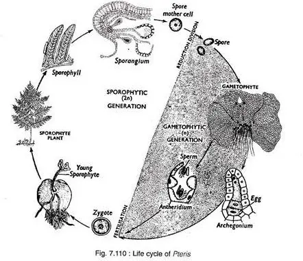
The reproductive process in Pteris, a homosporous fern, primarily involves spore formation and the development of gametophytes and sporophytes. This complex life cycle is characterized by alternation between the sporophyte and gametophyte generations, which are both integral to the fern’s reproduction and propagation.
- Spore-Producing Organ:
- Pteris reproduces through spores, specifically employing coenosori for spore production. These coenosori are marginally positioned, continuously borne on a vascular commissure connected with vein ends, resulting in a linear arrangement along the margin of the fronds.
- The individual identity of sori is obscured due to their continuous linear formation. Protection of the coenosori is provided by the reflexed margin of the pinnae, known as the false indusium.
- Within the coenosori, sporangia are intermixed with numerous sterile hairs, contributing to the overall structure.
- Development of Sporangium:
- The sporangium development in Pteris is of the leptosporangiate type. A single superficial cell on the receptacle serves as the sporangial initial, which divides transversely into an upper cell and a lower cell.
- The lower cell does not contribute to sporangium formation, while the upper cell differentiates into an apical cell with three cutting faces. This apical cell further divides, producing segments that develop into the sporangium structure.
- The jacket initial of the sporangium forms a single-layered jacket, while the archesporial cell divides into a tapetal initial and a primary sporogenous cell.
- The tapetal initial creates a two-layered tapetum through several divisions, supplying nutrition to the developing spores. The primary sporogenous cell subsequently divides to form twelve spore mother cells, which undergo meiosis to produce haploid spores.
- Structure of a Mature Sporangium:
- A mature sporangium features a long stalk that culminates in a capsule containing numerous spores. The capsule’s jacket is single-layered but consists of three types of cells: a thick-walled vertical annulus that incompletely arches over the sporangium, a thin-walled radially arranged stomium, and large parenchymatous cells with undulated walls.
- All spores within the capsule are structurally and functionally similar, indicating that Pteris is a homosporous pteridophyte. The spores are triangular, with a trilete aperture, and are bounded by two walls, where the outer wall, or exine, exhibits various ornamentation.
- The sporangium dehisces transversely along the stomium due to the shrinkage of annular cells, allowing for the dispersal of spores into the air.
- Gametophyte Development:
- Upon falling on a suitable substrate, the spores germinate. Initially, the spore wall (exine) ruptures, releasing the inner contents in the form of a germ tube. A transverse division in the germ tube produces the first rhizoid and the first prothallial cell.
- The prothallial cell subsequently divides to form a filament with an apical terminal cell that has two cutting faces, eventually developing into a spathulate prothallus. A mature prothallus forms, characterized by a cordate shape, dorsiventrally flattened structure, and photosynthetic capabilities.
- The prothallus comprises parenchymatous cells that vary in thickness; it is thinner toward the margin and thicker toward the center. The growing point is located in the apical notch, and rhizoids develop on the ventral surface.
- Pteris prothalli are monoecious and protandrous, with antheridia appearing first in the basal central or lateral regions among the rhizoids, while archegonia develop near the apical notch.
- Antheridium:
- The antheridial initial arises from a superficial cell on the ventral surface of the prothallus. This cell divides transversely, creating an outer upper cell and an inner lower cell.
- The upper cell, due to higher turgor pressure, bulges down, forming a dome shape. It further divides by an arched periclinal wall into a dome cell and a primary androgonial cell, which ultimately divides into 20-25 androcytes. Each androcyte transforms into a multiflagellated coiled antherozoid.
- Archegonium:
- The development of the archegonium in Pteris closely resembles that in Ophioglossum. A mature archegonium consists of a 5-6 celled projecting curved neck, a neck canal cell, a ventral canal cell, and an egg.
- Fertilization:
- Upon maturation, the antheridium absorbs water and swells. The increase in internal pressure causes the cover cells to split, releasing the antherozoids into a thin film of water present on the prothallus surface.
- Concurrently, disintegration of the ventral canal cell, neck canal cell, and neck cells creates an open passage for the antherozoids to reach the egg. Fertilization occurs when one antherozoid fuses with the egg, resulting in the formation of a zygote.
- New Sporophyte (Embryo):
- The first division of the zygote is vertical, followed by a transverse division that produces a quadrant, eventually forming a 32-celled embryo. The differentiation of the embryo begins at this 32-celled stage.
- No suspensor is formed; the hypobasal cells develop into the stem apex and foot, while the epibasal cells give rise to the cotyledon and root.
- As the embryo develops, the venter of the archegonium forms a protective layer called the calyptra around the embryo. The root and cotyledon grow rapidly, with the root penetrating the prothallus to establish the sporeling in the soil, followed by the development of the first leaf.

- https://www.studocu.com/in/document/mahatma-gandhi-university/botany/pteris-sem-4-botany-core/65600093/download/pteris-sem-4-botany-core.pdf
- https://www.biologydiscussion.com/botany/pteridophyta/pteris-structure-and-reproduction/45952