What is Nervous tissue?
- Nervous tissue, also known as neural tissue, is the main component of the nervous system, which is responsible for regulating and controlling body functions and activities. The nervous system consists of two main parts: the central nervous system (CNS), including the brain and spinal cord, and the peripheral nervous system (PNS), which consists of the branching peripheral nerves.
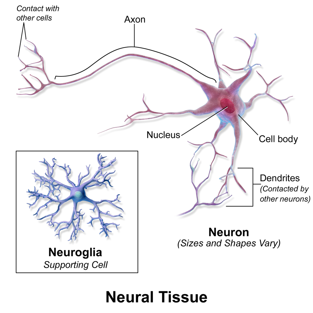
- Nervous tissue is composed of two primary types of cells: neurons and neuroglia (also known as glial cells or glia). Neurons are specialized cells that receive and transmit impulses, allowing for the communication and coordination within the nervous system. They have a long stem-like part called an axon, which sends action potentials to the next cell. Bundles of axons form nerves in the PNS and tracts in the CNS.
- Neuroglia, on the other hand, support and assist in the propagation of nerve impulses. They also provide nutrients to the neurons and maintain the extracellular environment around them. Glial cells play a vital role in signal conduction, protection against pathogens, and potential signaling functions.
- The functions of the nervous system include sensory input, integration, control of muscles and glands, homeostasis (maintenance of a stable internal environment), and mental activity such as emotions, memory, and thought processes. Nervous tissue enables the rapid transmission of stimuli from one part of the body to another, allowing for coordinated bodily functions and responses to external stimuli.
- Overall, nervous tissue is a specialized type of tissue that forms the foundation of the nervous system. It consists of neurons and glial cells, which work together to facilitate communication, regulate body functions, and support the overall functioning of the nervous system.

Definition of Nervous tissue
Nervous tissue is the specialized tissue that makes up the nervous system. It is composed of neurons and glial cells, and its main function is to transmit and process electrical signals, allowing for communication and coordination within the body.
Location of Nervous Tissue
- Nervous tissue is located in various parts of the body, primarily in the central nervous system (CNS) and peripheral nervous system (PNS).
- In the CNS, which includes the brain and spinal cord, nervous tissue is extensively present. The brain is responsible for higher cognitive functions, sensory processing, and motor coordination, while the spinal cord serves as a pathway for transmitting signals between the brain and the rest of the body.
- The peripheral nervous system (PNS) consists of the branching peripheral nerves that extend throughout the body. These nerves connect the CNS to the sensory organs, muscles, and other organs, enabling communication and control over bodily functions. The PNS is involved in sensory perception, motor control, and the regulation of various physiological processes.
- Within the nervous tissue, the key components are neurons, also known as nerve cells. Neurons are specialized cells that play a crucial role in transmitting electrical signals or impulses. These impulses are generated in response to stimuli and are propagated through elongated structures called axons. The axons extend from the cell body of the neuron and allow for communication with other cells and tissues.
- So, nervous tissue can be found in peripheral nerves throughout the body, as well as in the organs of the central nervous system, such as the brain and spinal cord. It is within these locations that neurons carry out their functions of receiving, integrating, and transmitting information, enabling the complex functioning of the nervous system.
Characteristics Of Nervous Tissue
- Composition: Nervous tissue is the main component of both the central nervous system (CNS) and peripheral nervous system (PNS) in the nervous system.
- Cell Types: Nervous tissue consists of two main types of cells: neurons and glial cells (also known as neuroglia). Neurons are specialized cells responsible for transmitting electrical signals, while glial cells provide support and protection to neurons.
- Components: Nervous tissue is composed of various structures, including dendrites, cell bodies (soma), axons, and nerve endings (terminals). Dendrites receive signals from other neurons, the cell body contains the nucleus and organelles, axons transmit signals away from the cell body, and nerve endings release neurotransmitters to communicate with other neurons.
- Neurotransmitters: Neurons release chemical substances called neurotransmitters. These neurotransmitters are responsible for transmitting signals between neurons in response to stimuli. They play a crucial role in the communication and functioning of the nervous system.
- Synapses: At the specialized endings of axons called synapses, there is a close connection between neurons. This is where the transmission of signals occurs. Neurotransmitters are released at the synapse and activate the dendrites of the adjacent neuron, allowing the signal to be propagated further.
- Longevity of Nerve Cells: Nerve cells, including neurons, have a long lifespan. Once mature, they cannot divide or be easily replaced (except for certain memory cells). This characteristic highlights the importance of protecting and maintaining the health of nervous tissue.
In summary, nervous tissue is characterized by its composition of neurons and glial cells, the presence of specialized structures like dendrites and axons, the release of neurotransmitters, the formation of synapses, and the long life and limited regenerative capacity of nerve cells. These characteristics enable the nervous tissue to carry out its essential functions in the coordination, communication, and regulation of the body’s activities.
Structure of Nervous tissue
The structure of nervous tissue is complex and composed of different types of cells. The main components of nervous tissue are neurons and neuroglial cells.
Neurons are the primary cells of the nervous system and are responsible for receiving and transmitting nerve impulses. They have a distinct structure that includes a cell body, dendrites, and an axon. The cell body contains the nucleus, cytoplasm, and cell organelles necessary for the neuron’s functioning. Dendrites are highly branched processes that receive information from other neurons and synapses, which are specialized points of contact. The axon is a long stem-like projection that carries the nerve impulse away from the cell body and communicates with other cells, known as target cells. Information in a neuron flows unidirectionally, from the dendrites through the cell body and down the axon.
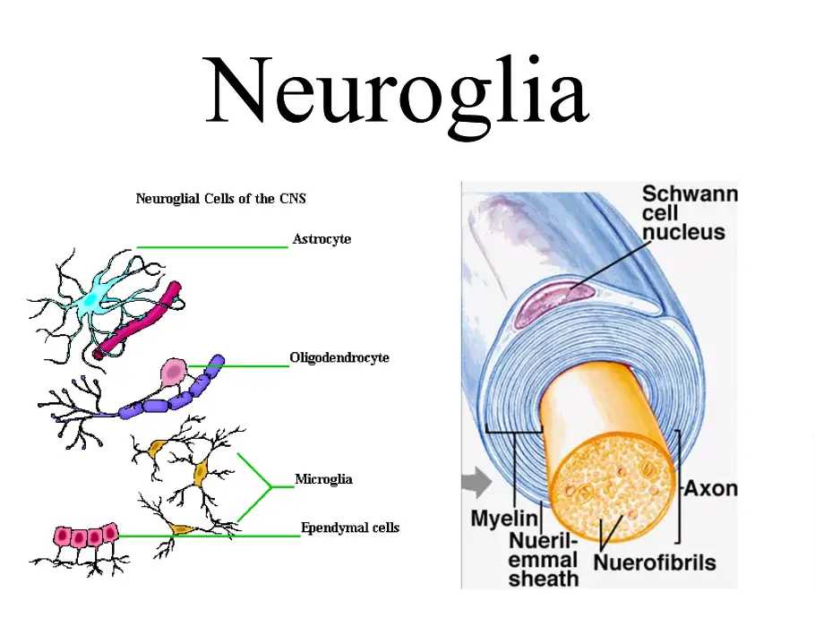
Neuroglial cells, also known as glial cells, are non-neural cells in the nervous tissue that provide support and various essential functions for neurons. There are several types of neuroglial cells found in the central nervous system (CNS) and peripheral nervous system (PNS). Some examples include:
- Astrocytes: These star-shaped cells have many processes and are the most abundant cell type in the brain. They provide support and maintain the extracellular environment around neurons.
- Microglial cells: These are the smallest neuroglial cells and act as the primary immune system for the CNS, protecting against pathogens and inflammation.
- Oligodendrocytes: Found in the CNS, these cells have few processes and are responsible for forming myelin sheaths around axons. Myelin sheaths act as insulation and increase the speed of nerve impulse conduction.
- Schwann cells: These are the PNS equivalent of oligodendrocytes and are responsible for maintaining axons and forming myelin sheaths in the peripheral nerves.
- Ependymal cells: These line the ventricles of the brain and help produce cerebrospinal fluid.
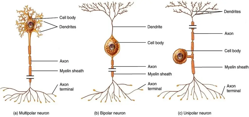
The structural classification of neurons includes multipolar neurons, bipolar neurons, pseudounipolar neurons, and unipolar brush cells. Each type has specific characteristics and functions within the nervous system.
Overall, the structure of nervous tissue is highly specialized, allowing for the transmission and processing of electrical signals essential for communication and coordination within the body.
Types of Nervous tissue
There are two types of Nervous tissue such as;
- Neurons
- neuroglia
1. Neuron
- Neurons are specialized cells that are fundamental to the functioning of the nervous system. The human nervous system contains approximately 100 billion neurons. These cells vary in shape and size, but they all consist of three main parts: the cell body (also known as the soma), dendrites, and an axon.
- The cell body of a neuron contains the nucleus and various organelles that are responsible for the cell’s metabolic functions. It plays a crucial role in integrating incoming signals and generating outgoing signals.
- Dendrites are branching extensions of the neuron that receive signals from other neurons or sensory receptors. They serve as the input sites for receiving information from the surrounding environment or from other neurons. The structure of dendrites, with their numerous branches and branches off branches, allows for a large surface area for receiving signals.
- The axon is a long, slender projection that carries electrical signals, known as action potentials, away from the cell body toward other neurons or target cells. Axons can vary in length, from a fraction of a millimeter to several feet long. They are covered by a fatty substance called myelin, which acts as an insulating layer and allows for faster transmission of electrical signals along the axon.
- At the end of the axon, there are specialized structures called axon terminals or synaptic terminals. These terminals form synapses, which are the points of communication between neurons or between neurons and other cells. When an action potential reaches the axon terminals, it triggers the release of chemical messengers called neurotransmitters. The neurotransmitters cross the synapse and bind to receptors on the receiving neuron or target cell, transmitting the signal to the next cell.
- The unique structure of neurons, with their complex branching and connectivity, enables the transmission of information and electrical signals throughout the nervous system. This intricate network of connections allows for the integration of sensory input, processing of information, and generation of appropriate responses. Neurons play a vital role in various functions such as sensation, movement, cognition, and behavior.

Structure of Neuron/Parts of a neuron
The structure of a neuron consists of several key components that allow it to function as a specialized cell in the nervous system.
- Cell Body (Soma): The cell body is the central part of the neuron and contains the nucleus, cytoplasm, and various cell organelles. It plays a vital role in the metabolism, growth, and repair of the neuron. Within the cell body, structures like Nissl bodies (composed of RNA, rough endoplasmic reticulum, and free ribosomes) aid in protein synthesis. Neurofilaments and neurotubules, thread-like proteins, provide structural support and assist in intracellular transport.
- Dendrites: Dendrites are numerous, branching extensions that arise from the cell body. They receive incoming signals and information from other neurons or sensory receptors. The dendrites conduct nerve impulses towards the cell body and play a crucial role in integrating and processing information.
- Axon: Neurons typically have a single long and unbranched extension called an axon. It is responsible for transmitting electrical signals, known as nerve impulses, away from the cell body and towards other neurons or target cells. The axon is covered by a lipid sheath called the myelin sheath, which is formed by specialized non-neuronal cells called Schwann cells in the peripheral nervous system (PNS) and oligodendrocytes in the central nervous system (CNS). The myelin sheath acts as insulation and facilitates faster transmission of nerve impulses. Nodes of Ranvier are gaps in the myelin sheath along the axon that play a crucial role in signal propagation.
- Axon Terminal: At the end of the axon, there are specialized structures called axon terminals or synaptic end bulbs. These terminals form synapses with other neurons or target cells. When an electrical impulse reaches the axon terminal, it triggers the release of chemical messengers called neurotransmitters. The neurotransmitters cross the synapse and bind to receptors on the receiving neuron or target cell, allowing for the transmission of the signal.
The structure of a neuron is characterized by its ability to receive, integrate, and transmit information through its dendrites, cell body, and axon. This organization allows for the flow of signals in one direction, from dendrites to axon terminals, enabling communication within the nervous system. The myelin sheath and nodes of Ranvier contribute to efficient signal conduction along the axon. Neurons’ specialized structure enables the immense complexity and connectivity within the nervous system, facilitating functions such as sensation, movement, and cognitive processes.
Shapes of neuron
Neurons exhibit different shapes based on the arrangement of their processes, such as dendrites and axons. The two main shapes of neurons are multipolar and bipolar.

- Multipolar Neurons: Multipolar neurons have multiple processes emerging from their cell bodies, hence their name. They typically have several dendrites attached to the cell body, along with a single, long axon. These neurons are commonly found in the central nervous system (CNS), including the brain. Motor neurons, which transmit signals from the CNS to the skeletal muscles, are an example of multipolar neurons. Their multiple processes allow for extensive connections and integration of information.
- Bipolar Neurons: Bipolar neurons have two opposing processes extending from each end of the cell body. One process is an axon, while the other is a dendrite. Bipolar cells are relatively rare compared to other types of neurons. They are primarily found in specialized sensory organs. In the olfactory epithelium, which is responsible for detecting smell stimuli, bipolar neurons play a crucial role in relaying sensory information to the brain. Additionally, bipolar cells are present in the retina of the eye, where they participate in the transmission of visual signals.
These two types of neurons, multipolar and bipolar, represent different structural arrangements that suit their specific roles in transmitting and processing information within the nervous system.
Types of neuron
Neurons, the fundamental cells of the nervous system, can be categorized into different types based on their structure and function.
I. Types of Neurons Based on Structure:

- Unipolar Neurons: Unipolar neurons have a single process extending from the cell body. This process acts as both an axon and a dendrite. Unipolar neurons are commonly found in the peripheral nervous system (PNS) and serve as sensory neurons, relaying sensory impulses from various parts of the body to the central nervous system (CNS).
- Bipolar Neurons: Bipolar neurons have two processes that extend from opposite ends of the cell body: one dendrite and one axon. These neurons are relatively less common and are primarily found in specialized sensory organs such as the retina of the eye, the cochlea of the ear, and the olfactory epithelium (responsible for detecting smell).
- Multipolar Neurons: Multipolar neurons possess multiple processes emanating from the cell body, consisting of numerous dendrites and a single axon. Most neurons in the CNS are multipolar neurons. They play various roles in the integration and processing of information within the brain and spinal cord.
II. Types of Neurons Based on Function:
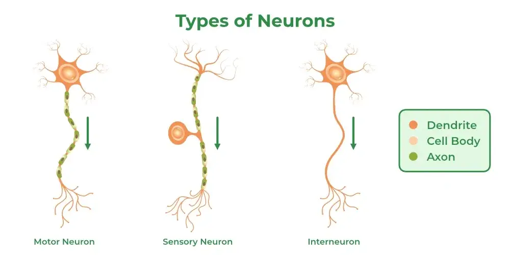
- General Somatic Afferent Neurons (Sensory Neurons): These neurons carry sensory impulses from the skin, skeletal muscles, joints, and connective tissues to the CNS. They provide information about touch, pain, temperature, and proprioception (awareness of body position).
- General Visceral Afferent Neurons: General visceral afferent neurons transmit sensory impulses from the internal organs, such as the heart, lungs, and gastrointestinal tract, to the CNS. They convey information related to the internal environment and visceral sensations.
- General Somatic Efferent Neurons (Motor Neurons): General somatic efferent neurons carry motor impulses from the CNS to the skeletal muscles, enabling voluntary movements.
- General Visceral Efferent Neurons: General visceral efferent neurons are responsible for transmitting motor impulses from the CNS to the visceral organs, controlling various involuntary functions such as digestion, heart rate, and glandular secretions.
- Special Visceral Efferent Neurons: Special visceral efferent neurons originate in the brain and innervate specific muscles involved in facial expressions, chewing, swallowing, and vocalization.
- Special Afferent Neurons: Special afferent neurons are receptor cells responsible for detecting specific sensory stimuli, including olfactory (smell), optic (vision), auditory (hearing), vestibular (balance), and gustatory (taste) stimuli. They transmit sensory information from these specialized sensory organs to the CNS.
These different types of neurons, based on their structure and function, work together to enable the complex functions of the nervous system, including sensory perception, motor control, and integration of information.
2. Neuroglia
Neuroglia, also known as glial cells or simply glia, are non-neuronal cells that provide crucial support and protection to neurons in the nervous system. While neurons are responsible for transmitting electrical signals and processing information, neuroglia have various supportive functions that contribute to the overall function and well-being of the nervous system.
There are several types of neuroglia in the central nervous system (CNS) and peripheral nervous system (PNS), each with specific roles and characteristics. The main types of neuroglia in the CNS include astrocytes, oligodendrocytes, microglia, and ependymal cells, while in the PNS, Schwann cells and satellite cells are the primary types.

Here are the classifications of neuroglial cells:
- Microglial cells: Microglia are the smallest neuroglial cells and act as the primary immune cells in the central nervous system (CNS). They are responsible for defending the CNS against infections, removing cellular debris, and regulating immune responses in the brain and spinal cord.
- Astrocytes: Astrocytes are star-shaped macroglial cells with multiple processes. They are the most abundant glial cell type in the CNS and have diverse functions. Astrocytes provide physical and metabolic support to neurons, regulate the extracellular environment, maintain the blood-brain barrier, and participate in processes like synapse formation, neurotransmitter regulation, and repair following injury.
- Oligodendrocytes: Oligodendrocytes are CNS cells with few processes. Their main function is to produce and maintain myelin, a lipid-based insulating substance that wraps around axons in the CNS. Myelin sheaths facilitate the rapid conduction of nerve impulses along axons, improving signal transmission efficiency.
- NG2 glia: NG2 glia are a distinct type of glial cell found in the CNS. They serve as developmental precursors of oligodendrocytes and are involved in the generation and maintenance of myelin-forming cells.
- Schwann cells: Schwann cells are the peripheral nervous system (PNS) counterparts of oligodendrocytes. They provide support and insulation to peripheral nerve fibers by forming myelin sheaths around axons. Schwann cells also participate in nerve regeneration and repair processes in the PNS.
- Satellite glial cells: Satellite glial cells are found in the PNS, specifically in ganglia where clusters of nerve cell bodies are located. They surround and support the neuronal cell bodies within ganglia, playing a role in regulating the microenvironment and metabolic processes.
- Enteric glia: Enteric glia are a specialized type of glial cell found in the enteric nervous system (ENS), which is responsible for regulating the gastrointestinal tract’s function. Enteric glia contribute to gut homeostasis, neuronal support, and communication within the ENS.
Each type of neuroglial cell has unique functions and plays a crucial role in maintaining the overall health and functioning of the nervous system.
Overall, neuroglia play essential roles in maintaining the structural integrity of the nervous system, regulating the extracellular environment, supporting neuronal function, and participating in immune responses. Although they are non-neuronal cells, neuroglia are indispensable for the proper functioning of neurons and overall nervous system function.
Function Of Nervous Tissue
The nervous tissue has several important functions in the body:
- Generation and transmission of nerve impulses: Neurons are specialized cells that generate and carry out nerve impulses. They produce electrical signals known as action potentials, which allow for the transmission of information across long distances within the nervous system. This is achieved through the release of chemical neurotransmitters that enable communication between neurons.
- Response to stimuli: Nervous tissue is responsible for detecting and responding to various stimuli from both the external and internal environment. Sensory neurons receive input from sensory receptors, such as those involved in vision, hearing, touch, and taste, and transmit this information to the central nervous system for processing and interpretation.
- Communication and integration: The nervous tissue facilitates communication between different parts of the body. It integrates incoming sensory information, processes it, and generates appropriate motor responses. This coordination of signals allows for the regulation and control of bodily functions, movements, and behaviors.
- Electrical insulation and debris removal: Glial cells, such as oligodendrocytes in the central nervous system and Schwann cells in the peripheral nervous system, play a crucial role in providing electrical insulation to nerve cells. They form myelin sheaths around the axons of neurons, which increases the speed and efficiency of nerve impulse conduction.
Additionally, glial cells help in removing debris and metabolic waste products from the nervous tissue, ensuring a clean and healthy environment for optimal neuronal function.
- Transmission of messages: Nervous tissue carries messages from one neuron to another and from neurons to other cells or organs in the body. This communication occurs through synapses, specialized junctions between neurons where neurotransmitters are released to transmit signals. These messages allow for the coordination of bodily functions, voluntary and involuntary movements, and the regulation of physiological processes.
Overall, the function of nervous tissue is to enable the detection of stimuli, generation and transmission of nerve impulses, integration and processing of information, and the coordination of various physiological and behavioral responses in the body.
Types Of Nerves
Signals are initiated as a result of any stimulation. They begin with the CNS (Central Nervous System), which means that impulses travel from the brain to the spinal cord in some situations. The signal travels from the CNS to the body’s exterior parts or edges, such as external organs and limbs, where it causes the necessary reaction. Muscle contraction or relaxation is the reaction to any stimulation. We develop goosebumps as an action as a result of the cold temperatures, which is a stimulant.
When the nerves get an electrochemical signal (neurotransmitter) or any impulse from the stimuli, they begin to operate by responding to the stimulus by receiving a signal from the brain. Nerves are classified according to their function.
1. Motor Nerves
- Motor neurons, also referred to as motor nerves, play a crucial role in transmitting signals from the brain and spinal cord to the muscles throughout the body. These signals enable the execution of basic activities such as speaking, walking, drinking water, blinking, sitting, and sleeping. Motor neurons are responsible for coordinating muscle contractions and movements.
- When motor neurons are damaged, it can result in muscle weakness or muscle atrophy, which is the shrinking of muscles due to lack of use or innervation. This can lead to difficulties in performing everyday tasks and movements.
- One prominent motor nerve is the sciatic nerve, which originates from the lower back and runs through the buttocks. The sciatic nerve is a composite of various nerves and is responsible for enabling movement in the entire leg. It branches out to innervate different regions, including the hamstring muscles, feet, thighs, and lower leg.
- The proper functioning of motor nerves is essential for maintaining coordination, balance, and mobility. Any impairment or damage to these nerves can significantly impact muscle control and movement abilities. Rehabilitation and therapeutic interventions may be necessary to restore or improve motor function in cases of motor nerve damage or dysfunction.
2. Sensory Nerves
- Sensory nerves, also known as sensory neurons, play a vital role in generating impulses or signals that travel in the opposite direction from motor neurons. They are responsible for gathering information from sensory receptors located in the muscles, skin, and internal organs, such as pressure, pain, temperature, and more. This information is then transmitted back to the brain and spinal cord for processing.
- Sensory nerves enable the body to perceive and interpret various sensations and stimuli from the external environment and internal body structures. They provide feedback to the central nervous system, allowing us to become aware of our surroundings and respond accordingly. However, it’s important to note that the eyes themselves are sensory organs responsible for vision and do not rely on sensory nerves for their function.
- Damage to sensory nerves can lead to a range of symptoms, including numbness, pain, tingling sensations (such as pins and needles), and hypersensitivity to touch or temperature. These sensory disturbances can impact one’s ability to perceive and interpret sensory information accurately, affecting daily activities and quality of life.
- Maintaining the health and function of sensory nerves is crucial for sensory perception and overall sensory-motor integration. In cases of sensory nerve damage or dysfunction, medical intervention and therapeutic approaches may be necessary to manage symptoms, promote nerve regeneration, and restore normal sensory function.
3. Autonomic nerves
The autonomic nervous system is responsible for regulating the actions of involuntary muscles, including the muscles of the heart, smooth muscles found in the stomach and other organs, and the glands. It controls various functions of the body that are not consciously controlled, such as heart rate, digestion, and metabolism.
The autonomic nervous system consists of two functional divisions:
- Sympathetic Nervous System: This division is responsible for activating the body’s “fight or flight” response in times of stress or danger. It prepares the body for action by increasing heart rate, dilating the airways, and redirecting blood flow to the muscles. The sympathetic nervous system helps mobilize the body’s resources to respond to threatening situations.
- Parasympathetic Nervous System: This division has a calming and relaxing effect on the body. It promotes activities such as digestion, excretion, and rest. The parasympathetic nervous system helps conserve and restore energy by slowing down heart rate, stimulating digestion, and promoting relaxation.
These two divisions of the autonomic nervous system work in a coordinated manner to maintain homeostasis and ensure the proper functioning of various bodily processes. They have opposing effects on different organs and systems, allowing for a balanced regulation of bodily functions.
The autonomic nervous system operates largely unconsciously and continuously to control and regulate vital functions throughout the body. It plays a crucial role in maintaining physiological balance and adapting to changing internal and external conditions. Disorders or dysfunctions of the autonomic nervous system can lead to various health issues and may require medical intervention to restore proper autonomic function.
4. Cranial nerves
The cranial nerves are a set of 12 pairs of nerves that emerge directly from the lower surface of the brain, primarily from the brainstem. Each cranial nerve has specific functions and innervates different regions of the head, neck, and some organs.
Here is a list of the cranial nerves in the order they are usually mentioned, from front to back:
- Olfactory Nerve (I): This nerve is responsible for the sense of smell and carries sensory information from the nose to the brain.
- Optic Nerve (II): The optic nerve is responsible for vision. It carries visual information from the eyes to the brain, allowing us to perceive visual stimuli.
- Oculomotor Nerve (III): This nerve controls the movement of most of the eye muscles, including the eyelid muscles, and plays a role in pupil constriction and accommodation (focusing).
- Trochlear Nerve (IV): The trochlear nerve is responsible for the movement of one of the eye muscles, the superior oblique muscle, which helps control eye movements.
- Trigeminal Nerve (V): The trigeminal nerve is a mixed nerve that has both sensory and motor functions. It is involved in facial sensation and controls the muscles of mastication (chewing).
- Abducens Nerve (VI): The abducens nerve controls the movement of the lateral rectus muscle, which is responsible for outward eye movement.
- Facial Nerve (VII): The facial nerve is a mixed nerve that controls the muscles of facial expression, taste sensation from the anterior two-thirds of the tongue, and helps with tear and saliva production.
- Vestibulocochlear Nerve (VIII): This nerve is responsible for hearing and balance. It carries auditory information from the inner ear to the brain and helps maintain equilibrium.
- Glossopharyngeal Nerve (IX): The glossopharyngeal nerve is involved in taste sensation from the posterior one-third of the tongue, as well as controlling the muscles involved in swallowing and salivation.
- Vagus Nerve (X): The vagus nerve is the longest cranial nerve and has widespread functions. It controls various organs, including the heart, lungs, and digestive system, and is involved in regulating heart rate, breathing, digestion, and other autonomic functions.
- Spinal Accessory Nerve (XI): The spinal accessory nerve controls certain neck muscles that are involved in head movement and shoulder elevation.
- Hypoglossal Nerve (XII): The hypoglossal nerve controls the movement of the tongue muscles, allowing for tongue mobility and speech articulation.
The cranial nerves play crucial roles in various functions such as smell, vision, eye movements, facial expressions, chewing, hearing, balance, taste, swallowing, and control of autonomic functions. Any dysfunction or damage to these nerves can result in specific sensory or motor deficits related to the areas they innervate.
Examples of Nervous Tissues in the Human Body
Nervous tissues are found throughout the human body, and they play a crucial role in transmitting and processing signals. Here are some examples of nervous tissues in the human body:
- Grey Matter: Grey matter is a type of nervous tissue found in the central nervous system (CNS). It is characterized by its greyish appearance (pinkish tan in a living brain) and contains cell bodies of neurons, along with other components. In grey matter, the cell bodies of neurons are the predominant feature. It also consists of capillaries, glial cells (such as microglia and astrocytes), dendrites, neuropils, and a few axon tracts. Grey matter is primarily located outside surrounding white matter in the cerebral cortex and cerebellum of the brain.
- White Matter: White matter is another type of nervous tissue in the CNS. It gets its name from its white appearance and is composed mainly of myelinated axons. Unlike grey matter, where cell bodies are dominant, white matter primarily consists of bundled axons, referred to as “tracts.” It also contains glial cells, specifically oligodendrocytes and astrocytes. White matter is located in the inside region of the cerebrum and cerebellum in the brain. In the spinal cord, however, white matter is located on the outside, while grey matter is situated towards the inner region.
These examples highlight the organization and distribution of nervous tissues in specific regions of the CNS. Grey matter and white matter work together to facilitate the transmission and processing of signals within the nervous system.
FAQ
What is nervous tissue?
Nervous tissue is a specialized type of tissue that makes up the nervous system. It consists of cells called neurons and neuroglia, which work together to transmit and process electrical and chemical signals in the body.
What are neurons?
Neurons are the primary cells of the nervous system responsible for transmitting electrical signals. They have a cell body, dendrites that receive signals, and an axon that carries signals to other neurons or target cells.
What is the function of nervous tissue?
The main function of nervous tissue is to transmit and process information throughout the body. It helps in sensory perception, motor control, coordination, and integration of various bodily functions.
What are neuroglial cells?
Neuroglial cells, or glial cells, are non-neuronal cells that provide support and protection to neurons. They include astrocytes, oligodendrocytes, microglia, and ependymal cells. Glial cells assist in maintaining the health and functionality of neurons.
How does nervous tissue transmit signals?
Nervous tissue transmits signals through electrical impulses called action potentials. When a neuron is stimulated, an action potential is generated and travels along the neuron’s axon. At the synapse, chemical neurotransmitters are released to transmit the signal to the next neuron or target cell.
What are the types of neurons?
Neurons can be classified into three types based on their structure: unipolar neurons, bipolar neurons, and multipolar neurons. Each type has a distinct arrangement of dendrites and axons.
What is the difference between grey matter and white matter?
Grey matter is composed of neuron cell bodies, dendrites, and other components. It is typically found on the outer regions of the brain and the inner region of the spinal cord. White matter consists mainly of myelinated axons and is located in the inner regions of the brain and the outer regions of the spinal cord.
What happens if there is damage to nervous tissue?
Damage to nervous tissue can result in various neurological disorders and conditions. Depending on the location and extent of the damage, it can lead to sensory loss, muscle weakness, impaired coordination, cognitive deficits, and other neurological symptoms.
How does nervous tissue develop?
During embryonic development, nervous tissue forms from the ectoderm layer of cells. Neural stem cells differentiate into neurons and glial cells, guided by various molecular signals and interactions. The development of the nervous system is a complex and highly regulated process.
Can nervous tissue regenerate?
Unlike other tissues in the body, nervous tissue has limited regenerative capacity. In some cases, certain neurons or neural connections can regenerate, especially in the peripheral nervous system. However, the central nervous system has limited regenerative abilities, and severe damage often leads to permanent loss of function.
References
- https://biologydictionary.net/nervous-tissue/
- https://open.oregonstate.education/aandp/chapter/12-2-nervous-tissue/
- https://pressbooks-dev.oer.hawaii.edu/anatomyandphysiology/chapter/nervous-tissue/
- https://www.biologyonline.com/dictionary/nervous-tissue
- https://courses.lumenlearning.com/wm-biology2/chapter/muscle-and-nervous-tissues/
- http://www.kgmu.org/download/virtualclass/anatomy/Nervous_Tissue.pdf
- https://www.vedantu.com/biology/nervous-tissue
- http://histologyguide.com/slidebox/06-nervous-tissue.html
- https://www.geeksforgeeks.org/nervous-tissue-definition-characteristics-functions-types/
- https://www.austincc.edu/histologyhelp/tissues/tx_nerv_tis.html
- https://ecampusontario.pressbooks.pub/neurosciencecdn/chapter/anatomy-physiology-the-nervous-system-and-nervous-tissue/
- https://www.onlinebiologynotes.com/nervous-tissue-neuron-neuroglia/