MIC Test Strip
- The Minimum Inhibitory Concentration (M.I.C.) of antimicrobial drugs against bacteria and the detection of resistance mechanisms can be found with the help of a MIC Test Strip, a quantitative assay.
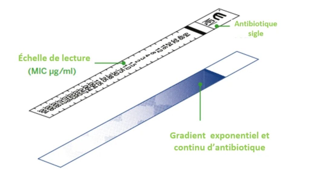
- Paper strips with predetermined antibiotic concentration gradients are known as minimum inhibitory concentration (MIC) strips (covering 15 double dilutions as for conventional MIC methods).
- The MIC Test Strip is a customised porous strip (International Patent) impregnated with an antibiotic concentration gradient through 15 two-fold dilutions of the standard M.I.C. method.
- The antimicrobial agent and its MIC (minimum inhibitory concentration) are both labelled on one side of the strip.
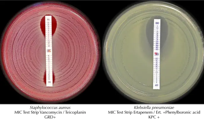
- The double-sided gradient incorporates the required diagnostic reagents for detecting ESBL, MBL, GRD, AmpC, and KPC.
- MIC Test Strips come in a wide variety of styles. We offer 10, 30, and 100 test runs of each setup.

Principle
- The preformed exponential gradient of antimicrobial agent is transferred instantly to the agar matrix upon application of the MIC Test Strip onto an inoculated agar surface.
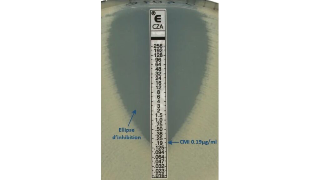
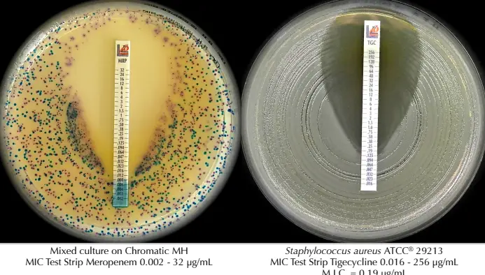
- A symmetrical inhibition ellipse, with its centre along the strip, forms after incubation for at least 18 hours.
- At the point where the edge of the inhibition ellipse meets the strip MIC Test Strip, the MIC is read straight from the scale in terms of g/mL.
- For resistance detection approaches, other growth/inhibition patterns may also be noticed.

Procedure
- To get a turbidity of 0.5 McFarland, lift well-isolated colonies from an agar plate (con fl uent or almost con fluent growth should be obtained after incubation).
- Dip a sterile swab into the inoculum suspension and squeeze it against the side of the test tube to get rid of any extra fluid.
- Before putting on the CMI strips, give the surface 15 to 20 minutes to soak up any extra moisture so that it is completely dry.
- Apply the strip to the agar surface with the scale facing up and the strip code facing the outside of the plate. Press the strip onto the agar surface with sterile forceps and make sure that the entire length of the antibiotic gradient is in full contact with the agar surface. Once the tape is on, you shouldn’t move it.
- If there are air bubbles under the strip, use sterile forceps to move them to the edge, being careful not to move the strip over the agar. Start with the least concentrated solution and work your way up to the most concentrated one.
- Incubate the agar plates at 35 2 °C while they are upside down.
- After 18 hours or more of incubation, a symmetrical inhibition ellipse forms in the middle of the strip. At the point where the edge of the inhibition ellipse meets the MIC test strip, the MIC is read directly from the g/mL scale.
Reading MIC strips
After incubation for at least 18 hours, a symmetrical inhibition ellipse forms in the centre of the strip. At the point when the edge of the inhibition ellipse meets the MIC test strip, the MIC is read straight from the g / mL scale.


References
- https://www.fishersci.com/us/en/browse/90222067/mic-test-strips?page=1#:~:text=Learn%20More-,MIC%20Test%20Strips%20are%20a%20quantitative%20assay%20for%20determining%20the,Ticarcillin%2DClavulanic%20Acid
- https://www.liofilchem.com/featured-products/mic-test-strip.html
- https://www.liofilchem.com/solutions/clinical/arm/mts-mic-test-strip
- http://www.liofilchem.net/en/pdf/mic_brochure.pdf
- https://www.launchdiagnostics.com/product/liofilchem-mic-test-strip-mts/
- https://www.rapidmicrobiology.com/products/mic-test-strip-2
- https://www.rapidmicrobiology.com/news/new-liofilchem-mic-test-strips
- https://bestbion.com/wp-content/uploads/2017/11/01-0001-LL-MIC-Test-Strip_small.pdf
- https://microbiologie-clinique.com/mic-test-strip.html