Taxonomy of Pests
The taxonomy of pests plays a crucial role in understanding and managing these organisms effectively. Taxonomy, as the science of classifying organisms, provides a systematic framework for organizing and categorizing pests based on their characteristics, relationships, and evolutionary history. By studying the taxonomy of pests, researchers and pest control professionals can gain valuable insights into their biology, behavior, and ecological interactions.
The process of taxonomy involves several key steps. One of the primary goals is the discovery and identification of new pest species. Through field surveys, observation, and collection of specimens, taxonomists can document and describe new pests, adding to the existing knowledge base. By carefully examining various characters, such as morphological features, molecular markers, and behavioral traits, taxonomists can differentiate and diagnose different pest taxa.
Formal description and naming of species are essential aspects of taxonomy. Each newly discovered pest species is given a scientific name that follows the rules of nomenclature. These scientific names are typically Latinized and consist of a genus and species epithet. The naming process ensures clarity and precision when referring to specific pest species, allowing for accurate communication among scientists and practitioners.
Taxonomy also involves placing pest species within a hierarchical classification system. This system, known as a taxonomic hierarchy, arranges organisms into various levels or ranks based on their evolutionary relationships. The hierarchy typically includes categories such as domain, kingdom, phylum, class, order, family, genus, and species. Each rank represents a distinct level of relatedness and helps organize and compare pests systematically.
By classifying pests into a hierarchical system, taxonomists provide a standardized and universal approach for discussing and studying these organisms. The classification system aids in information storage, retrieval, and communication, allowing scientists and researchers to share data about a pest’s morphology, genetics, distribution, hosts, ecology, and life cycles. This information is crucial for understanding pest biology, developing effective control strategies, and making informed decisions regarding pest management.
Moreover, taxonomy enables the identification of pests to a finer taxonomic level beyond the order. This finer-level identification can provide valuable insights into the specific traits, behaviors, and host preferences of individual pest species. It allows for more targeted and tailored pest management approaches, as different pest species may require different control methods or interventions.
In conclusion, the taxonomy of pests is a vital discipline that helps us understand and manage these organisms effectively. By classifying pests into hierarchical systems and providing accurate identification, taxonomy facilitates communication, research, and informed decision-making in pest control. Through the discovery, description, and classification of pest species, taxonomists contribute to our knowledge of these organisms and support efforts to mitigate their harmful effects.
Classification
Classification is a fundamental process in biology that involves organizing and categorizing organisms into groups based on their shared characteristics and evolutionary relationships. It provides a systematic framework for understanding and studying the diversity of life on Earth. The hierarchical nature of classification allows us to navigate the complex Tree of Life and place organisms in their appropriate taxonomic ranks.
At the core of classification is the concept of species. Species are the fundamental units of classification and are identified and named using binomial nomenclature. The scientific name of a species consists of two parts: the genus name and the species epithet. For example, the human bedbug is scientifically named Cimex lectularius, where Cimex represents the genus and lectularius indicates the species. The uniqueness of scientific names is crucial to avoid confusion, and in cases where duplication occurs, the younger name is replaced to maintain nomenclatural uniqueness.
Classification operates within a hierarchical system, with multiple categories above the species level. These categories indicate the placement of subordinate taxa within the broader classification scheme. Each taxonomic rank represents a specific level of relatedness among organisms. For instance, within the classification of the human bedbug, the hierarchy includes the phylum Arthropoda, subphylum Hexapoda, class Insecta, order Hemiptera, suborder Heteroptera, infraorder Cimicomorpha, superfamily Cimicoidea, family Cimicidae, genus Cimex, and species lectularius. Each rank builds upon the previous one, organizing organisms into progressively broader groups.
Establishing the correct placement of species within the hierarchy involves the use of systematics, a field of study within taxonomy. Systematics investigates the relationships among organisms by analyzing morphological characteristics, molecular data (such as DNA sequences), and other relevant information. Phylogenetics, a key aspect of systematics, determines the evolutionary history and relationships of organisms. It aims to identify monophyletic groups, which are natural taxa that include a common ancestor and all of its descendants. Artificial groups, on the other hand, do not conform to evolutionary history and can be polyphyletic or paraphyletic.
The results of systematics and phylogenetics are often represented as phylogenetic trees, which visually depict the genealogical relationships among the taxa under study. These tree-like diagrams aid in understanding the evolutionary history and relatedness of organisms, providing insights into their common ancestry and diversification over time.
In practical taxonomy, the important ranks typically considered are order, family, genus, and species, especially within the Class Insecta. These ranks help in organizing and classifying organisms effectively and are widely used in scientific research, communication, and practical applications.
In summary, classification is a systematic process that allows us to organize and categorize organisms based on shared characteristics and evolutionary relationships. It involves the identification and naming of species, placement within hierarchical taxonomic ranks, and the use of systematics and phylogenetics to determine evolutionary history. Classification provides a framework for understanding the vast diversity of life and facilitates communication and research in the field of biology.
PHYLUM Arthropoda
SUBPHYLUM Hexapoda
CLASS Insecta
ORDER Hemiptera
SUBORDER Heteroptera
INFRAORDER Cimicomorpha
SUPERFAMILY Cimicoidea
FAMILY Cimicidae
GENUS Cimex
SPECIES lectularius
Zoological nomenclature
Zoological nomenclature is the system of naming and classifying animal taxa, governed by a set of rules known as the International Code of Zoological Nomenclature (ICZN). The Code provides guidelines and regulations for the scientific naming of animal species and taxa at all levels of the taxonomic hierarchy. It ensures consistency, accuracy, and stability in the naming of animals in scientific literature and facilitates effective communication among researchers.
To become an officially recognized scientific name, a taxon must meet the requirements outlined in the Code. The Code specifies rules for the availability, formation, and usage of zoological names. It provides guidelines on the correct application of names, the priority of names, and the resolution of conflicts that may arise. By adhering to these rules, scientists ensure that names are unique, unambiguous, and valid within the context of zoological taxonomy.
In zoological nomenclature, there are specific conventions for the endings of names at different hierarchical levels above the genus level, known as family-group names. These conventions help users identify the level of taxonomic hierarchy being referred to. For example, superfamily names typically end with “-oidea,” such as Miroidea. Family names end with “-idae,” like Miridae. Subfamily names end with “-inae,” such as Mirinae. Tribe names end with “-ini,” like Mirini, and subtribe names end with “-ina,” such as Mirina.
In contrast, genus-group and species-group names do not follow standardized endings. These names are unique and have no prescribed suffixes. However, there are rules governing the formation of both genus-group and species-group names. Originally, these rules were based on Latin and Greek grammar, but the Code now allows for more flexibility in the formation of names.
The Code also addresses issues related to synonymy, homonymy, and the correction of incorrect names. It provides mechanisms for the recognition and designation of type specimens, which serve as reference specimens for each taxonomic name. These rules and regulations ensure stability and consistency in the usage of zoological names, avoiding confusion and facilitating accurate identification and classification of animal taxa.
Zoological nomenclature plays a critical role in scientific research, taxonomy, and communication within the field of zoology. By following the rules outlined in the Code, researchers can establish a standardized and internationally recognized naming system that contributes to the overall understanding and organization of animal biodiversity. The Code is regularly updated and revised to accommodate new discoveries, advancements in taxonomic knowledge, and changing perspectives in the field of zoology.
Classification of insects
The classification of insects is a fascinating field of study that helps us understand the incredible diversity and complexity of these small creatures. Insects belong to the Class Insecta, which is part of the subphylum Hexapoda within the Phylum Arthropoda. Arthropods, characterized by their bilaterally symmetrical bodies, rigid exoskeletons, and jointed limbs, encompass several subphyla, including Pycnogonida, Euchelicerata, Myriapoda, Crustacea, and Hexapoda.
Within Hexapoda, there are two distinct classes: the Entognatha and the Insecta. These classes are defined by specific characteristics. Hexapods have bodies divided into three distinct sections: the head, thorax, and abdomen. They possess one pair of antennae, three pairs of mouthparts, and three pairs of unbranched limbs known as uniramous limbs. Additionally, they respire through a tracheal system.
The Entognatha, a class within Hexapoda, have enclosed mouthparts. This class comprises three orders: springtails (Collembola), proturans (Protura), and diplurans (Diplura). Springtails are small arthropods that can be found in various habitats worldwide. Proturans are soil-dwelling organisms, and diplurans are primitive insects that also inhabit soil environments.
The Insecta, the larger class within Hexapoda, encompasses a diverse range of insects and is composed of 27-30 orders, depending on the classification system used. One notable example is the order Blattodea, which includes both cockroaches and termites. While some authors treat termites as a separate order (Isoptera), modern classification incorporates them within Blattodea. Papua New Guinea, a region rich in biodiversity, is home to 25 orders of insects. However, several orders, such as Raphidioptera, Mecoptera, Megaloptera, Grylloblattodea, and Mantophasmatodea, have not been reported in Papua New Guinea. The Zoraptera and Plecoptera are represented by a single species each in this region.
Studying the classification of insects provides us with a deeper understanding of their evolutionary relationships, ecological roles, and adaptations. This knowledge is crucial for various fields, including entomology, ecology, agriculture, and conservation. As scientists continue to explore and discover new species, the classification of insects will undoubtedly evolve, enriching our understanding of these remarkable creatures.
General Features of Insects
Insects, members of the class Insecta or hexapoda, are fascinating creatures with distinctive features that set them apart from other organisms. Here are some general features of insects:
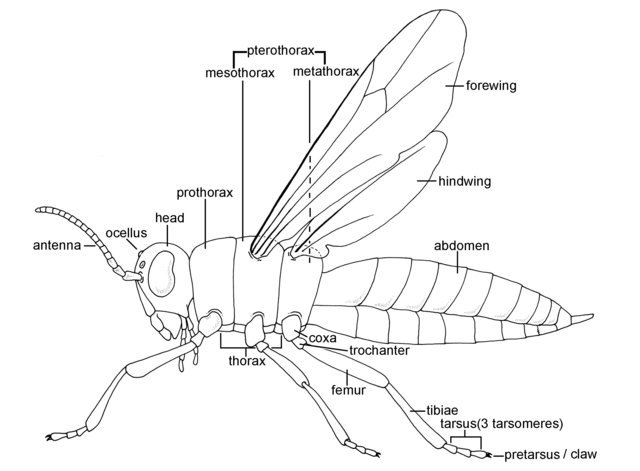
- Body Division: The body of insects is divided into three main parts: head, thorax, and abdomen. This characteristic segmentation is a defining feature of insects and is known as tagmosis.
- Exoskeleton: Insects possess an outer exoskeleton called the cuticle, which is primarily composed of a tough substance called chitin. This exoskeleton provides support and protection for the insect’s body.
- Jointed Legs: All insects have six legs, which are arranged in three pairs. These jointed legs enable insects to move and navigate their environment efficiently.
- Winged and Wingless Insects: Insects can be categorized into two main groups based on their wing structure. The group Pterygota consists of insects with wings, while the group Apterygota includes primitive wingless insects.
- Types of Development: Insect development can be classified into three main types: Ametabolous, Hemimetabolous, and Holometabolous. In Ametabolous development, insects undergo very little change during their development from larvae to adults. Hemimetabolous development involves insects that experience gradual changes in body form during their growth. Holometabolous development includes insects that undergo a complete metamorphosis, where they transform from wingless larvae to mature winged adults through an intermediate pupal stage.
- Nymphs and Larvae: During their life cycle, hemimetabolous insects progress through stages called nymphs, while holometabolous insects go through stages known as larvae. These immature stages show specific morphological characteristics that differ from their adult forms.
- Structure of Head, Thorax, and Abdomen: The head houses the insect’s sensory organs, such as antennae, compound eyes, and mouthparts, which are adapted to suit their feeding habits. The thorax is responsible for locomotion and carries the insect’s legs and wings (if present). The abdomen contains vital internal organs and plays a role in digestion, respiration, and reproduction.
In conclusion, insects exhibit remarkable diversity in their morphology and life cycle, making them a fascinating subject of study for entomologists and researchers. Understanding the general features of insects is essential for classifying and comprehending the intricacies of their biology and behavior.
Insect morphology
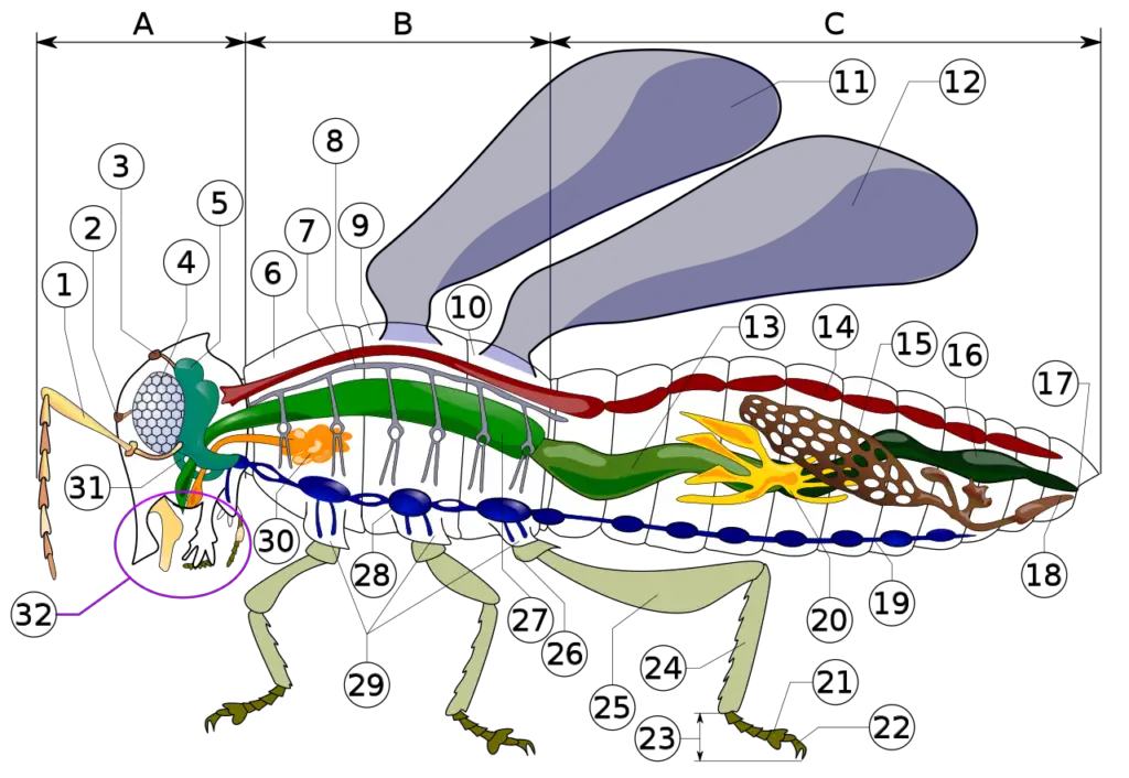
- antenna
- ocelli (lower)
- ocelli (upper)
- compound eye
- brain (cerebral ganglia)
- prothorax
- dorsal blood vessel
- tracheal tubes (trunk with spiracle)
- mesothorax
- metathorax
- forewing
- hindwing
- mid-gut (stomach)
- dorsal tube (heart)
- ovary
- hind-gut (intestine, rectum & anus)
- anus
- oviduct
- nerve cord (abdominal ganglia)
- Malpighian tubes
- tarsal pads
- claws
- tarsus
- tibia
- femur
- trochanter
- fore-gut (crop, gizzard)
- thoracic ganglion
- coxa
- salivary gland
- subesophageal ganglion
- mouthparts
Tagmosis
- Tagmosis is a process observed in insects that involves the grouping and combination of segments into functional units, resulting in the formation of three main body regions: head, thorax, and abdomen. In insects, each segment is identified by an area of sclerotization, which refers to the hardening and strengthening of the cuticle through the deposition of chitin. The segments are separated from each other by unsclerotized regions called intersegmental membranes.
- During tagmosis, the original 20 segments found in the embryonic stage are rearranged and divided into distinct tagmata. The head is composed of six or seven segments, the thorax contains three segments, and the abdomen consists of nine to eleven segments, along with a terminal tail-like structure called the telson.
- The body surface of insects has four primary regions: the dorsum (upper surface), the venteral (lower surface), and two lateral pleura, which separate the dorsum from the venter to which the bases of appendages are articulated if present. The sclerotization process gives rise to plates called sclerites on the body surface. Each segment typically has four sclerites: the tergum (dorsal plate), the sternum (ventral plate), and two pleura (one on each lateral side).
- The head region of insects houses essential sensory organs, including a pair of compound eyes, usually up to three ocelli (simple eyes), and a single pair of antennae, which are modified for various feeding habits. The thorax contains three pairs of legs and, in most adult insects, two pairs of wings. However, in some insects, one or both pairs of wings may be absent.
- The abdomen usually consists of 9 to 11 segments, along with a terminal pair of sensory cerci and external genitalia in certain insect species. The abdomen plays a crucial role in digestion, respiration, and reproduction in insects.
- Overall, tagmosis is a fundamental process that organizes the body of insects into distinct regions, enabling the specialization of structures and functions in different parts of the body. This segmentation and differentiation are essential for the diverse adaptations and behaviors observed in the insect world.
Body Wall (Integument)
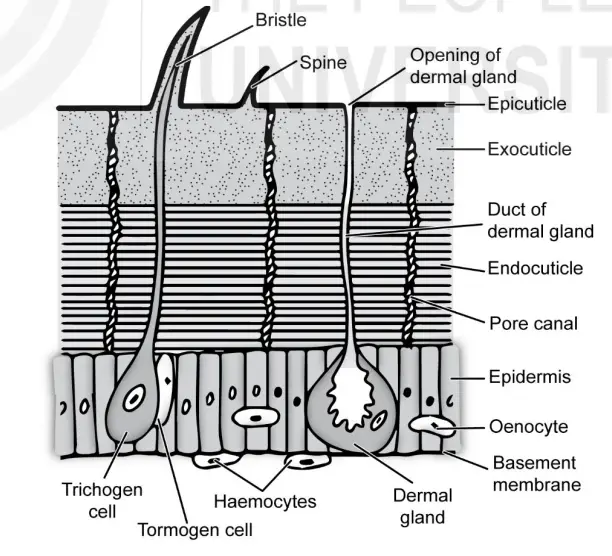
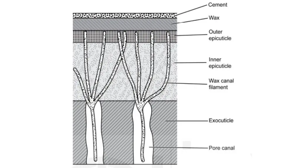
- The body wall, also known as the integument, is a vital structure that covers the entire body of insects. It is composed of two layers: the inner layer called the epidermis and the outer layer known as the cuticle. The cuticle serves multiple functions, including providing support, protection, and preventing water loss from the body surface. The integument is a complex structure with various specialized cells and glands that contribute to its functions.
- The cuticle consists of two main layers: the epicuticle and the procuticle. The epicuticle is a thin, waxy, and waterproof outer layer that acts as a barrier against external elements. Beneath the epicuticle lies the procuticle, which is thicker and chitinous. The procuticle is further divided into two layers: the exocuticle and the endocuticle. The exocuticle is rigid and sclerotized, providing strength and rigidity to the cuticle, while the endocuticle is tough and flexible, built from chitin and protein. The chitin is a long-chain polymer of N-acetylglucosamine, providing structural integrity to the cuticle.
- Insects have numerous ectodermal glands, which are groups of specialized cells in the epidermis that secrete various substances externally. Some common ectodermal glands include wax glands, lac glands, scent glands, poisonous glands, and mucous glands. These glands play essential roles in producing secretions that aid in waterproofing, protection, and communication.
- One unique feature of the integument is the process of moulting. Since the cuticle is hard and limits growth, insects must periodically shed their existing cuticle and replace it with a new, soft one that allows for expansion and growth. The shedding of the old cuticle is known as exuviae.
- The integument not only covers the external surface of the body but also lines various internal structures, including tracheal tubes, gland ducts, and parts of the digestive tract. It serves as a strong exoskeleton, providing muscular support to the insect’s body. The waterproofing properties of the cuticle are critical for the survival and success of insects in various habitats.
- Overall, the body wall, or integument, is a remarkable structure in insects, with its complex composition and functions. It is a key contributor to their survival, protection, and adaptation to diverse environmental conditions.
Body organisation
- The organization of the body in insects is a remarkable example of adaptation and specialization. It consists of distinct regions known as the head, thorax, and abdomen, each serving unique functions.
- The head, which appears as a single segment, is located at the anterior end of the insect’s body. It houses vital sensory organs such as the compound eyes, antennae, and mouthparts. The compound eyes provide insects with a wide field of vision, allowing them to detect movement and perceive their environment. The antennae serve as sensory organs, enabling insects to detect chemicals, vibrations, and other environmental cues. Additionally, the mouthparts vary greatly among different insect species and are adapted for various feeding habits such as biting, sucking, or siphoning.
- The thorax, positioned just behind the head, consists of three segments. These segments, known as the prothorax, mesothorax, and metathorax, are often highly modified to accommodate the insect’s legs and wings. Insects typically possess three pairs of legs, each attached to one of the thoracic segments. These legs allow insects to move, walk, jump, or swim, depending on their ecological niche and adaptations. The wings, when present, are usually attached to the mesothorax and metathorax. Insects exhibit a wide range of wing types, including membranous wings, elytra (hardened forewings), and modified structures like halteres (reduced wings used for balance in flies).
- The abdomen, located posterior to the thorax, is composed of eleven segments, although some segments may be lost or fused in certain species. The abdomen plays a crucial role in housing internal organs such as the digestive, reproductive, and excretory systems. It also contains specialized structures such as stingers or ovipositors, depending on the species. The abdominal segments often bear external structures like spiracles, which are openings that allow insects to respire. In some insects, the abdomen can be highly adapted for specific functions, such as sound production or defense mechanisms.
- To effectively describe and understand the morphology of an insect, it is important to be familiar with orientation descriptors. These descriptors aid in communication and facilitate the study of insect body structure. Orientation descriptors include terms like anterior (towards the head), posterior (towards the rear), dorsal (on the upper surface), ventral (on the lower surface), and lateral (on the sides). Mastering these descriptors is essential for accurate anatomical descriptions and effective scientific communication.
- The intricate body organization of insects reflects their incredible diversity and adaptability. It allows them to occupy a wide range of ecological niches and has contributed to their successful radiation and colonization of various habitats. Understanding the organization and morphology of insect bodies provides valuable insights into their biology, behavior, and evolutionary history.
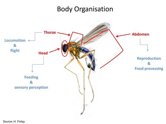
Head
The head of insects is a highly specialized structure consisting of six fused body segments that form a compact head capsule. The orientation and position of the head with respect to the rest of the body vary among different insect species. In beetles, the mouthparts are directed forward, a condition known as prognathous. In cockroaches and grasshoppers, the mouthparts point downward, a condition referred to as hypognathous. In bugs, there is an opisthorhynchous condition where the mouthparts are directed backward between the coxae of the fore-legs.
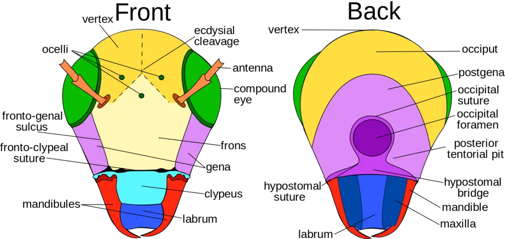
The head is enclosed within a sclerotized capsule that lacks outward appearance of segments but is marked by grooves called sutures or sulci. These sutures divide the head into distinct regions. Some of the areas of the head marked by these sutures have specific names. The posterior region, known as the occipit, is horse-shoe-shaped and joins the vertex dorsally and the genae laterally. The vertex contacts the frons anteriorly, and the clypeus lies anterior to the frons, often fused into a frontoclypeus.
The head of insects has several important features, including:
- Mouthparts: Insects have specialized mouthparts adapted for different feeding habits. These can include mandibles, maxillae, and labium, among others, each designed for specific functions such as cutting, chewing, piercing, or sucking.
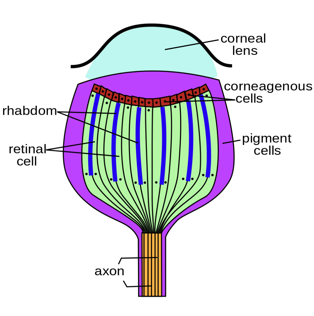
- Compound Eyes: Paired compound eyes are located dorsolaterally on the head, between the vertex and genae. Compound eyes are composed of numerous individual visual units called ommatidia, which allow insects to see a wide field of view and detect movement effectively.
- Antennae: Insects possess sensory antennae located above and anterior to the genae. These antennae play a crucial role in detecting environmental cues, such as chemical signals and vibrations.
- Ocelli: In addition to compound eyes, insects often have simple eyes called ocelli, located between the compound eyes. Ocelli are light-sensitive and provide information about the intensity and direction of light.
The head is essential for various functions, including feeding, sensing the environment, and detecting potential mates. Its distinct adaptations and structures allow insects to thrive in diverse habitats and fulfill their ecological roles within ecosystems.
1. Mouth parts
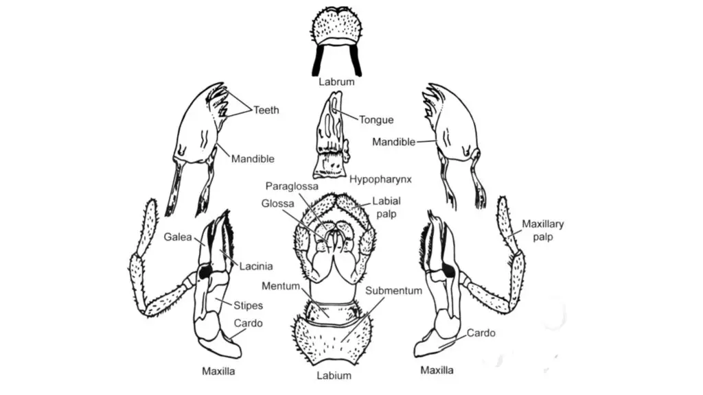
The mouthparts of insects are essential structures located on the head, which are formed by the fusion of six segments. These mouthparts are crucial for feeding and are usually the appendages of all head segments except the second one. The mouthparts consist of five basic components:
- Labrum: The labrum forms the upper lip and is part of the roof of the preoral cavity and mouth. It helps protect the mouth and aids in food manipulation.
- Hypopharynx: The hypopharynx is a tongue-like structure that projects anteriorly and forms the back of the preoral cavity.
- Mandibles: Insects typically have a pair of mandibles that are located at the sides of the mouth. The mandibles are strong and usually adapted for cutting and piercing. They are essential for biting and chewing food.
- Maxillae: The maxillae are found behind the mandibles and consist of two lobes-like structures, the inner lacinia, and the lateral galea. They also have five-segmented maxillary palps located laterally to the galea. The maxillae play a role in holding and macerating food. The maxillary palps contain chemoreceptors and mechanoreceptors that help in selecting suitable food before ingestion.
- Labium: The labium is formed by the fused appendages of the 6th segment with the sternum. It has a postmentum at the base, which is subdivided into submentum and mentum. At the distal end, the labium typically bears three-segmented palpi on each side. In some insects, the labium may have lobes known as ligula, consisting of outer lobes called paraglossae and inner lobes called glossae. The labial palps are sensory structures, similar to the maxillary palps.
The mouthparts of insects can vary in structure and function depending on the insect order and its specialized diet. Modifications in mouthparts are associated with the specific feeding habits of different insect species. For example, some insects have adapted mouthparts for piercing and sucking plant juices or blood, while others have developed mouthparts for chewing and grinding solid food. These adaptations play a vital role in the ecological niche and survival of insects in their respective environments.
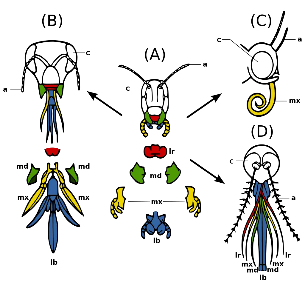
Legend: a – antennae
c – compound eye
lb – labium
lr – labrum
md – mandibles
mx – maxillae (Evolution_insect_mouthparts_coloured.png: Xavier Vázquezderivative work: ecelan, CC BY-SA 3.0, via Wikimedia Commons)
Mouth Part and its Modifications
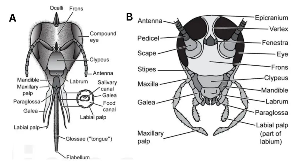
i) Chewing-lapping type
The “chewing-lapping type” of mouthparts is a specialized adaptation found in honey bees (Apis species). These mouthparts are modified to suit the bee’s feeding habits, which involve collecting nectar and pollen from flowers and molding wax.
In honey bees, the mouthparts undergo specific modifications:
- Maxillary Lacinia & Palps: These parts are rudimentary or reduced in size and not actively involved in feeding behavior.
- Paraglossae: These structures are present at the base of the glossae, which are elongated and united to form a tongue-like structure.
- Labrum: The labrum lies below the clypeus and functions as a chewing organ, primarily used in molding wax.
- Mandibles: The mandibles are short, spoon-like structures located on either side of the labrum. They play a role in manipulating and shaping wax during nest construction.
- Labium: The labium has reduced paraglossae, but the glossae are well-developed and elongated to form a tongue-like structure. At the tip of the glossae, there is a small structure called the labellum or honey spoon.
- Labial Palps: The labial palps are elongated and serve as sensory structures.
The mouthparts of honey bees exhibit a combination of chewing and lapping functions. The mandibles and labrum are of the chewing type and are used for manipulating wax. On the other hand, the labium and maxillae are of the lapping type.
The feeding process in honey bees involves the use of their modified mouthparts. The glossae and labial palps form a tube around the nectarines of flowers, and the glossae move up and down in the tube to collect nectar from the flowers. This process allows the honey bees to efficiently gather nectar and pollen from flowers, which are essential for their survival and the production of honey. The specialized chewing-lapping mouthparts in honey bees are a remarkable example of adaptation to a specific ecological niche in their environment.
ii) Biting and chewing type
The “biting and chewing type” of mouthparts are commonly found in insects like cockroaches. These mouthparts are adapted for biting and chewing solid food.
The mouthparts in insects with biting and chewing type include:
- Labrum: The labrum is a rectangular, flap-like structure located at the front of the mouth. It serves as an upper lip and helps in manipulating and guiding the food into the mouth.
- Mandibles: The mandibles are a pair of strong, toothed structures located on either side of the mouth. They are the primary chewing organs and are used to bite and break down solid food into smaller pieces.
- Maxillae: One pair of maxillae lies behind the mandibles. Each maxilla possesses a tactile maxillary palp, which acts as a sensory organ to assess the texture and suitability of the food.
- Labium: The labium is the lower lip and lies behind the maxillae. It helps in holding the food and pushing the masticated food into the mouth for further processing.
In insects with biting and chewing mouthparts, the feeding process involves biting off pieces of solid food with the mandibles and then further chewing and masticating the food using the other mouthparts. The mandibles play a crucial role in breaking down the food into smaller, more manageable pieces. The maxillary palps and labium assist in holding and manipulating the food during the feeding process.
For example, in cockroaches, the salivary channel opens at the base of the hypopharynx, which appears like a tongue. The hypopharynx aids in lubricating the food with saliva, making it easier to swallow.
The biting and chewing type of mouthparts are well-suited for consuming a wide range of solid foods, such as plant matter, other insects, and decaying organic material. These mouthparts allow insects to adapt to various ecological niches and food sources, making them successful and diverse in their feeding habits.
iii) Piercing and sucking type
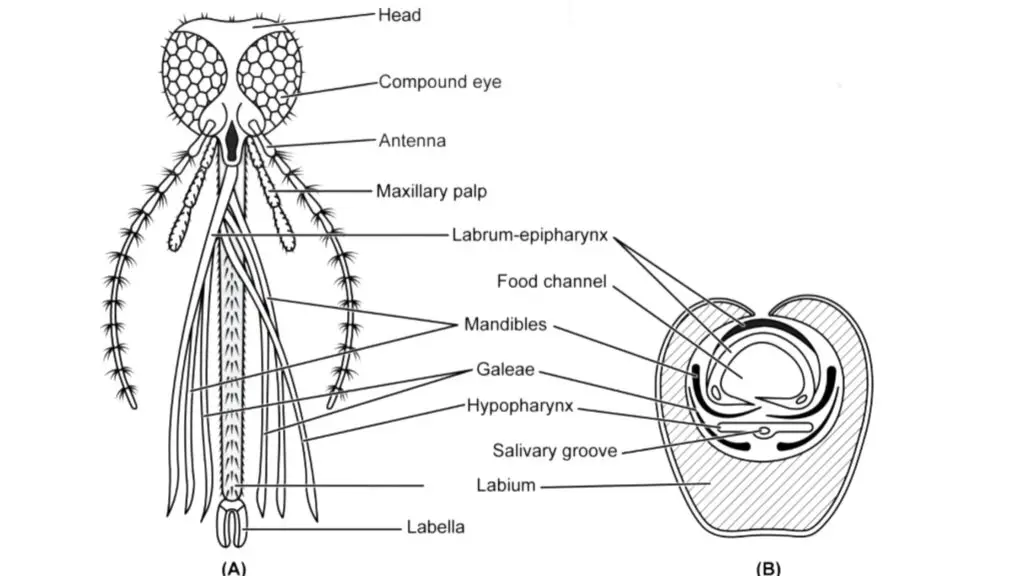
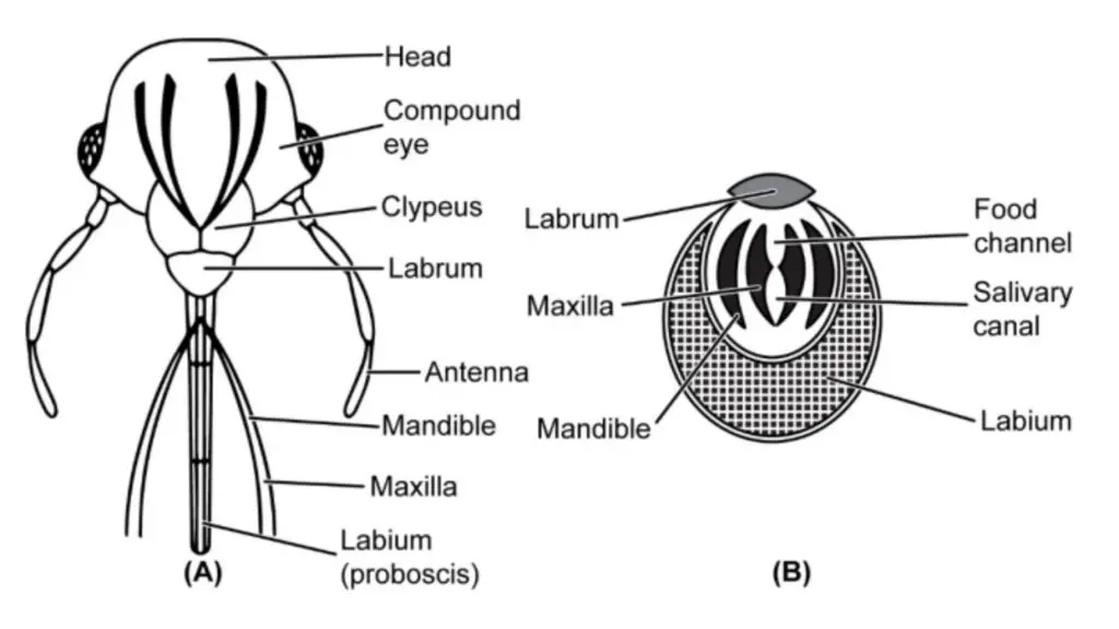
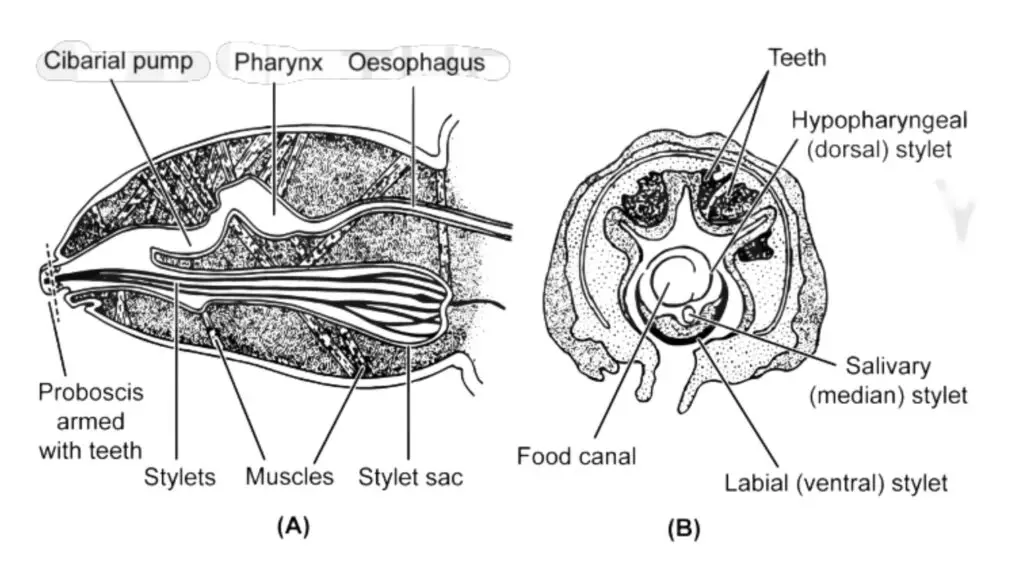
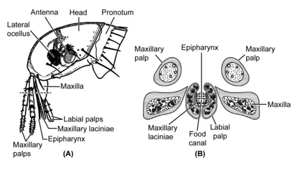
The “piercing and sucking type” of mouthparts are specialized adaptations found in various insects, such as mosquitoes, bed bugs, lice, and fleas. These mouthparts are designed for piercing the skin of hosts and sucking their blood or other fluids for feeding.
- Mosquitoes:
- The labium is modified to form an elongated, dorsally grooved, fleshy tube called a proboscis.
- Labial palps are transformed into labella at the tip of the proboscis.
- The labrum is needle-like and covers the labial groove dorsally.
- The mandibles, maxillae, and hypopharynx are elongated and placed in the labial groove.
- The stylets in the labial groove move apart to form a food channel through which the mosquito sucks blood.
- Female mosquitoes use their specialized mouthparts to pierce the host’s skin and inject saliva, which prevents blood coagulation.
- Bed Bugs:
- Bed bugs have a three-segmented labium.
- The mandibles and maxillae are modified into elongated stylets with blade and saw-like tips, respectively.
- The labrum covers the labial groove at the base.
- The maxillae are grooved to form upper food channels and lower salivary channels.
- Hypopharynx is absent in bed bugs.
- Lice:
- Lice have three stylets: one dorsal and two ventral, which lie in the ventral sac of the head.
- Mouthparts consist of a sucking snout-like projection called haustellum, with a circle of teeth on its inner surface.
- The stylets are protrusible and used to pierce the host’s skin for blood-feeding.
- Saliva is injected into the wound to prevent blood coagulation.
- The blood oozed on the surface is sucked in by the suctorial pharynx.
- Fleas:
- Fleas have a single stylet derived from the epipharynx and laciniae of the maxillae.
- The stylet forms two long cutting blades enclosed by the labial palps.
- Epipharynx and laciniae form the food channel through which the flea sucks blood.
In all these insects, the piercing and sucking type mouthparts are specialized for obtaining blood or other fluids from their hosts. These adaptations allow them to survive and thrive in their respective habitats and fulfill their nutritional needs. The ability to pierce the host’s skin and extract blood efficiently is crucial for their survival and reproduction.
iv) Sponging type (House fly)
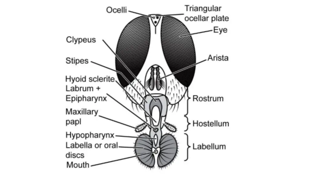
The mouthparts of house flies (Musca domestica) are adapted for sponging and sucking liquid or semi-liquid food. These mouthparts allow the house fly to feed on a wide variety of food sources, including decaying organic matter, plant juices, and other liquid substances.
The mouthparts of house flies consist of several structures:
- Labrum-Epipharynx: The labrum and epipharynx together form a structure that covers the oral groove dorsally. This helps in guiding the food into the mouth and prevents spillage.
- Maxillae: The maxillae are reduced in size and are present as maxillary palps. They lie on the rostrum, which is a conical-shaped structure articulated with the head.
- Haustellum: The haustellum is the middle part of the proboscis and has a mid-dorsal oral groove. It plays a crucial role in sucking up liquids and semi-liquids.
- Hypopharynx: The hypopharynx is a blade-like structure located deep inside the oral groove. It covers the labrum and epipharynx groove ventrally and has a salivary channel.
- Labium: The labium is modified and forms a long, fleshy, and retractile proboscis. The distal part of the proboscis consists of a pair of oval-shaped fleshy lobes called labella.
The proboscis is divided into three parts: the rostrum, haustellum, and labella. The rostrum is proximally articulated with the head and forms the base of the proboscis. The haustellum, with its oral groove and hypopharynx, is responsible for collecting and channeling the food into the mouth.
The distal part of the proboscis, the labella, contains fine channels called pseudotracheae, which open externally and allow the house fly to suck up fluids. These pseudotracheae meet in the mouth, lying between the two lobes of the labella, and lead into the food canal.
With their sponging and sucking mouthparts, house flies can easily feed on liquid or semi-liquid substances, making them successful scavengers and vectors for various diseases as they come into contact with decaying matter and potentially contaminated food sources.
v) Siphoning type
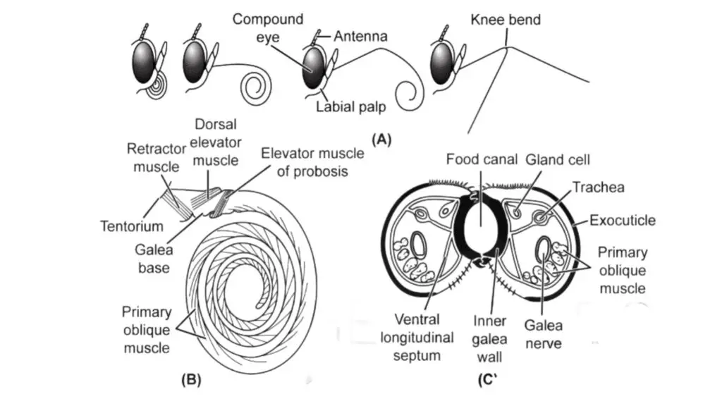
The siphoning type of mouthparts is found in butterflies and moths, and it is specialized for sucking flower nectar and fruit juices. These mouthparts allow these insects to feed on liquid substances and play a crucial role in pollination.
The mouthparts of butterflies and moths consist of the following structures:
- Labrum: The labrum is small and triangular in shape. It is articulated to the clypeus, which is a part of the insect’s face.
- Proboscis: The proboscis is the key component of the siphoning mouthparts. It is well-developed and elongated, formed from the modified galeae of the maxillae. Each galea has a lateral longitudinal groove. The galeae are opposed to each other and together form a food channel.
- Hypopharynx and Epipharynx: Unlike other mouthparts, the siphoning type lacks the hypopharynx and epipharynx.
During resting, the proboscis is coiled beneath the head of the butterfly or moth. However, during feeding, it becomes straight and is placed on the food source, such as a flower. The food is drawn up through the proboscis by capillary action, similar to how a liquid is drawn up through a thin tube.
The siphoning type mouthparts are well-adapted for sipping nectar from flowers. When a butterfly or moth lands on a flower, it uncoils its proboscis and inserts it into the flower’s nectar-producing structures. The grooves in the proboscis allow the insect to draw up the liquid nectar, which serves as a source of energy for the insect.
The siphoning type mouthparts are a remarkable adaptation that allows butterflies and moths to thrive on liquid food sources, making them important pollinators for many flowering plants. Their feeding behavior plays a crucial role in the pollination of various plant species, contributing to the diversity and sustainability of ecosystems.
2. Antenna
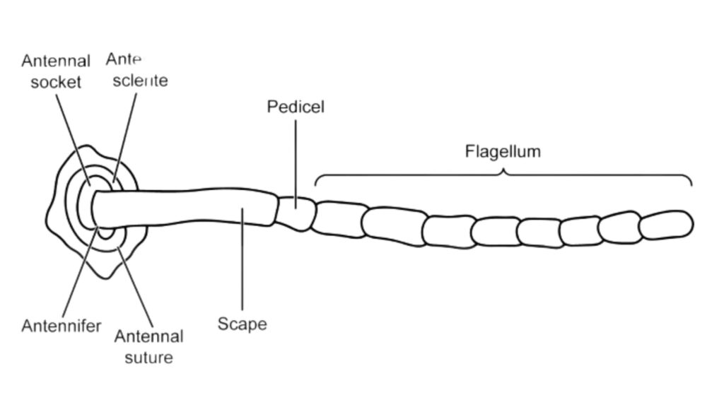
Antennae are sensory appendages found in all insects except in the order Protura. They are a crucial part of the insect’s sensory system and play a vital role in perceiving their environment. A typical antenna is composed of three main parts: the scape, the pedicel, and the flagellum.
- Scape: The scape is the basal segment of the antenna and is articulated with the insect’s head. It serves as the main connection between the antenna and the head.
- Pedicel: The pedicel is the second segment of the antenna, and it is usually single-segmented. It connects the scape to the flagellum.
- Flagellum: The flagellum is the elongated and segmented part of the antenna. It is divided into a series of similar annuli or units. The number of annuli can vary depending on the insect species.
The antenna is pivotally attached to the head capsule at a single point called the antennifer. This pivotal attachment allows the antenna to move and explore the surrounding environment.
There are two main types of antennae based on their musculature:
- Annulated Antenna: In Pterygota and Thysanura, the scape and pedicel are supplied with a pair of muscles that control the movement of the antennae. The flagellum, on the other hand, lacks muscles and is purely sensory. This type of antenna is called annulated.
- Segmented Antenna: In Diplura and Collembola, both the scape and pedicel have similar musculature as found in Pterygota insects. Additionally, each unit or annulus of the flagellum has intrinsic musculature, making each annulus a true segment. This type of antenna is called segmented.
Antennae are highly specialized sense organs. In annulated antennas, the pedicel houses Johnston’s organ, a chordotonal organ that responds to the movement of the flagellum. The flagellum of the antenna contains numerous sensory organs or sensilla, which function as chemoreceptors (detecting chemicals), mechanoreceptors (responding to mechanical stimuli), thermoreceptors (sensing temperature changes), and hygroreceptors (detecting humidity levels). These sensory structures allow insects to perceive various environmental cues, including the presence of food, mates, predators, and environmental conditions.
The diverse sensory capabilities of insect antennae are essential for their survival, navigation, and communication within their ecosystems. Antennae are a remarkable adaptation that enables insects to thrive and play their vital roles in the natural world.

Modifications in antennae
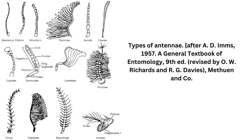
Antennae in insects show a remarkable diversity of forms, and they often undergo various modifications that are adapted to the specific needs and behaviors of different species. Here are some of the common modifications observed in insect antennae:
- Setaceous: Found in cockroaches, setaceous antennae gradually decrease in width from the base to the apex, giving them a tapering appearance.
- Filiform: Grasshoppers have filiform antennae, where all the annuli of the flagellum are of the same width, resulting in rod-like structures.
- Moniliform: Termites exhibit moniliform antennae, with ovoid or bead-like annuli on the flagellum.
- Pectinate: This type of antenna is seen in sawflies, beetles, and moths, where the annuli of the flagellum have a row of projections on one side, resembling a comb.
- Serrate: Found in the pulse beetle, serrate antennae have tooth-like projections on one side of the annuli.
- Clavate: Butterflies and moths have clavate (club-like) antennae, where the distal annuli of the flagellum gradually broaden and form a club-like structure.
- Capitate: The Khapra beetle has capitate antennae, where the end (distal annuli) of the flagellum is abruptly clubbed.
- Geniculate: Ants exhibit geniculate antennae, where the pedicel joins the flagellum in an elbow joint manner.
- Lamellate: Ber beetles have lamellate antennae, where the distal annuli are drawn out into leaf-like compact plates.
- Plumose: Female mosquitoes have plumose antennae, where the bases of the annuli of the flagellum have thick, large, and long whorls of hairs.
- Pilumose: Also found in female mosquitoes, pilumose antennae have short, thin, and few whorls of hair at the bases of the annuli of the flagellum.
- Flabellate: Stylops (male) exhibit flabellate antennae, where the basal annulus is modified into a fan-like structure.
- Bi-pectinate: Silk moths have bi-pectinate antennae, with long stiff projections on both sides of the annuli of the flagellum.
- Aristate: Houseflies have aristate antennae, with the flagellum being single-segmented and bearing a single stiff hair called an arista. The arista has hair on both lateral sides.
These various modifications in insect antennae reflect the incredible adaptability of insects to their ecological niches, allowing them to sense and respond to specific environmental cues and behaviors. The diversity of antennae plays a crucial role in the survival and success of insects in their respective habitats.
3. Visual system (Eyes)
Light is sensed by many sensory organs in insects, the most important of which are simple eyes or dorsal ocelli and compound eyes.
Ocelli (Simple eyes)
- Ocelli, commonly known as simple eyes, are a type of photoreceptive organs found in many adult insects and some nymphs. Typically, three ocelli are arranged in a triangle between the compound eyes on the head. Each ocellus consists of a transparent area of the cuticle, which acts as a lens. The epidermis beneath the lens is transparent and colorless. Beneath this transparent layer, nerve cells are grouped in two or more clusters. Within each cluster, cells form structures called rhabdomeres, and pigment may be present between these groups of cells. Nerve fibers extend from the cells, passing out at the back of the ocellus and running at least halfway down the ocellar cells.
- Unlike the compound eyes, the ocelli do not form images. Instead, they are specialized for perceiving changes in light intensity. They can detect variations in brightness and serve as light sensors, helping insects to respond to changes in their environment, particularly in relation to light and shadows. Ocelli are not designed for high-resolution vision or complex image formation like the compound eyes. Instead, they complement the compound eyes, providing additional information about light conditions.
- In some insect larvae, lateral ocelli, also known as stemmata, are present. These ocelli are considered purely visual organs and serve a similar purpose as the ocelli in adult insects. Lateral ocelli are usually found in the larvae of certain insect orders and play a role in their visual perception and responses.
- Overall, ocelli play a vital role in the sensory abilities of insects, contributing to their orientation and behavior in the environment. While they may not offer detailed vision like compound eyes, they are essential for detecting changes in light and aiding insects in their daily activities and survival.
Compound eyes
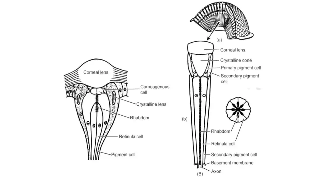
- Compound eyes are the specialized organs of vision found in insects and crustaceans. They are characterized by their unique structure, which consists of numerous repeating units called ommatidia. Each ommatidium functions as an independent photoreceptive unit, with its own optical system and light-sensitive photoreceptor cells.
- The number of ommatidia in a compound eye can vary widely, ranging from just a few to thousands, depending on the species. Ommatidia appear as tiny facets on the surface of the compound eye and are typically arranged in a hexagonal pattern. Each ommatidium has its own focusing system, a rigid transmitting system, a cluster of light-sensitive cells, and a screening pigment to distinguish one ommatidium from another. Additionally, each ommatidium has its own field of vision.
- The exposed, transparent, and colorless distal end of each ommatidium is known as the cornea or lens. The cornea plays a crucial role in the focusing system of the compound eye. It is rigid and fixed in shape, with a fixed focal length. The cornea is epidermal in origin and is produced by two cells known as cornea cells, which also form the primary pigment cells around the ommatidium.
- When light enters the compound eye, each ommatidium forms an image of a single point of light from an object in the insect’s visual field. All the individual points of light from all the ommatidia are then combined by the insect’s brain to form a composite image. This mosaic-like image consists of numerous spots of light, providing the insect with a wide field of view and the ability to detect motion.
- There are two main types of compound eyes: appositional and superpositional. In appositional compound eyes, each ommatidium has its own lens and receives light from a specific direction, making them well-suited for bright light conditions and providing high-resolution vision. In superpositional compound eyes, multiple ommatidia share a single lens, allowing for a larger field of view, but typically resulting in lower resolution. Superpositional eyes are better adapted for dim light conditions.
- Compound eyes are highly effective for vision and detecting motion, which are essential for the survival and behavior of insects and crustaceans in their environments. Their unique structure and capabilities contribute to the success of these organisms in various ecological niches.
Apposition eyes
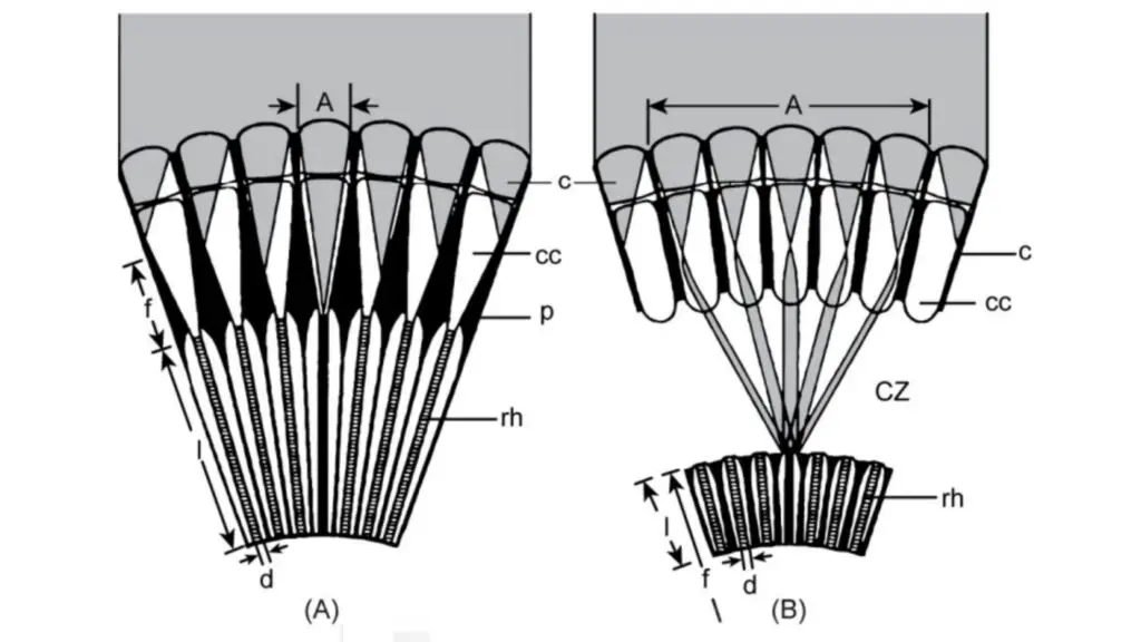
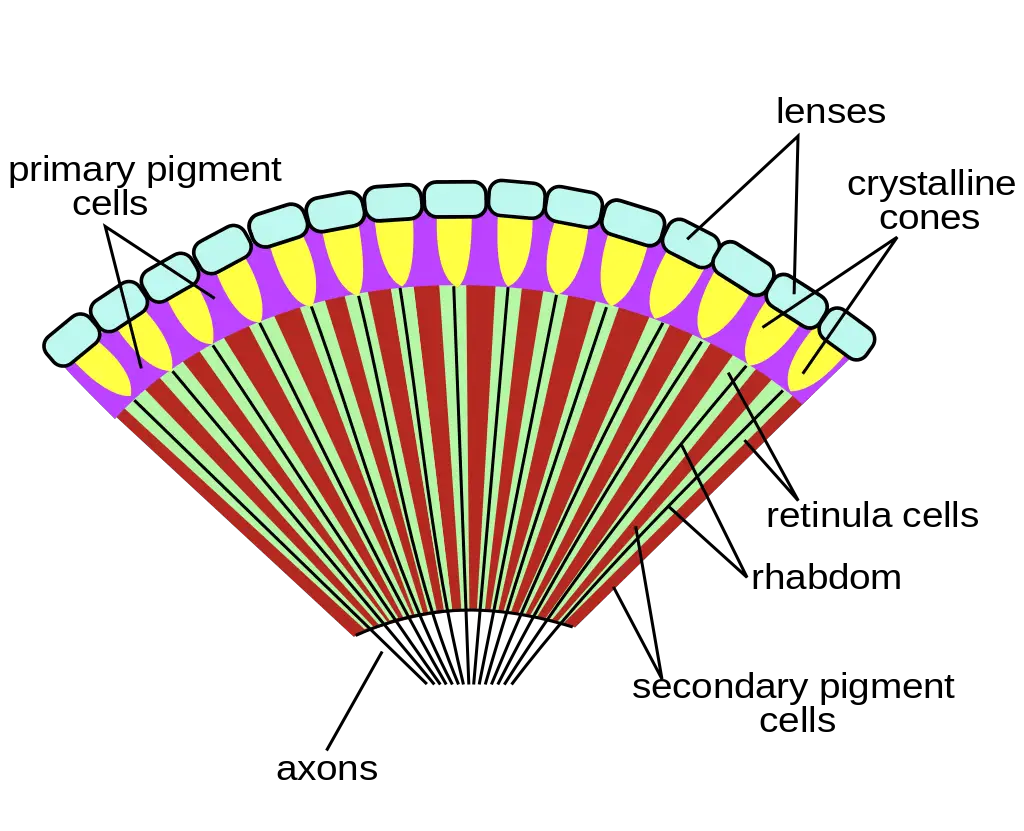
- Apposition eyes are a type of compound eyes found in some insects, such as honeybees and locusts. These eyes are well-suited for bright light conditions and provide high-resolution vision. Each ommatidium in the apposition eye functions independently, receiving light from a specific direction. This separation is achieved by maximizing the dispersion of screening pigments within the cells of each ommatidium. As a result, neighboring ommatidia are isolated from each other, and light entering the cornea stimulates only one ommatidium without affecting nearby ones.
- The apposition eyes work in a manner where each ommatidium is stimulated by a single point of light, sending one signal to the brain. This results in the brain registering the incoming signals as individual points in a mosaic image. The ability to discern and process each point of light separately allows these insects to have detailed and precise vision.
- The structure of an apposition eye includes a cornea, which is the transparent and colorless lens covering each ommatidium. Beneath the cornea are four cone cells forming the crystalline cone, which acts as a second lens in the focusing system. Immediately behind the lens, there are 6 to 8 elongated light-sensitive nerve cells known as retinular cells. These cells form the sensory complex of the ommatidium, with each retinular cell containing microvilli running along its length. The microvilli nearest the ommatidial axis form the rhabdomeres, which are specialized light-receiving structures. The rhabdomeres are packed with rhodopsin, a light-sensitive pigment.
- The axons from each corneal cell pass out through the basement membrane at the back of the eye and synapse with the optic ganglion. The optic ganglion receives information from all the ommatidia of the eye and then transmits it to the brain through the optic nerve. The brain processes the signals from the individual ommatidia to form a coherent and detailed visual image of the insect’s surroundings.
- Apposition eyes are highly effective for tasks requiring accurate vision, such as locating food sources, recognizing other individuals, and navigating complex environments. These specialized eyes contribute significantly to the survival and success of insects that possess them.
Superposition eyes
Superposition eyes are a type of compound eyes found in some nocturnal insects, such as moths, that are adapted to function in dim light conditions. These eyes work differently from apposition eyes, which are more suited for bright light conditions and provide high-resolution vision. In superposition eyes, the screening pigments are contracted in poor light, allowing light to pass obliquely through the eyes without being absorbed. As a result, the rhabdomes (light-receiving structures) in superposition eyes may receive light from multiple lenses, creating a superposition image.
The main differences between apposition eyes and superposition eyes are as follows:
- Light Reception: In apposition eyes, each ommatidium receives light entering its own cornea. In contrast, in superposition eyes, the rhabdomes receive light from several corneas, allowing the eyes to make more effective use of available light.
- Cone Stalk: Apposition eyes lack a cone stalk, while superposition eyes have a cone stalk present.
- Screening Pigments: In apposition eyes, screening pigments are well developed and form a curtain of pigment separating adjacent ommatidia. In superposition eyes, screening pigments are concentrated and do not form a curtain of pigment between adjacent ommatidia.
- Crystalline Cone: The length of the crystalline cone in apposition eyes is approximately equal to its focal length, while in superposition eyes, it is about twice its focal length.
- Connection Between Cone and Rhabdom: In apposition eyes, the lower end of the cone and the upper end of the rhabdom are contiguous. In superposition eyes, the cone and rhabdom are discontiguous and connected by a cone stalk.
- Length of Reticular Cells: Reticular cells in apposition eyes are quite long, extending from the crystalline cone to the basement membrane. In superposition eyes, reticular cells are short and confined to the bases of the ommatidia.
- Image Formation: In apposition eyes, a single point of light is associated with a single point in a mosaic image. In superposition eyes, the single point of light is not associated with a single point in a mosaic image, and a complete image of the visual field is not formed. Instead, a blurred image or no image is formed, but the brain is aware that there is light in the environment.
- Adaptation: Apposition eyes are adapted to bright light and are found in diurnal insects like honey bees. Superposition eyes are adapted to weak or dim light and are found in nocturnal insects like moths.
In summary, superposition eyes are specialized for low-light conditions, allowing nocturnal insects to be active and navigate in the dark. Their unique structure and function make them well-suited for survival and successful nocturnal activities.
Thorax
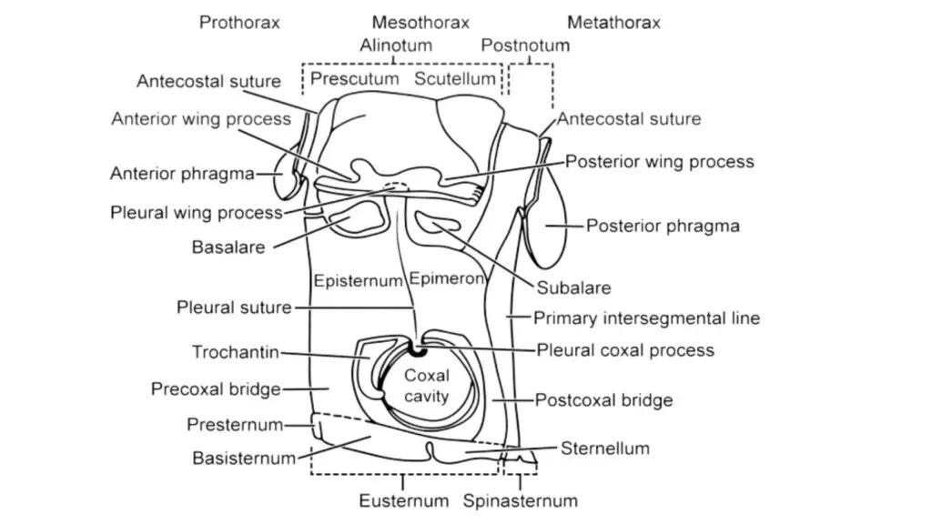
- The thorax is a critical body segment in insects, located between the head and the abdomen. It is composed of three segments: the prothorax (first segment), mesothorax (second segment), and metathorax (third segment). In winged insects, the mesothorax and metathorax are enlarged in comparison to the prothorax, collectively forming the pterothorax, which bears the wings.
- The pterothorax is the site of attachment for the wings, with each segment carrying one pair of wings. In total, insects have three pairs of legs, one pair on each thoracic segment. These legs serve primarily for locomotion, but they may also have other functions depending on the insect’s species and lifestyle.
- The second and third thoracic segments have spiracles, which are respiratory openings. These spiracles are found on the lateral sides of the thorax and allow air to enter the insect’s tracheal system.
- Each thoracic segment is divided into four distinct regions: the tergum (dorsal region or notum), the two lateral regions called pleura, and the ventral region called the sternum. In winged insects, the notum of each segment is divided into three sclerites: the prescutum, scutum, and scutellum. The pleuron is divided into two sclerites, the anterior episternum, and the posterior epimeron. The sternum is further divided into four sclerites: the prosternum, basisternum, sternellum, and spina-sternum.
- The pronotum, which is the notum of the prothorax, is generally smaller and simpler in structure compared to the other segments. However, in some insects like cockroaches, the pronotum is expanded and modified as a shield, covering the head anteriorly and the mesothorax posteriorly.
- The head is connected to the thorax by a membranous structure called the cervix or neck. The notum, pleura, and sternum of each thoracic segment have a variety of sclerites, which can vary greatly from one insect order to another.
- The thorax plays a crucial role in insect movement and locomotion. The powerful muscles attached to the thoracic segments allow insects to move their legs and wings, enabling them to fly, walk, jump, or perform other specialized movements essential for their survival and behavior.
1. Legs
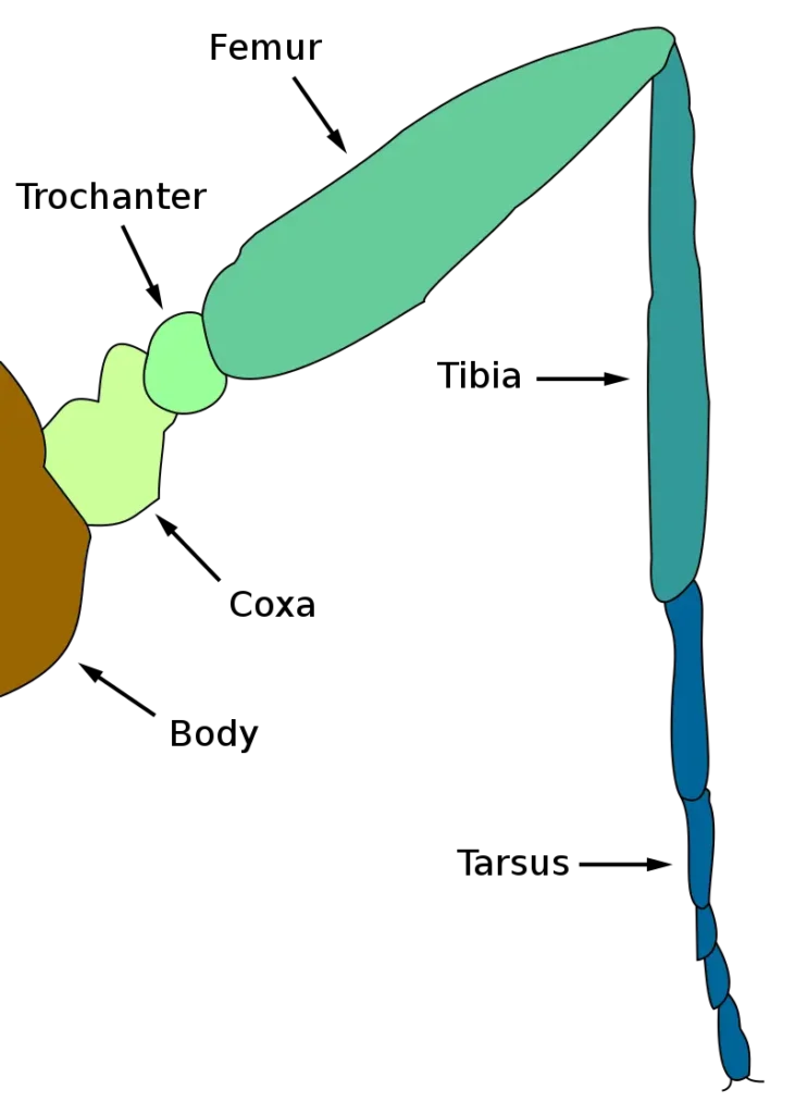
Insects are characterized by their hexapod nature, possessing three pairs of jointed legs that are articulated to the pleura of the thorax. Each leg is composed of six segments, arranged in sequence from the body: coxa, trochanter, femur, tibia, tarsus, and pretarsus. The tarsus, in particular, is usually comprised of 3 to 7 segments called tarsomeres.
- Coxa: The coxa is the proximal segment of the leg, and it articulates with the pleuron of the thorax. It is believed to be derived from an ancestral precoxa, though in some insects, it includes the sternite as well.
- Trochanter: The trochanter is the segment that has dicondylic articulation with the coxa and is rigidly jointed to the femur. In certain insects, such as Odonata (dragonflies and damselflies) and some hymenopterans (ants, bees, and wasps), there are two trochanters.
- Femur: The femur is the largest and stoutest segment of the leg in most insects. It is firmly attached to the trochanter.
- Tibia: The tibia has dicondylic articulation with the femur and is relatively slender compared to the femur. At the distal end of the tibia, there is often a spur or projection on the inner edge.
- Tarsus: The tarsus is a crucial segment, and the ancestral form was single-segmented. However, in modern insects, the tarsus is typically divided into five subsegments, known as tarsomers. The number and arrangement of tarsomers play an essential role in the identification of different insect species.
- Pre-tarsus: The distal segment of the leg is called the pretarsus. While some insects like Collembola, Protura, and certain larvae of Endopterygota have a single claw, most insects have a pair of claws and an unguis between them. Additionally, there is a pre-tarsus supporting plate known as unguitractor, located above the unguitractor is a forward lob-like extension from the distal end of the pretarsus called the arolium. In some insects, instead of the arolium, there is a median bristle present between the two lobes called the empodium. Both the arolium and pulvilli (lobes) act as adhesive organs, allowing insects to climb on smooth and steep surfaces. In certain insects, like house flies, pulvilli play an essential role in picking up pathogens from infected areas.
Modification in legs
Insects exhibit a variety of leg modifications based on their adaptation to different habitats and lifestyles. Some of the common modifications in insect legs are as follows:
- Ambulatory: Found in insects like black field crickets, the legs are long, cylindrical, and adapted for walking.
- Cursorial: Seen in cockroaches, these legs are cylindrical, long, and equipped with well-developed coxae, enabling the insects to run swiftly.
- Saltatorial: Found in grasshoppers, these legs have greatly enlarged hind femurs with strong muscles, facilitating powerful jumping movements.
- Scansorial (Clinging): Observed in lice, the tibia is stout and possesses a thumb-like process called a tibial thumb. The single-segmented tarsus bears a curved claw known as the tarsal claw, allowing the insect to cling to hair or fibers.
- Fossorial: Seen in mole crickets, these legs are adapted for digging into the ground, with flattened tibia and tarsus bearing blunt spines, resembling a shovel.
- Raptorial: Found in mantises, these legs are adapted for catching and holding prey between the grooved femur and sickle-shaped tibia of the forelegs.
- Web-Spinning: Present in Embioptera (Webspinners), the metatarsus of the leg is enlarged to accommodate silk glands, allowing the insect to spin webs.
- Natatorial: Occurring in certain aquatic bugs like Belostoma indica, the legs are broad and flattened with fringed dense hair, resembling oars, which are adapted for swimming.
- Scooping: Found in dragonflies and damselflies, the anterior legs are long, have rows of stiff bristles closely placed together, and are used to seize prey during flight.
- Bladder-Footed: Seen in thrips, the distal tarsomeres bear vesicles that help the insect cling firmly to the surfaces they feed on.
- Antennae Cleaning: Found in honey bee workers, the fore-legs have a semicircular bristle notch on the metatarsus’s inner edge and a spur on the tibia’s inner edge. This helps in cleaning pollen grains adhered to the antennae.
- Pollen Brushing: Also observed in honey bee workers, the middle legs have rows of bristles on their inner surfaces to brush pollen into a heap, and spurs are used to remove pollen and wax from the pollen basket and ventral surface of the abdomen, respectively.
- Pollen Collecting: Present in the hind legs of honey bee workers, the outer surface of the tibia has a concave cavity fringed marginally with hair called a pollen basket. This cavity is used to store pollen grains, aided by the rows of hair pollen combs present on the inner surface of the metatarsus.
These leg modifications enable insects to excel in various ecological niches, assisting them in their diverse behaviors, locomotion, and feeding habits.
2. Wings
- Wings are one of the most remarkable features of adult insects, providing them with the unique ability to fly. They are specialized lateral cuticular projections attached to the pterothorax, the middle segment of the thorax. Most adult insects possess two pairs of wings, making them the only invertebrates capable of sustained flight. The ability to fly has played a crucial role in their dispersal and colonization of various habitats.
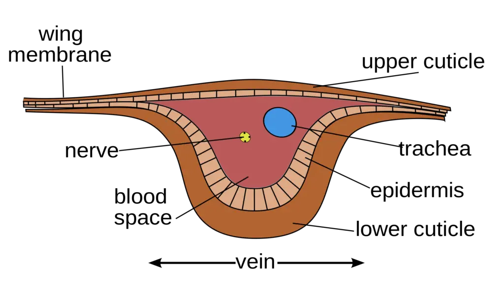
- The wings are composed of a thin, transparent membrane supported by a network of veins. The veins are formed by the separation of two layers of cuticle, and they play a crucial role in providing strength, rigidity, and shape to the wings. The veins also contain nerves and tracheae, allowing for sensory input and oxygen supply during flight.
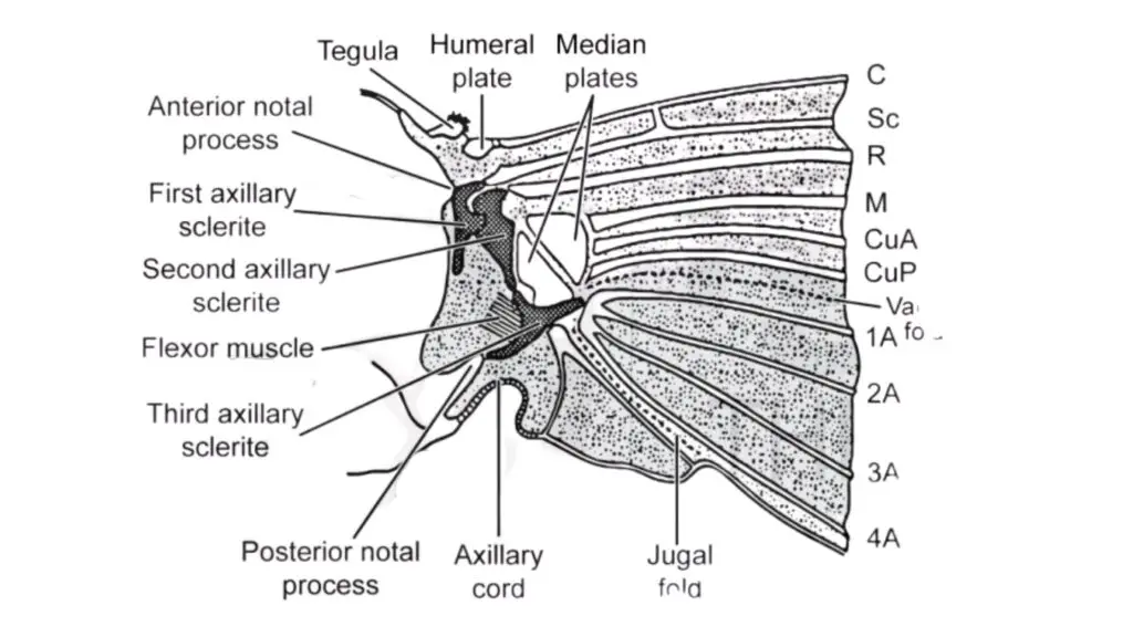
- At the base of each wing, there are three sclerites and two median plates that articulate with the thorax. These structures aid in the movement of the wings during flight and allow the wings to be folded back neatly over the body when at rest. The muscles responsible for wing movement are either directly inserted into the base of the wings or indirectly move the wings by distorting the thorax.
- The wings are well-vascularized, as they have a network of hollow veins through which hemolymph (the insect equivalent of blood) flows. The presence of hemolymph in the wings provides additional support during flight.
- In some insects, such as trichopterans (caddisflies), small hairs are present on the wings, providing additional stability and aerodynamic advantages. In contrast, lepidopterans (butterflies and moths) have scales covering their wings, which contribute to their brilliant colors and patterns.
- Wings allow insects to explore vast distances, locate mates, search for food, and escape from predators. Their ability to fly has made insects highly successful in colonizing various habitats and has played a critical role in their ecological significance. The diverse adaptations and characteristics of insect wings are a testament to the incredible complexity and efficiency of nature’s design.
Venation
Venation refers to the pattern of veins present on the wings of insects. It plays a crucial role in providing structural support, shape, and rigidity to the wings during flight. The venation of insect wings can vary significantly between different species and even within the same species, reflecting their adaptation to specific ecological niches and lifestyles.
In some insects, venation may be greatly reduced, as seen in some wasps, where only a few major veins are present. On the other hand, in certain insects, venation can increase through the branching of existing veins or the development of additional veins between the original ones. An example of increased venation can be observed in the wings of grasshoppers.
Dragonflies and damselflies are known for their unique venation pattern, which includes cross veins forming a reticulum-like structure on their wings.
The concept of the archedictyon is a hypothetical scheme proposed for the very first winged insect. It suggests the presence of 6-8 longitudinal veins in the wings of this primitive insect.
Veins on insect wings are named according to the Comstock Needham system, devised by Comstock and Needham, which includes the following major veins:
- Precosta (P): Located anterior to the costa.
- Costa (C): The anterior-most vein or leading marginal vein.
- Sub Costa (S): The second longitudinal and unbranched vein.
- Radius (R): The third longitudinal and branched vein, with one to five branches reaching the wing margin.
- Media (M): The fourth longitudinal and branched vein, with one to four branches reaching the wing margin.
- Cubitus: The fifth longitudinal and branched vein, with one to three branches reaching the wing margin.
- Anal veins: Unbranched veins located behind the cubitus.
- Jugal veins: The jugal lobe of the wings is often occupied by a network of irregular veins called jugal veins.
In modern insects, the precosta is usually fused with the costa, and the jugal vein is rarely visible due to evolutionary modifications.
The diverse venation patterns found in insects highlight the remarkable adaptability and versatility of these creatures in different environmental conditions and ecological roles. The variations in venation contribute to the incredible diversity of insect species and their ability to occupy various habitats and niches around the world.

Areas of the wings
The wings of insects are divided into specific areas based on the arrangement of veins, fold lines, and flexion lines. These areas play an essential role in the structure and function of the wings during flight. Here are the main areas of the wings:
- Remigium: The remigium is the anterior area of the wing where most of the veins concentrate. It is responsible for providing structural support and rigidity to the wing during flight.
- Clavus: The clavus is the area posterior to the remigium. It is characterized by a concentration of veins and is divided into two distinct regions:a. Anterior Anal Area (Vannus): This area is located in the anterior part of the clavus and is defined by specific veins and fold lines. It plays a role in wing stability and maneuverability during flight.b. Posterior Membranous Jugal Area: This area is situated in the posterior part of the clavus and is mainly composed of a membranous region with fewer veins. It contributes to the overall shape and flexibility of the wing.
- Fold Lines: Fold lines are specific areas along the wings where the wing can be folded. These fold lines enable insects to tuck their wings neatly and efficiently when at rest or not in use.
- Flexion Lines: Flexion lines are locations on the wings where the wing can bend during flight. These lines allow the wings to change shape and adjust their curvature during different phases of flight.
Two important lines on the wings are:
a. Claval Furrow: This is a flexion line on the wing, almost constant in position across different insect species. It contributes to the wing’s flexibility and bending ability.
b. Jugal Fold: The jugal fold is a fold line on the wing, also relatively constant in position across various insects. It allows the wings to be folded and unfolded efficiently.
- Median Flexion Line and Anal (Vanal) Fold Line: These lines form variable and less defined area boundaries on the wings. They are less constant in position across different insect species and may not be as well-defined as the claval furrow and jugal fold.
The division of wings into these specific areas and the arrangement of veins and lines play a crucial role in the insect’s ability to fly, control its flight, and maintain stability during various flight maneuvers. The diverse wing structures and areas contribute to the vast range of flight capabilities observed among different insect species.

Coupling mechanism
The coupling mechanism in insects refers to the way the forewings and hindwings are linked and work together during flight. The coupling mechanism can vary among different insect species, and it plays a crucial role in the efficiency and effectiveness of their flight. Here are some examples of coupling mechanisms in insects:
- Independent Wing Movement: In some insects, such as those in the order Orthoptera, the forewings and hindwings are not physically linked to each other. As a result, they beat completely independently of each other during flight. While this arrangement provides some degree of flight control, insects with two wings are generally more efficient flyers compared to those with four wings.
- Lobs, Spines, or Hooks: In certain insect species, there may be specialized structures like lobs, spines, or hooks at the base of the wings. These structures play an important role in the coupling of the wings, ensuring coordinated movement during flight.
- Hamulate Coupling: In honey bees, the coupling of the wings is achieved through a mechanism called “hamulate coupling.” This involves catching a fold along the posterior margin of the hindwing using hooks or hamuli present along the costal margin of the forewing. This coupling mechanism ensures that the wings move together, allowing the bee to achieve more precise control over its flight.
- Frenulum and Reticulum: In many Lepidopterans (butterflies and moths), the hindwings are firmly coupled to the forewings during flight. This coupling is achieved through a specialized structure called the frenulum, which is one or more hind wing bristles. These bristles engage with a catch or reticulum (a network of hooks or spines) present on the underside of the forewing. The coupling mechanism ensures that the wings work in unison, enabling the butterfly or moth to achieve stable and controlled flight.
The coupling mechanism in insects is a fascinating adaptation that allows them to achieve remarkable flight capabilities. Whether it’s independent wing movement for agile flight or precise coupling mechanisms for stable flight, each insect’s wing arrangement is uniquely suited to its specific flight requirements and ecological niche.
Frenate coupling
- Frenate coupling, also known as jugal coupling or amplexiform coupling, is a specific type of wing coupling mechanism found in certain insects. This coupling mechanism is observed in various groups, including mecopterans (scorpionflies), Lepidopterans (butterflies and moths), and trichopterans (caddisflies).
- In insects with frenate coupling, a strong structure called the jugal lobe is present beneath the costal margin of the hind wing. The jugal lobe acts as a catch or hook, engaging with a reticulum or specialized structure on the forewing to hold the wings together during flight. This coupling allows the forewing and hind wing to move in sync, ensuring coordinated and stable flight.
- In some insects, such as the citrus butterfly and silkmoth, frenate coupling is particularly prominent. During coupling, the posterior margin of the forewing and the anterior margin of the hind wing overlap broadly. This overlapping arrangement creates a secure and stable connection between the wings, enhancing the insect’s ability to fly efficiently and maneuver in the air.
- Frenate coupling is a remarkable adaptation that provides insects with precise control over their flight movements. By ensuring that the wings work together harmoniously, frenate coupling allows these insects to perform intricate aerial maneuvers, navigate through complex environments, and efficiently carry out tasks like foraging, mating, and migration.
- Overall, the presence of frenate coupling in certain insect groups showcases the diverse and specialized adaptations that have evolved to meet the unique flight requirements of different species. Each coupling mechanism, including frenate coupling, is a remarkable example of the fascinating adaptations that enable insects to conquer the skies and thrive in their respective habitats.
Modification in wings
Insects display various modifications in their wings that are adapted to meet specific ecological and functional needs. These modifications contribute to the diverse forms and abilities of winged insects. Some of the notable modifications in wings are as follows:
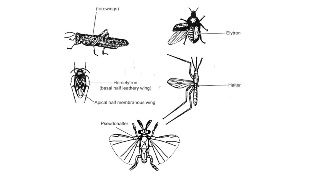
- Tegmina: In grasshoppers and cockroaches, the forewings are thickened and leathery, and they are called tegmina. Tegmina serve as protective covers for the hind wings and the body. They are partially or completely overlapped with each other when at rest.
- Elytra: Beetles have hardened forewings known as elytra. Elytra are often colorful and patterned and provide protection for the hind wings and abdomen. When not in use, elytra meet along the mid-dorsal line, covering the abdomen. They do not overlap with each other.
- Hemelytra: In certain insects like plant bugs, the basal part of the forewing is leathery (hemelytron), while the apical part is membranous. This combination of leathery and membranous regions allows for a flexible yet protective wing structure.
- Halters: In dipteran insects (flies), the hind wings are modified into small, knob-like structures called halters. Halters serve as gyroscopic organs, providing feedback on the insect’s position and movement in flight, helping it maintain balance and stability.
- Pseudohalters: Strepsipteran insects have modified forewings known as pseudohalters. Unlike true halters, pseudohalters do not act as balance organs and have a different function.
- Thrips Wings: Thrips have slender wings with reduced venation. They also have a fringe of long cilia along the wing edges, which aids in flight and may play a role in sensory perception.
- Wing Reduction: In certain insect groups, wings are either reduced in size (brachypterous) or absent altogether (apterous). This reduction may be a secondary adaptation to specific habitats or lifestyles.
- Wing Margins and Angles: In the study of insect wings, specific names are given to the margins and angles of the wings. The costal, anal, and apical margins refer to the anterior, posterior, and outer edges, respectively. The apical angle, anal angle, and humeral angle refer to the angles formed between these margins.
- Axillary Sclerites: The proximal region of the wing contains three axillary sclerites that articulate the wing to the thorax. These sclerites play a crucial role in allowing the wings to move freely during flight.
Overall, the diverse modifications in wings demonstrate the incredible adaptability and specialization of insects, enabling them to thrive in various habitats and ecological niches. These modifications have evolved over time to cater to the specific needs and challenges faced by different insect species, enhancing their survival and reproductive success.
Abdomen
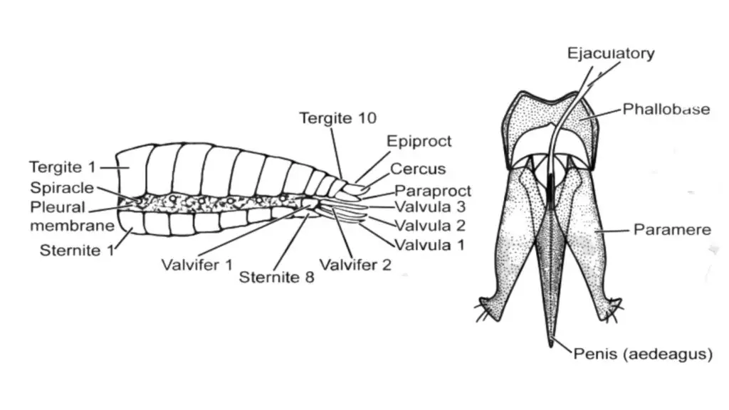
- The abdomen is the posterior region of the insect body and is typically composed of 6-11 segments, along with a post-segmental telson that bears the anus. The number of visible segments in the abdomen varies among different insect species. For example, in Acrididae (grasshoppers), all eleven segments are visible, while in Muscidae (house flies), only segments 2-5 are visible, and the remaining segments (6-9) are telescopic and hidden inside the body.
- Abdominal segments are marked off from each other and joined together by membranes, though some segments may be fused in certain insects. Each typical abdominal segment consists of a dorsal tergum, a lateral pleural region on each side, and a ventral sternum. The posterior part of each segment typically overlaps the anterior part of the next segment. The more anterior segments of the abdomen usually lack appendages, but some immature aquatic insects and apterygotes (wingless insects) possess abdominal appendages.
- Spiracles, which are openings for the respiratory system, are found in the pleural membrane of the more anterior segments of the abdomen. The anus is located at the posterior end of the abdomen and is surrounded by three protective sclerites: one dorsal epiproct and two lateral paraprocts. Just beneath the anus, the genital opening is situated, which is surrounded by specialized sclerites forming the external genitalia.
- In female insects, the appendages of the eighth and ninth abdominal segments collectively form an egg-laying structure called an ovipositor. This ovipositor consists of four valvifers and six valvulae, which facilitate the laying of eggs. In males, the genital opening is usually found within a tube-like structure called the aedeagus (penis), which is inserted into the female’s body during copulation.
- Apart from these typical abdominal structures, various other modifications and specialized appendages may be present in different insect species. Some examples include pincers, median caudal filaments, cornicles, abdominal prolegs, stings, abdominal gills, furcula, and more. These structures play diverse roles in the reproductive, defensive, and locomotory functions of insects. Overall, the abdomen, with its various adaptations and appendages, plays a crucial role in the survival and reproduction of insects across a wide range of habitats and lifestyles.
beetle (After Snodgrass 1957.)
Insect life stages and development
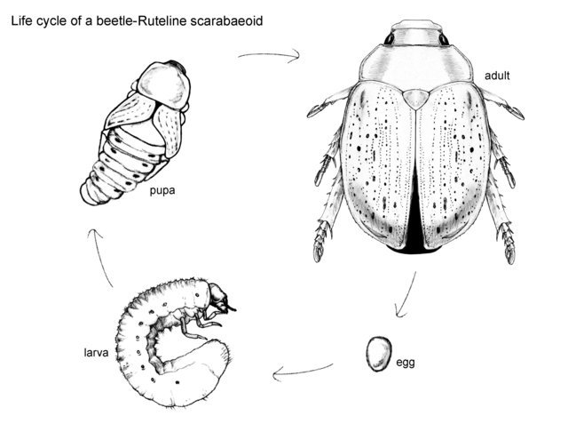
Insects display a diverse range of life stages and developmental patterns, categorized into three main types: ametabolous, hemimetabolous, and holometabolous development. The growth of insects is characterized by discontinuous periods of growth and moulting, with each growth phase known as an instar or stadium.
- Ametabolous development is observed in basal or “primitive” insect orders such as Archaeognatha (bristletails) and Zygentoma (silverfish). In these insects, growth is indeterminate, meaning individuals continue to moult throughout their lives, albeit with an increase in size upon reaching adulthood. Ametabolous larvae closely resemble adult forms but lack genitalia.
- Hemimetabolous development, also known as incomplete metamorphosis, involves a series of moulting stages and growth. Nymphs, the immature forms of these insects, closely resemble adults in morphology but lack wings and genitalia. The development of wings occurs externally, with nymphs possessing wing buds, earning them the name “exopterygotes.” Hemimetabolous orders include Ephemeroptera (mayflies), Odonata (dragonflies), Polyneoptera (Orthopteroid orders), and Paraneoptera (Hemipteroid orders).
- The majority of insects undergo holometabolous development, also known as complete metamorphosis. This developmental process involves a profound reorganization of the body, resulting in distinct body forms between larvae and adults of the same species. The dramatic transformation occurs during the pupal stage, where the wings develop internally, giving rise to the term “endopterygotes.” Holometabolous development is characteristic of major insect orders such as Coleoptera (beetles), Diptera (flies), Hymenoptera (bees and wasps), and Lepidoptera (butterflies and moths). Other orders exhibiting holometabolous development include Neuroptera (lacewings), Siphonaptera (fleas), Trichoptera (caddisflies), Raphidioptera (snakeflies), Megaloptera (dobsonflies), Mecoptera (scorpionflies), and Strepsiptera (strepsipterans).
These distinct life stages and developmental patterns contribute to the remarkable diversity and success of insects, with each type of development playing a crucial role in their respective ecological niches.
FAQ
What is the morphology of pests?
The morphology of pests refers to their physical characteristics and structures. It includes features such as body shape, size, coloration, appendages (e.g., antennae, wings, legs), mouthparts, and other distinctive traits.
How do pests’ morphology help in their identification?
By studying the morphology of pests, experts can identify and classify different pest species. Morphological characteristics serve as important diagnostic features, allowing pest control professionals to determine the specific pest species present and develop appropriate control strategies.
What are some common morphological characteristics of pests?
Common morphological characteristics of pests include body segmentation, presence or absence of wings, shape and arrangement of antennae, mouthpart adaptations for feeding, color patterns, and distinctive body structures like spines, hairs, or scales.
Are there differences in morphology among different pest species?
Yes, there are significant differences in morphology among different pest species. Each species has unique characteristics that can help distinguish them from others. Pest identification often relies on observing and comparing these morphological differences.
How can the morphology of pests help in pest management?
Understanding the morphology of pests is crucial for effective pest management. It allows professionals to identify pests accurately, assess their behavior and habits, and develop targeted control strategies. Morphological knowledge can also aid in identifying potential entry points and vulnerabilities in structures to prevent pest infestations.
Are there any specialized structures in pest morphology related to feeding or reproduction?
Yes, pests often have specialized structures related to feeding and reproduction. For example, some pests have piercing-sucking mouthparts adapted for blood-feeding, while others have chewing mouthparts for feeding on plant material. Reproductive structures can also be distinctive, such as the ovipositor in female insects for depositing eggs.
Can the morphology of pests provide information about their life cycle?
Yes, studying the morphology of pests can provide valuable insights into their life cycles. Changes in morphology during different life stages (e.g., egg, larvae, pupae, adult) can be observed and documented, helping in the identification and understanding of pest life cycles.
How is the knowledge of pest morphology used in integrated pest management (IPM)?
In integrated pest management, knowledge of pest morphology is used alongside other pest identification techniques. By accurately identifying pests through their morphology, IPM practitioners can choose the most appropriate combination of control methods, minimizing the use of pesticides and implementing sustainable pest management practices.
Can pest morphology be used to differentiate between similar-looking species?
Yes, detailed examination of pest morphology can often help differentiate between similar-looking species. Subtle differences in body shape, coloration, antennae structure, or wing patterns can be important in distinguishing closely related pest species that may have similar habits or cause similar damage.
How can one learn more about the morphology of pests?
To learn more about the morphology of pests, individuals can refer to entomology textbooks, online resources, and field guides specific to pests of interest. Additionally, seeking guidance from entomologists or pest control professionals can provide valuable insights and practical knowledge on pest morphology and identification.
- Text Highlighting: Select any text in the post content to highlight it
- Text Annotation: Select text and add comments with annotations
- Comment Management: Edit or delete your own comments
- Highlight Management: Remove your own highlights
How to use: Simply select any text in the post content above, and you'll see annotation options. Login here or create an account to get started.