What is Hyphae?
- Hyphae, the plural form of hypha, represent the intricate, filamentous structures predominantly observed in fungi and actinobacteria. These structures are pivotal for the vegetative growth of these organisms. Originating from the Ancient Greek term “ὑφή” (huphḗ), which translates to ‘web’, a hypha is characterized by its elongated, branching nature. Collectively, an aggregation of hyphae is termed as mycelium.
- Each individual hypha is composed of one or more cells, which are safeguarded by a robust cell wall primarily made up of chitin.
- As these structures grow, they extend both their internal cellular components and their external cell walls, predominantly from their tips. To further facilitate growth and division, septa, or internal partitions, may form within the hyphae.
- These septa not only demarcate individual cells within a hypha but also play a role in the organism’s overall growth dynamics. As the hypha continues its growth trajectory, it can either branch out from an existing tip or initiate the formation of a new tip from a pre-existing hypha, thereby diversifying its growth patterns.
- In essence, hyphae are fundamental to the growth and development of fungi and certain bacteria, serving as the primary mode of vegetative expansion and playing a crucial role in the life cycle of these organisms.
Definition of Hyphae
Hyphae are the elongated, branching filamentous structures of fungi and actinobacteria, collectively forming the mycelium, which is essential for their growth and development.
Hyphae Types
Fungal hyphae, the filamentous structures integral to the growth and development of fungi, exhibit a diverse range of types based on various criteria. Here, we delve into the classification of hyphae, elucidating each type with scientific precision:
- Classification Based on Cell Division:
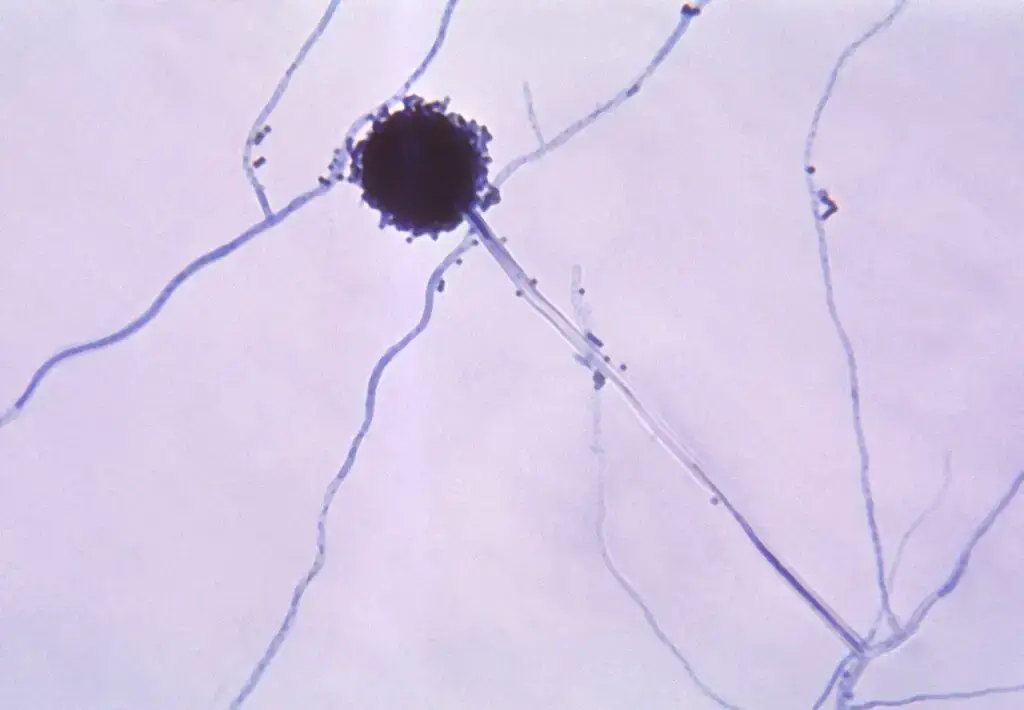
- Septate Hyphae: These hyphae possess septa, which are internal partitions that demarcate individual cells within the hyphal structure. Organisms like Aspergillus are typical examples. The septa, while compartmentalizing the cytoplasm, contain pores that facilitate the movement of materials and organelles between cells. Notably, septa can prevent the flow of nutrients when certain cells within the hyphae perish, courtesy of septal plugs known as Woronin bodies.
- Primary Septa: Formed during nuclear division, they segregate the newly formed nuclei from the parent nucleus.
- Adventitious Septa: These septa emerge independently of nuclear division and are prevalent when there are protoplasm concentration changes.
- Aseptate (or Coenocytic) Hyphae: Characterized by their absence of septa, these hyphae are continuous elongated cells. Typically shorter and more erect than their septate counterparts, they are less branched. Mucor and certain zygomycetes are examples of organisms with aseptate hyphae.
- Septate Hyphae: These hyphae possess septa, which are internal partitions that demarcate individual cells within the hyphal structure. Organisms like Aspergillus are typical examples. The septa, while compartmentalizing the cytoplasm, contain pores that facilitate the movement of materials and organelles between cells. Notably, septa can prevent the flow of nutrients when certain cells within the hyphae perish, courtesy of septal plugs known as Woronin bodies.
- Classification Based on Morphology:
- Plectenchyma: These are hyphae that appear bundled or fused together.
- Rhizomorph: Resembling branched cords, they play a role in nutrient acquisition.
- Appressorium: Certain fungi employ these specialized hyphal cells to infect plants.
- Classification Based on Cell Wall and Overall Form:
- Generative Hyphae: Recognized by their thin cell walls and a higher frequency of septa, these hyphae can develop reproductive structures.
- Binding Hyphae: These possess a thicker cell wall compared to generative hyphae and exhibit extensive branching.
- Skeletal Hyphae: Defined by their long, thick cell walls and fewer septa, they provide structural integrity to the fungal body.
- Classification Based on Refractive Appearance:
- Gloeoplerous (or Gloeohyphae): These hyphae exhibit an oily or granular appearance under microscopy due to their high refractive index. They can be either yellowish or clear (hyaline) and can be selectively stained using specific reagents.
- Classification Based on Growth Location:
- Vegetative Hyphae: These are primarily involved in nutrient absorption and growth.
- Aerial Hyphae: These hyphae extend above the substrate and are often involved in spore production.
Hyphae Structure
The structure of hyphae, the filamentous branches inherent to fungi, is intricate and tailored to support the growth and functionality of these organisms. Here, we elucidate the structural intricacies of hyphae with scientific precision:
- Cellular Composition: A hypha is essentially a tubular structure, each segment of which is composed of one or more cells. These cells are enveloped by a protective cell wall, which ensures the integrity and rigidity of the hyphal structure.
- Septa and Pores: Many fungi possess hyphae that are partitioned by internal cross-walls termed “septa.” These septa play a pivotal role in demarcating individual cells within the hyphal filament. Intriguingly, these septa are not impermeable barriers; they are perforated with pores sizable enough to allow the passage of cellular organelles such as ribosomes and mitochondria, and occasionally even nuclei, between adjacent cells. This facilitates cellular communication and resource sharing within the hyphal structure.
- Cell Wall Composition: Distinct from plants, which predominantly have cellulosic cell walls, the primary structural polymer in fungal cell walls is chitin. Chitin is a long-chain polysaccharide containing nitrogen, specifically N-acetylglucosamine. This chitinous cell wall offers a combination of strength and flexibility, enabling the hyphae to grow and navigate through various substrates.
- Hyphal Diameter: On average, hyphal structures exhibit a diameter ranging between 4 to 6 microns, underscoring their microscopic nature.
- Growth Dynamics: The genesis of a hypha begins from a spore or germ. The inaugural hyphal cell emerges and perpetuates its growth predominantly at the apex. While in aggregated forms, hyphae might be discernible to the naked eye, individual hyphae are microscopic tubular entities encapsulating a multinucleate cytoplasm, all safeguarded by a plasma membrane.
- Cell Wall Dynamics during Growth: As hyphae elongate, the cell wall at the hyphal tip undergoes lysis, facilitating the addition of new cells at the apex. Concurrently, the synthesis of a new cell wall occurs to shield these nascent cells, ensuring the continuous and directional growth of the hyphae.
Fungal Hyphae Cells
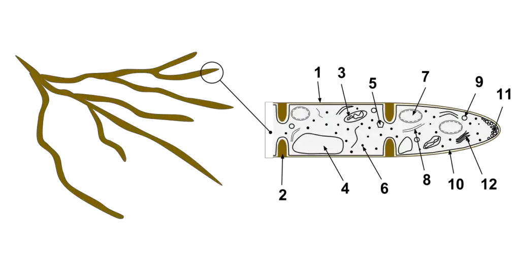
Fungal hyphae cells are the fundamental structural units of multicellular fungi, forming an intricate network known as the mycelium. These cells are elongated and tubular, facilitating the rapid growth and spread of the fungus in various substrates. A closer examination of the fungal hyphae cell reveals a complex organization of organelles and structures, each with a distinct function.

- Hyphal Wall: This is the outermost protective layer of the hyphal cell, primarily composed of chitin. It provides structural support and protects the cell from external threats.
- Septum: Found in septate hyphae, the septum is an internal cross-wall that divides the hypha into individual cells. It often contains pores, allowing for the exchange of organelles and nutrients between cells.
- Mitochondrion: The powerhouse of the cell, mitochondria are responsible for producing energy through the process of cellular respiration.
- Vacuole: A membrane-bound organelle, the vacuole is involved in waste disposal, storage of nutrients, and maintaining cellular turgor pressure.
- Ergosterol Crystal: Ergosterol is a sterol found in fungal cell membranes. It plays a role similar to cholesterol in animal cells, providing fluidity and integrity to the membrane.
- Ribosome: These are the sites of protein synthesis in the cell, translating the genetic code into functional proteins.
- Nucleus: The control center of the cell, the nucleus houses the genetic material (DNA) and coordinates cell growth, metabolism, and reproduction.
- Endoplasmic Reticulum (ER): An extensive network of membranes, the ER is involved in protein and lipid synthesis, as well as the detoxification of certain chemicals.
- Lipid Body: These are storage organelles for lipids, providing energy reserves for the cell.
- Plasma Membrane: This semi-permeable barrier surrounds the cell, regulating the entry and exit of substances. It plays a crucial role in maintaining the cell’s internal environment.
- Spitzenkörper: A unique organelle in fungi, the Spitzenkörper regulates tip growth in hyphae. It organizes and releases vesicles containing cell wall components to the growing tip.
- Golgi Apparatus: Often referred to as the cell’s “post office,” the Golgi apparatus modifies, sorts, and packages proteins and lipids for transport within or out of the cell.
Hyphae Production
Hyphae production in fungi is a meticulously orchestrated process, deeply intertwined with the organism’s life cycle and environmental factors. Here, we elucidate the genesis and development of hyphae with scientific precision:
- Initiation via Spores: The life cycle of fungi is initiated with the formation of spores, which are generated within specialized structures known as fruiting bodies. These spores serve as the primary reproductive units for fungi.
- Spore Dispersal and Germination: Upon maturation, spores are liberated and disseminated into the surrounding milieu, facilitated by various vectors such as wind or animals. The fate of these spores is contingent upon the environment they encounter. In the presence of optimal conditions, encompassing adequate nutrients, appropriate temperature, and requisite moisture, these spores commence the germination process, giving rise to the preliminary hyphal structures.
- Hyphal Growth and Mycelium Formation: The nascent hyphae, emerging from germinating spores, undergo elongation predominantly at their tips. This growth mechanism, termed “apical elongation,” ensures the continuous addition of new cells at the hyphal apex, facilitating its extension. As these hyphae proliferate and intertwine, they collectively form a network known as the mycelium, which represents the vegetative phase of the fungal organism.
- Cellular Segmentation in Hyphae: Not all hyphae are homogenous in their cellular structure. Some exhibit internal partitions, known as septa, which demarcate individual cells within the hyphal filament. Conversely, certain fungi produce aseptate or coenocytic hyphae, which lack these internal divisions, resulting in continuous, multinucleate structures.
Hyphae Growth
The growth of hyphae, the filamentous structures in fungi, is a meticulously orchestrated process that hinges on cellular mechanisms and specific organelles. Here, we delve into the intricacies of hyphal growth with scientific precision:
- Apical Growth: Hyphal elongation predominantly occurs at the tips. This tip growth is facilitated by the extension of cell walls through the external assembly and polymerization of cell wall components, coupled with the internal synthesis of new cell membranes.
- Role of the Spitzenkörper: Central to the mechanism of tip growth is an intracellular organelle known as the Spitzenkörper. This organelle is essentially an aggregation of membrane-bound vesicles laden with cell wall components. Originating from the endomembrane system of fungi, the Spitzenkörper receives vesicles from the Golgi apparatus. These vesicles, traversing the cytoskeleton, reach the cell membrane and release their contents via exocytosis. This process not only contributes to the expansion of the cell membrane but also facilitates the formation of the new cell wall. Intriguingly, the movement of the Spitzenkörper along the hyphal apex is directly proportional to the apical growth rate, underscoring its regulatory role in hyphal elongation.
- Organelles Involved in Hyphal Growth:
- Actin: A protein that plays a pivotal role in maintaining cell shape and facilitating movement.
- Microtubules: Cytoskeletal structures essential for intracellular transport and cell division.
- Vesicles: Membrane-bound sacs that transport materials within the cell.
- Golgi Apparatus: An organelle involved in the modification, packaging, and transport of proteins and lipids.
- Spitzenkörper: As previously mentioned, this organelle is instrumental in organizing and regulating hyphal growth.
- Cell Wall Dynamics: As the hypha elongates, vesicles move towards the hyphal tip, releasing enzymes and other compounds. These enzymes are pivotal for the lysis of the existing cell wall and the subsequent synthesis of a new one. Post lysis, cellular growth at the hyphal tip facilitates elongation. Concurrently, the newly formed cell is enveloped by a nascent cell wall, synthesized by the enzymes and compounds at the tip. This nascent wall is then fortified, ensuring the structural integrity of the elongating hypha.
- Cellular Segmentation: As the hypha continues its growth trajectory, septa may form behind the growing tip, partitioning the hypha into discrete cells. Moreover, hyphae can undergo branching either by bifurcating a growing tip or by sprouting a new tip from an existing hypha.
Hyphae Modifications
Hyphal structures in fungi are not monolithic but can undergo various modifications to adapt to specific ecological roles and environmental challenges. These modifications underscore the versatility and adaptability of fungi. Here, we elucidate the diverse modifications of hyphae with scientific precision:
- Haustoria: Certain parasitic fungi evolve specialized hyphal structures known as haustoria. These structures penetrate host cells, facilitating the absorption of nutrients from the host. Haustoria are pivotal for the survival and propagation of parasitic fungi as they enable them to extract sustenance from their hosts.
- Arbuscules: In a mutualistic relationship, mycorrhizal fungi form structures called arbuscules. Analogous to haustoria, these structures play a crucial role in nutrient exchange between the fungus and its plant host. By aiding in nutrient and water absorption, arbuscules enhance the overall health and growth of plants.
- Ectomycorrhizal Extramatrical Mycelium: This modification amplifies the soil area accessible to plant hosts. By channeling water and nutrients to ectomycorrhizas, specialized fungal organs situated on plant root tips, the extramatrical mycelium augments the nutrient uptake capacity of plants.
- Lichen Hyphae: Lichens, symbiotic associations between fungi and algae or cyanobacteria, exhibit hyphae that envelop the photosynthetic partner, termed gonidia. This envelopment forms a significant portion of the lichen’s structure, ensuring the stability and functionality of the symbiotic relationship.
- Nematode-Trapping Structures: Some fungi have evolved specialized hyphal modifications to trap nematodes. These modifications can manifest as constricting rings or adhesive nets, designed to ensnare and immobilize nematodes, which the fungus then consumes.
- Mycelial Cords: To transport nutrients across more considerable distances, fungi can form mycelial cords. These structures act as conduits, ensuring efficient nutrient distribution within the fungal network.
- Bulk Fungal Tissues: Structures like mushrooms and the tissues of lichens predominantly comprise interwoven and often interconnected hyphae. This felted arrangement provides structural integrity and facilitates the specific functions of these fungal tissues.
Hyphae Behavior
Hyphal behavior in fungi is a manifestation of their adaptive responses to environmental cues and their intrinsic growth mechanisms. Here, we delve into the intricacies of hyphal behavior with scientific precision:
- Tropism and Directional Growth: The trajectory of hyphal growth is not arbitrary but can be modulated by external environmental stimuli. One such influential factor is the application of an electric field, which can guide the direction of hyphal elongation. This phenomenon, where organisms grow in response to external cues, is termed tropism.
- Chemotropism: Beyond physical cues, hyphae exhibit a remarkable ability to sense reproductive units, even from considerable distances. In response, they exhibit directed growth towards these units. This behavior, where growth is directed by chemical gradients, is a form of chemotropism. It underscores the fungi’s ability to detect and respond to chemical signals in their environment, facilitating reproductive success.
- Penetrative Growth: Hyphae are not just surface dwellers. They possess the capability to navigate through permeable surfaces, weaving their way through the matrix to penetrate deeper layers. This behavior is crucial for fungi as it allows them to access nutrients buried within substrates, colonize new environments, and establish themselves in diverse ecological niches.
Hyphae Functions
Hyphae, the filamentous structures inherent to fungi and certain bacteria, are multifunctional and adapt to the specific needs of individual fungal species. Here are the primary functions of hyphae, elucidated with scientific precision:
- Nutrient Absorption from Host Organisms: Certain parasitic fungi possess specialized hyphae equipped with haustoria, which are unique tips designed to penetrate the cell walls of host organisms, be it plants or other species. This adaptation allows these fungi to efficiently extract nutrients from their hosts.
- Symbiotic Nutrient Absorption from Soil: A notable symbiotic relationship exists between mycorrhizal fungi and vascular plants. In this association, the fungi produce specialized hyphae known as arbuscules that infiltrate the roots of vascular plants. These arbuscules are adept at absorbing water and nutrients from the soil, thereby enhancing the nutrient uptake for the plant. Concurrently, the fungi benefit by obtaining sugars and other organic compounds from the plant, promoting their own growth.
- Predatory Structures for Nematode Capture: Evolution has led certain fungal species to develop hyphae that act as nematode-trapping structures. These specialized hyphae form intricate nets or ring-like configurations to ensnare nematodes, providing an additional nutrient source for the fungi.
- Nutrient Transportation Mechanisms: Some fungi, including those forming lichens and mushrooms, have developed mycelial chords, which are chord-like hyphal structures. These chords facilitate the transport of nutrients over considerable distances, ensuring the distribution of essential compounds throughout the fungal organism.
- Enzymatic Secretion for Nutrient Breakdown: Hyphae are instrumental in secreting enzymes and other biochemicals that aid in the decomposition of organic matter. This enzymatic breakdown simplifies complex molecules, making nutrient absorption more efficient for the fungi.
Examples of Hyphae
Hyphae are the thread-like structures that make up the body of multicellular fungi. They play a crucial role in nutrient absorption and growth. Here are some examples of fungi that possess hyphae, along with a brief description of their unique hyphal characteristics:

- Aspergillus: A common mold found in the environment. Its hyphae are septate, meaning they have cross-walls that divide the individual cells.
- Mucor: Often found in soil, animal feces, and decaying organic matter. Mucor has aseptate (or coenocytic) hyphae, meaning they lack cross-walls and appear as continuous tubes.
- Penicillium: A genus of fungi known for producing the antibiotic penicillin. Its hyphae are septate and branch extensively.
- Rhizopus: Commonly known as bread mold. Like Mucor, it has aseptate hyphae.
- Candida: While primarily known as a yeast, under certain conditions, Candida can produce pseudohyphae, which are elongated cells that resemble true hyphae.
- Mycorrhizal fungi: These fungi form symbiotic relationships with plants. Their hyphae, called arbuscules, penetrate plant roots to facilitate nutrient exchange.
- Trichophyton: A genus of fungi that can cause skin infections in humans and animals. Its hyphae can invade keratinized tissues, such as skin and nails.
- Armillaria: Known as honey fungus, it forms rhizomorphs, which are bundles of hyphae that look like black shoestrings and help the fungus colonize new areas.
- Claviceps purpurea: The fungus responsible for ergot disease in rye and other cereals. Its hyphae invade the plant tissues, leading to the production of toxic alkaloids.
- Ophiocordyceps unilateralis: This parasitic fungus infects ants, leading them to climb vegetation and attach there before they die. The fungus then produces long stalk-like hyphae that emerge from the back of the ant’s head to release spores.
These examples showcase the diversity of fungi and the various ways in which their hyphae adapt to different ecological roles and environments.
Quiz
What is the primary structural polymer in fungal cell walls?
a) Cellulose
b) Peptidoglycan
c) Chitin
d) Lignin
[expand title=”Show answer” swaptitle=”Hide answer”] c) Chitin [/expand]
Which of the following describes a long, branching, filamentous structure of a fungus?
a) Spore
b) Mycelium
c) Hypha
d) Haustoria
[expand title=”Show answer” swaptitle=”Hide answer”] c) Hypha [/expand]
Hyphae that are not partitioned by septa are termed:
a) Septate
b) Aseptate
c) Biseptate
d) Triseptate
[expand title=”Show answer” swaptitle=”Hide answer”] b) Aseptate [/expand]
Which specialized hyphal structure is formed by parasitic fungi for nutrient absorption within host cells?
a) Arbuscule
b) Mycorrhiza
c) Haustoria
d) Appressorium
[expand title=”Show answer” swaptitle=”Hide answer”] c) Haustoria [/expand]
The average diameter of hyphae is approximately:
a) 1-2 µm
b) 4-6 µm
c) 10-12 µm
d) 15-20 µm
[expand title=”Show answer” swaptitle=”Hide answer”] b) 4-6 µm [/expand]
The intracellular organelle associated with tip growth in fungi is called:
a) Golgi apparatus
b) Spitzenkörper
c) Endoplasmic reticulum
d) Lysosome
[expand title=”Show answer” swaptitle=”Hide answer”] b) Spitzenkörper [/expand]
Which type of fungi forms a mutualistic relationship with plants and assists in nutrient and water absorption?
a) Mycorrhizal fungi
b) Yeast
c) Mold
d) Rust fungi
[expand title=”Show answer” swaptitle=”Hide answer”] a) Mycorrhizal fungi [/expand]
In which type of hyphal growth do new cells form at the tip of the hyphae?
a) Lateral growth
b) Apical elongation
c) Radial expansion
d) Basal extension
[expand title=”Show answer” swaptitle=”Hide answer”] b) Apical elongation [/expand]
Which of the following is NOT a type of hyphal modification?
a) Haustoria
b) Arbuscules
c) Mycelial cords
d) Chloroplasts
[expand title=”Show answer” swaptitle=”Hide answer”] d) Chloroplasts [/expand]
The collective network of hyphae in fungi is termed:
a) Sporulation
b) Mycelium
c) Conidia
d) Fruiting body
[expand title=”Show answer” swaptitle=”Hide answer”] b) Mycelium [/expand]
FAQ
What are hyphae?
Hyphae are the long, branching, filamentous structures of fungi, which collectively form the mycelium.
How do hyphae grow?
Hyphae grow at their tips through a process known as apical elongation, where new cells are continually formed at the tip of the hyphae.
What is the primary component of the fungal cell wall in hyphae?
The primary structural polymer in fungal cell walls is chitin.
What is the difference between septate and aseptate hyphae?
Septate hyphae have internal cross-walls called “septa” that divide the hyphae into cells, while aseptate hyphae lack these divisions and appear as continuous tubes.
Why are hyphae important for fungi?
Hyphae are essential for nutrient absorption, growth, and reproduction in fungi. They also play a role in the formation of reproductive structures and fruiting bodies.
What are haustoria in the context of hyphae?
Haustoria are specialized hyphal structures formed by some parasitic fungi to penetrate host cells and absorb nutrients.
How do hyphae respond to environmental stimuli?
The direction of hyphal growth can be influenced by environmental stimuli, such as the presence of nutrients, light, or even the application of an electric field.
What is the role of the Spitzenkörper in hyphal growth?
The Spitzenkörper is an intracellular organelle associated with tip growth in fungi. It organizes and regulates the delivery of vesicles containing cell wall components to the growing tip.
Can hyphae be seen with the naked eye?
Individual hyphae are microscopic. However, when they form a dense network, as in the mycelium, they can be visible to the naked eye.
Do all fungi have hyphae?
While most fungi have hyphal structures, some, like yeasts, primarily exist as single cells and do not typically form extensive hyphal networks.
References
- Fricker et al. (2017). The Mycelium as a Network. Microbiol Spectr. 5(3): doi: 10.1128/microbiolspec.FUNK-0033-2017.
- Lew, R. (2011). How does a hypha grow? The biophysics of pressurized growth in fungi. Nat Rev Microbiol. 9(7): 509-18.
- Steinberg et al. (2017). Cell Biology of Hyphal Growth. Microbiol Spectr. 5(3): doi: 10.1128/microbiolspec.FUNK-0034-2016.
- Charles Drechsler. (1953) Botany: Morphological features of some fungi capturing and killing amoebae.
- Martin Tegelaar and Han A. B. Wosten (2017) Functional distinction of hyphal compartments. Scientific Reportsvolume 7, Article number: 6039 (2017).
- http://www.davidmoore.org.uk/Assets/fungi4schools/Reprints/Mycologist_articles/Isaac_answers/v13pp089-090fungal_septa.pdf
- http://eagri.org/eagri50/PATH171/lec03.pdf