A human cell is the basic unit that makes up your body, kind of like tiny rooms in a massive mansion working together to keep everything running. Picture it as a microscopic bubble with a flexible outer layer—the membrane—that decides what enters or exits. Inside, there’s a gooey mix called cytoplasm, packed with tiny machines that handle different jobs. The nucleus sits in the center, acting like a control room. It holds DNA, the blueprint that tells the cell how to build proteins, repair itself, or even when to split into two new cells.
Human cells aren’t one-size-fits-all. They adapt to their roles. Red blood cells, for example, ditch their nucleus to carry oxygen better, while nerve cells stretch like wires to send signals from your toe to your brain. Some cells, like skin cells, are disposable—they’re born, work, and flake off in weeks. Others, like heart cells, stick around for decades.
Here’s the kicker: every cell knows its job without being told. Muscle cells contract, immune cells hunt germs, and fat cells store energy—all guided by DNA. They even chat with each other using chemicals, coordinating everything from healing a papercut to growing hair. Trillions of these tiny workers silently keep you alive, proving that big things really do come in small packages.
What is Human Cell?
- Considered a eukaryotic cell as they include membrane-bound organelles, a human cell is the fundamental structural and functional unit of the human body.
- Comprising a phospholipid bilayer with attached proteins, the cell membrane controls molecular entrance and departure and preserves internal environment.
- Considered the control centre of the cell, the nucleus stores the genetic material (DNA) guiding cell functions like development, differentiation, and reproduction.
- By means of cellular respiration, mitochondria—the powerhouses of the cell—generate adenosine triphosphate (ATP), therefore providing the energy needed for cellular operations.
- Divided into rough (containing ribosomes) and smooth areas, the endoplasmic reticulum correspondingly is engaged in lipid metabolism and protein synthesis.
- Received from the endoplasmic reticulum for usage either inside or outside the cell, the Golgi apparatus further sorts, bundles proteins and lipids.
- Key in cellular cleansing and defence, lysosomes include hydrolytic enzymes that break down and recycle foreign particles and cellular trash.
- Filling the cell, the semi-fluid matrix known as the cytoplasm serves as the medium for organelles to be suspended from and where metabolic events take place.
- Human cells show specialisation, producing varied kinds including neurones, muscle cells, epithelial cells, blood cells, each suited to serve certain purposes vital for survival.
- Beginning with Robert Hooke’s 1665 observations of cork cells, historical breakthroughs in cell biology were further advanced by Anton van Leeuwenhoek’s discovery of microbes, therefore laying the basis for cell theory.
- Refined in the 19th century by scientists such Matthias Schleiden, Theodor Schwann, and Rudolf Virchow, cell theory holds that all living entities are made of cells and that all cells develop from pre-existing cells.
- Because it offers understanding of developmental biology, disease causes, and the generation of new treatment options including regenerative medicine, the study of human cells is fundamental to biomedical science.
- The Human Cell Atlas and other ongoing initiatives seek to map every kind of cell in the human body, therefore improving our knowledge of cellular variety and interactions—qualities essential for disease diagnosis and treatment.
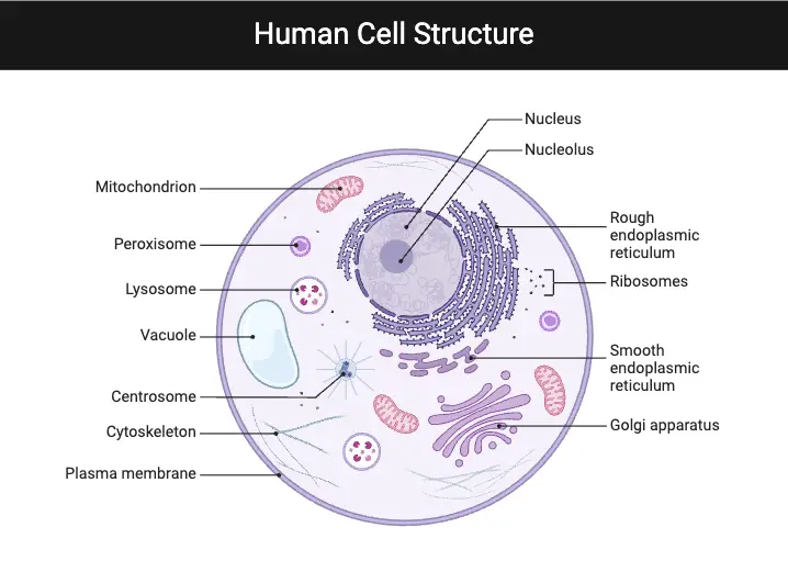
Human Cell Structure
- Human cells are eukaryotic, that is to say they have a distinct nucleus and many membrane-bound organelles allowing specialised operations.
- Comprising a phospholipid bilayer mixed with interspersed cholesterol and proteins, the cell membrane acts as a dynamic, semi-permeable barrier controlling molecular access and departure.
- Stored as chromatin and coordinates cellular activity through gene expression, the nucleus serves as the control centre for storing genetic material.
- Essential for the synthesis of ribosomal RNA and assembly of ribosomes, the nucleolus resides within the nucleus.
- Comprising all organelles and serving as the medium for metabolic processes, structural support, and intracellular movement, the cytoplasm is a gel-like matrix.
- Often referred to as the “powerhouses of the cell,” mitochondria create ATP by oxidative phosphorylation, therefore supporting several cellular activities.
- Two divisions define the endoplasmic reticulum:
- Processing and synthesising proteins depend critically on the rough endoplasmic reticulum, studded with ribosomes.
- Involved in lipid production, detoxification, and calcium storage is the smooth endoplasmic reticulum.
- Receiving proteins and lipids from the endoplasmic reticulum, the Golgi apparatus changes, sorts, and packs them for distribution to certain cellular sites.
- Important in cellular recycling and defence, lysosomes include hydrolytic enzymes that break down waste products, damaged organelles, and foreign compounds.
- Participating in the breakdown of fatty acids and the detoxification of reactive oxygen species, peroxisomes
- Comprising microfilaments, intermediate filaments, and microtubules, the cytoskeleton preserves cell structure, promotes intracellular mobility, and helps cells divide.
- Accurate distribution of genetic material depends on other structures like centrioles, which also help to arrange the mitotic spindle during cell division.
- The basis for tissue and organ development in multicellular animals, these cellular components cooperate to support activities like metabolism, growth, reaction to external stimuli, and reproduction.
1. Cell membrane
- Comprising a dynamic, semi-permeable barrier, the cell membrane encloses the cell and divides the internal from the exterior environments.
- Mostly made of a phospholipid bilayer, the hydrophilic (water-attracting) phosphate heads face outward and the hydrophobic (water-repelling) fatty acid tails face inside.
- Different proteins found in this lipid bilayer carry out necessary roles like signal reception, chemical transport, and cell-to—cell adhesion facilitation.
- By means of their interspersion in the membrane, cholesterol molecules help to maintain its fluidity and structural stability, therefore enabling the membrane to stay flexible under various situations.
- The cell membrane is described in the fluid mosaic model as a continually shifting, flexible structure where lipids and proteins can migrate laterally, therefore facilitating quick adaptation and interactions.
- Through processes including passive diffusion, assisted diffusion, and active transport, it controls the flow of molecules hence preserving cellular homeostasis.
- Receptor proteins that link to hormones, neurotransmitters, and other signalling molecules to start intracellular signalling paths also find a platform on the membrane.
- Apart from regulating signal transmission and material flow, the cell membrane participates in endocytosis and exocytosis, which enable the cell to absorb nutrients and waste materials.
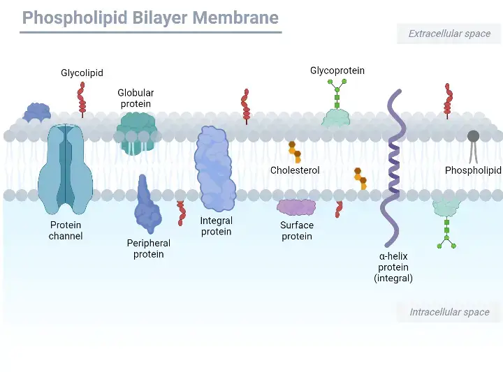
Cell membrane Structure
- Historically called the plasmalemma, the cell membrane—also referred to as the plasma membrane or cytoplasmic membrane—separates the inside of the cell from the external environment.
- It consists of a lipid bilayer wherein hydrophobic (water-repelling) tails face each other and phospholipids create two layers with hydrophilic (water-attracting) heads facing outward and inward.
- Various proteins found in this bilayer have roles related to transport, signal reception, and cell-to-cell adhesion, therefore augmenting the selective permeability of the membrane.
- Interspersed throughout the bilayer, cholesterol molecules assist to control the fluidity of the membrane and preserve its structural integrity under various situations.
- Key functions in cell identification and communication are performed by carbohydrates coupled to proteins and lipids on the extracellular surface of the membrane.
- Protecting the cell from its surroundings and regulating the flow of ions and organic molecules in and out of the cell and its organelles is the main purposes of the cell membrane.
- By means of its selective permeability, the membrane helps to preserve the internal balance and functionality of the cell by facilitating important activities like waste elimination, nutrient absorption, and the reception of external signals.
Cell membrane Functions
- The cell membrane acts as a physical barrier that maintains the cell’s structural integrity by mechanically enclosing its contents.
- It regulates the movement of ions and molecules into and out of the cell, ensuring that essential substances are selectively permitted to cross the membrane.
- Through passive diffusion and osmosis, small molecules such as oxygen (O₂) and carbon dioxide (CO₂) can move across the membrane along their concentration gradients.
- The membrane plays a critical role in mediating the exchange of materials with the surrounding environment, which is essential for maintaining cellular homeostasis.
- It is involved in bulk transport processes such as exocytosis, where the contents of secretory vesicles are expelled from the cell, and endocytosis, where external substances are internalized via vesicular uptake.
2. Cytoplasm
- Except for the nucleus, cytoplasm is the semi-fluid, gel-like material that permeates the cell and offers a stage for cellular activities.
- It mostly consists of cytosol, a water-based fluid including ions, enzymes, and organic molecules—along with cytoskeletal components and suspended organelles.
- Many metabolic processes, like glycolysis, which helps transform nutrients into energy, find their location in cytoplasm.
- Apart from preserving the form of the cell, the cytoskeleton inside the cytoplasm helps organelles and intracellular movement to be more easily moved.
- Through its transmission of signals from the cell membrane to interior components, it is fundamental in signal transduction.
- The cytoplasm helps to distribute organelles and cellular components to daughter cells during cell division hence maintaining appropriate cell operation.
- Maintaining homeostasis and sustaining the general metabolic activities of the cell depend on its dynamic and controlled surroundings.
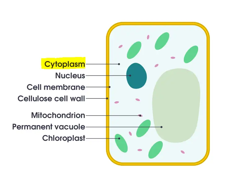
Cytoplasm Structure
- Comprising the cytosol, a water-based solution including ions, enzymes, and many tiny molecules, the cytoplasm is a complex, dynamic matrix filling the cell.
- Organelles with specialised cellular activities suspended within the cytosol include lysosomes, the endoplasmic reticulum, Golgi apparatus, and mitochondria.
- Integrated into the cytoplasm, the cytoskeleton—a network of microfilaments, intermediate filaments, and microtubules—offers structural support as well as helps intracellular transport and cell division.
- It is arranged in microdomains indicating a non-homogeneous and highly controlled environment by which particular biological events take place.
- The cytoplasm’s general architecture lets it function as a scaffold for preserving the cell’s shape and internal organisation as well as a medium for metabolic events.
Cytoplasm Functions
- Most of the metabolic activities in a cell occur in the gel-like matrix known as the cytoplasm, which supports essential biochemical pathways including glycolysis and protein synthesis.
- It hangs organelles in place such that structures such the Golgi apparatus, the endoplasmic reticulum, and mitochondria are suitably arranged for effective cellular activity.
- The cytoskeleton—a network of microfilaments, intermediate filaments, and microtubules—that preserves the cell’s form, ensures mechanical stability, and enables intracellular movement and transport is housed in the cytoplasm.
- It provides a means for the waste products, ions, and nutrients to be distributed and exchanged throughout the cell, therefore maintaining general cellular equilibrium.
- The cytoplasm is divided into daughter cells during cell division such that every new cell inherits the required organelles and molecular components for survival.
- Signal transduction also depends on the dynamic environment of the cytoplasm, which distributes signals from the cell membrane to interior structures to coordinate cellular activity.
3. Nucleus
- A membrane-bound organelle, the nucleus controls eukaryotic cells from their inside.
- It arranges the genetic material of the cell—mostly deoxyribonucleic acid (DNA)—in a chromatin-like fashion.
- Regulating cellular functions like growth, division, and differentiation, the nucleus controls gene expression.
- The nuclear envelope, a bilayer membrane, encloses it and has nuclear pores to control molecule movement between the nucleus and the cytoplasm.
- The nucleolus is a well-known area inside the nucleus in charge of synthesis of ribosomal RNA (rRNA) and ribosome assembly.
- Because it guarantees proper replication and distribution of genetic information during cell division, the nucleus is very important for cell reproduction.
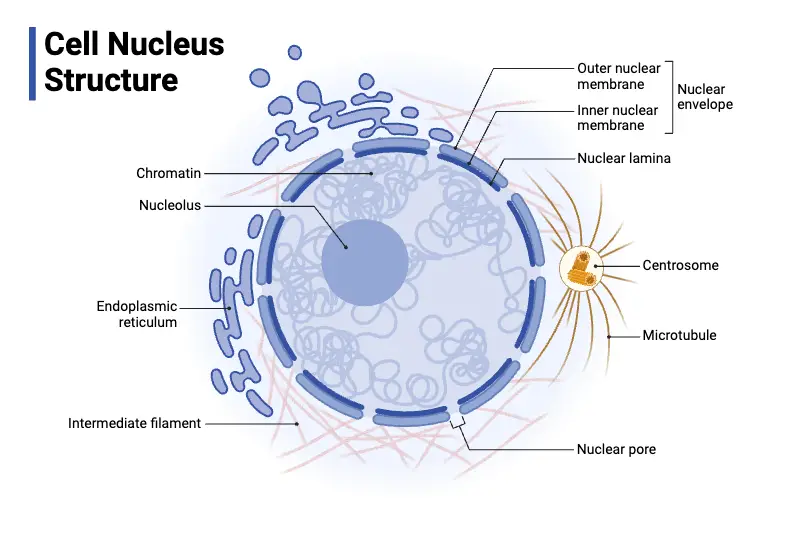
Structure of Nucleus
- The big, membrane-bound organelle known as the nucleus stores and regulates the genetic material of a cell.
- It is surrounded by a double lipid bilayer known as the nuclear envelope, which divides an outer from an inner membrane across a perinucleational gap.
- Nuclear pores, sophisticated protein structures controlling the bidirectional flow of molecules between the nucleus and the cytoplasm, are found within the nuclear envelope.
- The nuclear lamina, a supporting meshwork of intermediate filament proteins (lamins) that helps preserve nuclear shape and organise chromatin, lines the inside surface of the nuclear envelope.
- Inside the nucleus, the nucleoplasm forms a gel-like matrix for chromatin and other nuclear elements.
- Two kinds of chromatin, euchromatin, which is less compacted and transcriptionally active, and heterochromatin, which is more condensed and generally inactive. Chromatin is the complex of DNA wrapped around histone proteins
- Prominent substructure inside the nucleus, the nucleolus is devoted to the synthesis of ribosomal RNA (rRNA) and the assembly of ribosomal subunits—necessary for the synthesis of proteins.
- Other nuclear components guarantee exact control over gene expression and cell cycle development by include many regulating proteins and elements involved in DNA replication, transcription, and repair.
Functions of Nucleus
- Through regulation of gene expression and direction of all cellular activity, the nucleus serves as the control centre for the cell.
- Between the nucleus and the cytoplasm, many nuclear pores in the nuclear envelope enable chemicals—including regulatory factors, energy molecules, DNA, and RNA—to be selectively exchanged.
- Essential for protein synthesis, the nucleolus—a separate substructure inside the nucleus—is devoted to ribosome generation by synthesis of ribosomal RNA and assembly of ribosomal subunits.
- Comprising chromatin, long, thin strands of DNA complexed with proteins, chromosomes contain the genetic material encoding the directions for all cellular activities.
- Messenger RNA is generated in the nucleus, the main site of transcription, to transport genetic information for protein synthesis.
- To guarantee correct replication and segregation of genetic material, the chromatin condenses into clearly defined chromosomes during cell division.
- Apart from genetic storage, the nucleus contains many proteins and RNA molecules that help to regulate and sustain cellular activities by means of regulated molecular transport.
4. Mitochondrion
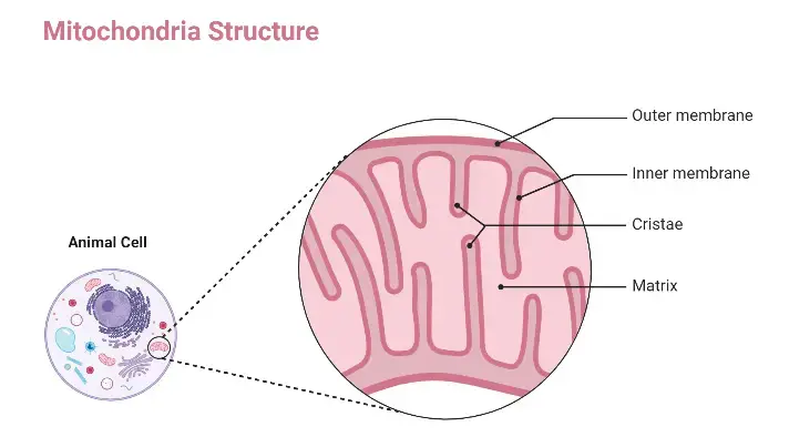
- Mitochondria are double membrane-bound organelles regarded as the cell’s powerhouse because they produce the majority of the adenosine triphosphate (ATP) through oxidative phosphorylation.
- They include an exterior membrane enclosing the organelle and an inner membrane folding into cristae, hence augmenting the surface area available for energy-generating processes.
- Enzymes necessary for the citric acid cycle are found in the mitochondrial matrix, the area within the inner membrane; moreover, mitochondrial DNA (mtDNA) and ribosomes help mitochondria to synthesis some of their own proteins.
- Not just in ATP generation but also in control of cellular metabolism, calcium homeostasis, and the natural course of death (programmed cell death).
- Supported by their own genome and features reminiscent of prokaryotic cells, mitochondria are thought to have started from an old symbiotic association with free-living bacteria.
- Cellular energy balance and general equilibrium depend on proper mitochondrial activity; dysfunctions in these organelles are linked to a spectrum of metabolic and degenerative disorders.
Structure of Mitochondrion
- Dynamic organelles, mitochondria differ greatly depending on the kind of cell; red blood cells, for instance, lack them whereas liver cells might have more than 2000.
- Their structure is determined by a double-membrane system generating five separate zones:
- a phospholipid bilayer buried with proteins that offers a very porous barrier on the outside of the mitochondria.
- Establishing the proton gradient utilised during oxidative phosphorylation depends critically on the intermembrane space—that little compartment between the outer and inner membranes.
- Extensively folded into structures known as cristae, the inner mitochondrial membrane—also a phospholipid bilayer enhanced with proteins—is rich in surface area for metabolic processes.
- These infordings create a cristae space that improves electron transport and ATP production capability.
- The matrix, the deepest compartment including ribosomes, mitochondrial DNA, and citric acid cycle enzymes, is the one with enzymes.
- The two membranes’ different characteristics mirror their respective functions in energy generation and molecular movement.
- Remaining structure is called a mitoplast after the outer membrane is removed, therefore stressing the functional significance of the double-membrane arrangement.
- Reflecting its evolutionary beginnings as a symbiotic bacteria, this compartmentalised structure is essential for the mitochondrion’s function in producing ATP and controlling cellular metabolism.
Functions of Mitochondrion
- Generating ATP, the energy currency of a cell, mitochondria mostly transform nutrients into chemical energy.
- Their integration of signals to modify metabolic pathways helps to control cellular metabolism.
- Essential for many kinds of cellular activities, calcium ions are stored and released by the organelles.
- Their aid preserves the mitochondrial membrane potential, which is necessary for effective ATP generation.
- Calcium signalling depends on mitochondria, which also help to coordinate intracellular communication.
- By means of steroid production and hormonal signalling, they integrate endocrine actions with energy metabolism.
5. Endoplasmic reticulum (ER)
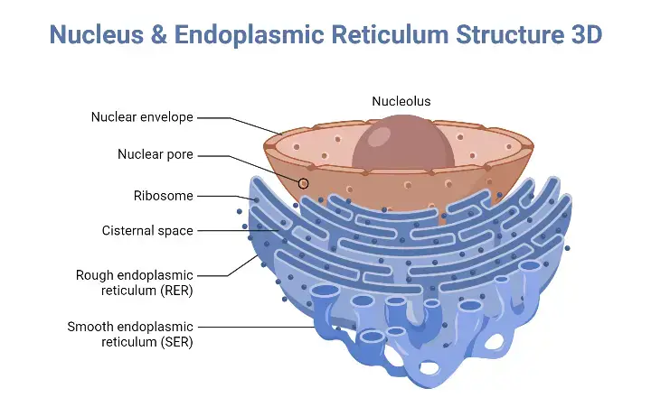
- Comprising a vast network of membrane tubules and sacs, the endoplasmic reticulum (ER) is present in eukaryotic cells continuous with the nuclear envelope.
- It appears primarily in two forms: the smooth ER (SER), without ribosomes, and the rough ER (RER), studded with ribosomes.
- The rough ER mostly helps proteins intended for secretion, integration into membranes, or usage in lysosomes synthesise, fold, and change themselves.
- The smooth ER is involved in the production of lipids and steroids, drug and toxic chemical detoxification, calcium ion storage and control.
- By acting as locations for macromolecule synthesis and processing as well as by helping intracellular transport and signalling, both types of the ER together are very vital for preserving cellular homeostasis.
Structure of Endoplasmic reticulum (ER)
- Comprising an enormous network of linked membranous tubules and flattened sacs, the endoplasmic reticulum runs continuously with the nuclear envelope throughout the cytoplasm of the cell.
- There are two basic types to it: the smooth ER, which is made of a more tubular network without ribosomes, and the rough ER, which contains ribosomes attached to its cytoplasmic surface and seems to be studded, sheet-like.
- With its large surface area giving plenty of sites for ribosome binding and nascent protein entry into its lumen, the rough ER’s shape is optimised for protein synthesis and processing.
- Reflecting its specialised functional tasks, the smooth ER with its highly branching tubular shape is engaged in lipid synthesis, detoxification activities, and calcium ion storage.
- Both types of the ER provide a dynamic and continuous membrane system that helps proteins and lipids be transported and changed as well as signals from the nucleus to the rest of the cell be integrated.
Functions of Endoplasmic reticulum (ER)
- Protein quality control depends on the endoplasmic reticulum (ER), which folds protein molecules into their correct three-dimensional forms inside its cisternae.
- It helps synthesised proteins be transported by packing them into vesicles that move these molecules to the Golgi apparatus for further sorting and modification.
- The smooth ER specialises in the production of lipids, including phospholipids and oils, which are essential for building and preserving cellular membranes.
- It is also crucial in endocrine signalling as it helps sex hormones and steroids to be synthesised.
- Furthermore engaged in glycogen hydrolysis—that is, breaking down glycogen to release glucose—which is vital for cellular energy metabolism—the ER
6. Ribosome
- Found in every living cell and the location of protein synthesis, ribosomes are big, sophisticated molecular machinery.
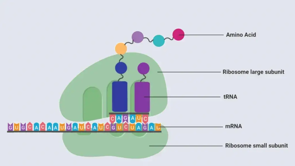
- Mostly consisting of proteins and ribosomal RNA (rRNA), they create two different subunits—a small subunit and a big subunit—which come together during translation.
- While in prokaryotic cells they are usually free-floating in the cytoplasm, in eukaryotic cells ribosomes either freely reside within the cytoplasm or are connected to the rough endoplasmic reticulum.
- By catalysing the synthesis of peptide bonds between amino acids, ribosomes essentially convert messenger RNA (mRNA) sequences into polypeptide chains.
- Highly conserved throughout all species, the process of protein synthesis by ribosomes—also known as translation—showcases its basic importance in cellular function and development.
- With rRNA not only providing structural support but also directly catalytic action in the creation of the peptide bonds during protein building, ribosomes also function as ribozymes.
Structure of Ribosome
- Large macromolecular complexes made of two separate subunits joined together during protein synthesis form ribosomes.
- Every subunit consists of several ribosomal proteins and ribosomal RNA (rRNA), which taken together provide a highly ordered structure necessary for catalytic activity.
- Whereas the big subunit (60S) has the peptidyl transferase centre that creates peptide linkages between amino acids, the small subunit (40S) binds and decodes messenger RNA (mRNA) in eukaryotic cells.
- Though they have a 30S small subunit and a 50S big subunit, prokaryotic ribosomes are somewhat smaller and have a similar general layout and purpose.
- The two subunits only combine during translation, when their complementary surfaces and contacts guarantee appropriate alignment of the mRNA and transfer RNA (tRNA). They stay apart in the cytoplasm.
- Cryo-electron microscopy and other structural investigations have exposed complex aspects of ribosomal architecture and highlighted conserved areas vital for correct and effective protein production.
- The exact structure of the ribosome explains its function as a molecular machine: it coordinates the production of proteins essential for cell operation and the decoding of genetic information.
Functions of Ribosome
- Translating messenger RNA (mRNA) sequences into polypeptide chains, ribosomes function as the cellular machinery for protein synthesis.
- Driven mostly by ribosomal RNA (rRNA) functioning as a ribozyme, they catalyse the creation of peptide bonds between amino acids during translation.
- They guarantee the correct alignment of mRNA and transfer RNA (tRNA) molecules therefore guaranteeing the precise decoding of the genetic code.
- Producing proteins necessary for structural support, enzyme activity, and regulatory activities, ribosomes are crucial for cellular development and maintenance.
- Reflecting their participation in both cytosolic and secretory protein production, they are either freely present in the cytoplasm or bound to the rough endoplasmic reticulum in eukaryotic cells.
7. Golgi apparatus
- Found in eukaryotic cells, the Golgi apparatus—also called Golgi complex or Golgi body—is a membrane-bound organelle.
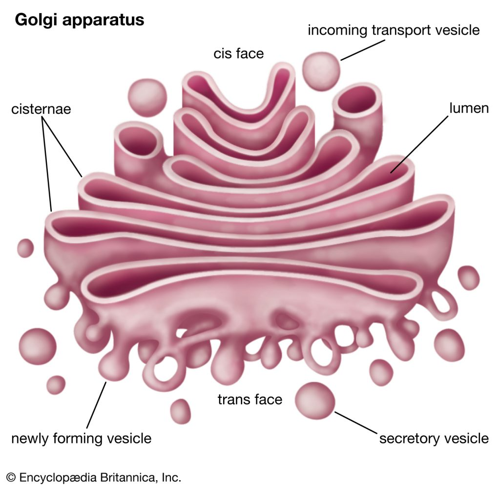
- It is made of a sequence of flattened, stacked cisternae acting as a central hub for protein and lipid modification, sorting, and packing.
- Transported to the Golgi apparatus, endoplasmic reticulum’s produced proteins and lipids undergo further changes including glycosylation, phosphorylation, and proteolytic cleavage.
- These processed molecules are guided by the Golgi apparatus into vesicles for movement to either secretion outside the cell, delivery to the cell membrane, or targeting to lysosomes.
- Its importance is in preserving cellular organisation and function as it guarantees appropriate distribution and processing of biomolecules for appropriate cellular activity.
Structure of Golgi apparatus
- The Golgi apparatus is set up in stacks of flattened, membrane-bound sacs called cisternae.
- It is strongly polarised, with a cis face pointing towards the endoplasmic reticulum to grab freshly produced proteins and lipids and a trans face guiding altered molecules to their intended sites.
- While the medial cisternae perform consecutive metabolic changes including glycosylation, the cis-Golgi network (CGN) serves as the entrance site where arriving vesicles combine.
- Acting as a sorting station, the trans-Golgi network (TGN) packages the produced molecules into vesicles headed for different cellular sites including secretory routes, lysosomes, or the cell surface.
- Embedded in the Golgi membranes, structural proteins and resident enzymes guarantee that every cisterna fulfils certain roles during the maturation process.
- The Golgi apparatus shows dynamic behaviour; cisternial maturation helps to continuously process and migrate biomolecules across the cell.
Functions of Golgi apparatus
- For proteins and lipids produced in the endoplasmic reticulum, the Golgi apparatus serves as the principal processing and packaging hub.
- At its cis face, it gets transport vesicles loaded with recently synthesised biomolecules, which undergo post-translational changes.
- To guarantee appropriate folding and function, proteins are altered within the Golgi by glycosylation, phosphorylation, and proteolytic cleavage.
- These altered molecules are sorted and guided by the organelle into certain vesicles that carry them to their appropriate locations—cell membrane, lysosomes, extracellular space, or another organelle.
- Crucially for cell-cell identification and communication, complex carbohydrates and glycolipids are synthesised via this mechanism.
- The dynamic character of the Golgi apparatus—including vesicular trafficking and cisternial maturation—allows it to effectively meet the metabolic needs of the cell and preserve cellular homeostasis.
8. Lysosome
- Membrane-bound organelles with a variety of hydrolytic enzymes tailored for macromolecule breakdown are lysosomes
- Through processes like autophagy and phagocytosis, they break down damaged organelles, cellular trash, and foreign materials, therefore acting as the recycling centre for the cell.
- Lysosomes’ inside is kept acidic, which maximises the action of their enzymes for effective breakdown.
- Through recycling the breakdown products for usage in several metabolic activities, they help to maintain cellular homeostasis.
- Many illnesses, including lysosomal storage disorders brought on by the buildup of undigested substrates within the cell, have their roots in dysfunction in lysosomal activity.
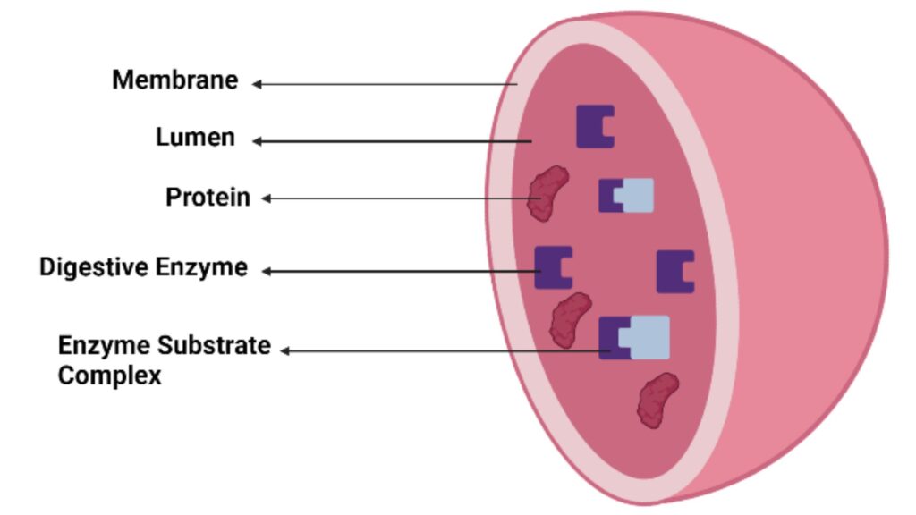
Structure of Lysosome
- Found in eukaryotic cells, lysosomal solitary membrane-bound organelles are spherical or oval.
- A lipid bilayer encloses them and preserves an acidic interior, which is necessary for the action of enzymes.
- Their lumen includes a range of hydrolytic enzymes used to break down macromolecules: proteases, lipases, nucleases, glycosidases
- Proton pumps (vacuolar ATPases) buried in the lysosomal membrane help to preserve lysosomal acidity.
- On lysosomes, membrane proteins help to fuse with vesicles carrying cellular trash, damaged organelles, or engulfed particles.
- The lysosomal membrane also helps to keep the strong enzymes within intact, therefore shielding other cellular components from damage.
- Their dynamic shape helps lysosomes to adapt and combine with other compartments, therefore facilitating cellular recycling and homeostasis.
Functions of Lysosome
- Using hydrolytic enzymes, lysosomes break down damaged organelles, macromolecules, and foreign materials, therefore serving as the recycling centre for the cell.
- They help autophagy, the process by which cells break down and recycle their own components to preserve cellular homeostasis and react to metabolic demand.
- They contribute to the defence systems of the cell by merging with endocytic vesicles carrying extracellular trash or pathogens, therefore facilitating phagocytosis.
- Lysosomes guarantee effective enzymatic breakdown by keeping an acidic interior environment, therefore helping to eliminate cellular waste and stop harmful accumulation.
- Recycled and used for new cellular biosynthesis, the breakdown products from lysosomal digestion help to provide energy and regulate metabolism generally.
- By releasing enzymes that help destroy cellular components when the cell is injured or no longer needed, lysosomes are involved in inducing planned cell death—apoptosis.
9. Centrosome
- The major microtubule organising centres (MTOCs) in animal cells are centrosomes, fundamental cellular structures.
- Usually composed of two centrioles—cylindrical configurations of microtubules surrounded by an amorphous matrix called the pericentriolar material—they reflect
- Maintaining cell structure, intracellular movement, and organelles’ location depends on the microtubule network being organised by the centrosome.
- Centrosomes double and assist in the formation of the mitotic spindle during cell division, therefore guaranteeing correct separation of chromosomes to the daughter cells.
- Although most animal cells include centrosomes, plant cells often lack a clear centrosome and arrange their microtubules differently.
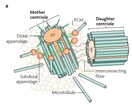
Structure of Centrosome
- In animal cells, the main microtubule organising centre is a non-membranous organelle called the centrosome.
- Two centrioles make up its basic structure; each is structured as a cylindrical array of nine microtubule triplets in a perpendicular direction.
- Around the centrioles lies the pericentriolar material (PCM), an amorphous matrix high in proteins that nucleate and fix microtubules.
- Maintaining cell form, intracellular movement, and organelle location depends on the microtubule network of the cell being assembled and stabilised, so its ordered structure helps to achieve these goals.
- The centrosome doubles during cell division to create two separate spindle poles, therefore guaranteeing correct chromosomal separation to the daughter cells.
- Though most plant cells lack centrioles, other species have comparable microtubule organising centres even if centrosomes are a trademark of animal cells.
Functions of Centrosome
- In animal cells, centrosomes are the main microtubule organising centre (MTOC; they nucleate and attach microtubules to form the internal framework of the cell).
- Their arrangement of the microtubule network directs the location and movement of organelles and vesicles, hence preserving cell shape and polarity.
- Crucially in forming the mitotic spindle that guarantees correct chromosomal separation to daughter cells, centrosomes replicate and create the spindle poles during cell division.
- They guarantee that organelles and the nucleus are precisely arranged by helping to control cell cycle development and provide spatial organisation of the cell.
- Additionally active in cell signalling pathways influencing activities like cell migration and differentiation are centrosomes.
10. Peroxisomes
- Nearly all eukaryotic cells have tiny, membrane-bound organelles called peroxisomes, which are very essential for cellular metabolism.
- They include oxidases and catalase, which catalyse processes to break down fatty acids and detoxify toxins such hydrogen peroxide.
- Peroxisomes guard cells from oxidative damage and support redox equilibrium by turning hydrogen peroxide into water and oxygen.
- They help to synthesise plasmalogens, which are vital phospholipids in the brain and heart, and participate in lipid metabolism including the beta-oxidation of extremely long-chain fatty acids.
- Linked to several hereditary diseases, including Zellweger syndrome, abnormalities in peroxisome activity emphasise its significance in human health and development.
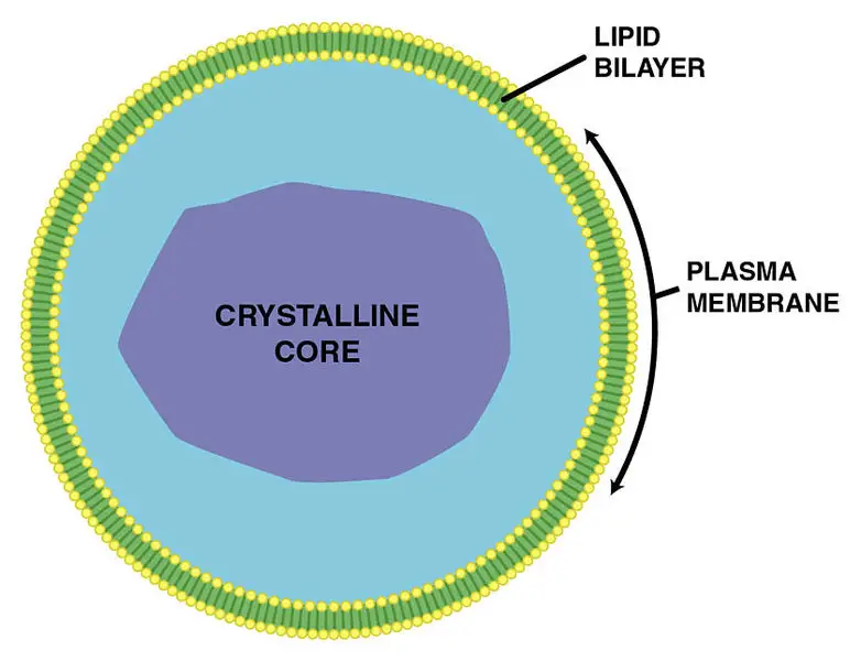
Structure of Peroxisomes
- Small single-membrane-bound organelles with a phospholipid bilayer enclosing their interior matrix define peroxisomes.
- Enzyme and metabolite selective import from the cytosol depends critically on the peroxisomal membrane.
- Inside, the matrix is crammed with enzymes including catalase and many oxidases needed to break down fatty acids and detoxify reactive oxygen species.
- High enzyme concentrations allow the matrix to show paracrystalline inclusions—seen under electron microscopy.
- Unlike mitochondria, peroxisomes synthesise all peroxisomal proteins in the cytosol and transported into the organelle; they lack ribosomes or their own genetic material.
- Specific membrane proteins ensure their correct function and distribution throughout the cell, therefore facilitating the synthesis, maintenance, and dynamics of peroxisomes.
- Usually round or oval in form and ranging in size from roughly 0.1 to 1 µm in diameter, peroxisomes help to effectively control cellular metabolic processes.
Functions of Peroxisomes
- Peroxisomes function in the detoxification of reactive oxygen species by converting hydrogen peroxide into water and oxygen through the enzyme catalase.
- They play a pivotal role in the beta-oxidation of very long chain fatty acids, breaking them down into shorter fatty acids for further metabolic processing.
- Peroxisomes are integral to lipid metabolism, including the synthesis of plasmalogens, which are essential phospholipids for cell membrane integrity and nervous system function.
- They participate in the metabolism of amino acids and other biomolecules, contributing to overall cellular energy balance and metabolic homeostasis.
- Peroxisomes help regulate the cellular redox state by processing reactive oxygen species, thereby protecting cells from oxidative damage.
- They are involved in the breakdown of purines and polyamines, which aids in the removal of potentially harmful metabolic byproducts.
11. Centrioles
- Found largely in animal cells and certain lower plant cells, centrioles are cylindrical, microtubule-based structures.
- Usually housed in pairs inside the centrosome, the major microtubule-organizing centre of the cell, they are
- Crucially for its structural integrity, each centriole consists of nine triplet microtubules arranged in a round, cartwheel configuration.
- Their organisation of the mitotic spindle fibres that guarantees correct chromosomal segregation during mitosis helps to define a basic function in cell division.
- Beyond cell division, centrioles help create cilia and flagella—qualities necessary for cell movement and signalling.
- Tightly controlled during the cell cycle, the duplication of centrioles guarantees that every daughter cell inherits the correct count, therefore preserving cellular integrity and function.
- Linked to many diseases, including certain malignancies and ciliopathies, abnormalities in centriole number or structure emphasise their importance in both health and illness.
- Originally noted in the late 19th century, centrioles have become more important for establishing cell polarity and control of the cell cycle.
- Investigating centriole biogenesis and control constantly helps us to understand their function in development and possible treatments for disorders caused by mistakes in cell division.
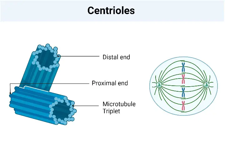
Structure of Centrioles
- Nine pairs of microtubule triplets grouped in a circular arrangement underlie the structural integrity of centrioles, cylindrical organelles.
- A cartwheel structure exists at the proximal end that provides the ninefold symmetry; this cartwheel is composed of a central hub with radial spokes linking to each microtubule triplet.
- Every triplet consists of two partial microtubules and one whole microtubule, a configuration that improves organelle stability and stiffness.
- Found at the distal end of centrioles, distal and subdistal appendages are crucial for both organising the microtubule network during cell division and attaching the centriole to the cell cortex.
- Important in the assembly and maintenance of the centriole’s structure are certain proteins such as SAS-6 and Cep135, therefore guaranteeing appropriate formation and function.
- The complex structure of centrioles is essential for their functions in aiding proper chromosomal segregation during mitosis and in the biogenesis of cilia and flagella, which are absolutely necessary for cell movement and signalling.
Functions of Centrioles
- Centrioles are pivotal in organizing the microtubule network by forming the core of the centrosome, which directs the assembly of the spindle apparatus necessary for accurate chromosome segregation during mitosis
- They duplicate once per cell cycle, ensuring that each daughter cell inherits a pair of centrioles, which is crucial for maintaining genomic stability and proper cell division
- Serving as basal bodies, centrioles initiate the formation of cilia and flagella, thereby enabling cell motility, sensory perception, and signal transduction processes
- In addition to their role during cell division, centrioles contribute to the spatial organization of the cytoskeleton during interphase, influencing cell shape, polarity, and intracellular transport
- The regulation and structural integrity of centrioles are critical for proper cell cycle progression, and any aberrations in their function or number are linked to developmental disorders, ciliopathies, and certain types of cancer
12. Cytoskeleton
- Structurally supporting, maintaining cell form, and enabling intracellular organisation, the cytoskeleton is a complex network of protein filaments.
- Microtubules, actin filaments (microfilaments), and intermediate filaments—each with unique structural and dynamic purposes—make up its mostly three-type filament composition.
- While actin filaments help to produce cell motility and shape changes and intermediate filaments give tensile strength to the cell, microtubules serve as tracks for the movement of organelles and vesicles.
- Cell division, intracellular transport, and the creation of cell polarity all depend on the cytoskeleton, therefore influencing activities like mitosis, migration, and signal transduction.
- With the introduction of electron microscopy, which revealed its complicated organisation and dynamic character, its discovery and later research increased dramatically in the middle of the 20th century.
- In biomedical research, knowledge of cytoskeletal dynamics is crucial as changes in these structures are linked to many illnesses, including cancer, neurodegenerative diseases, and other developmental defects.
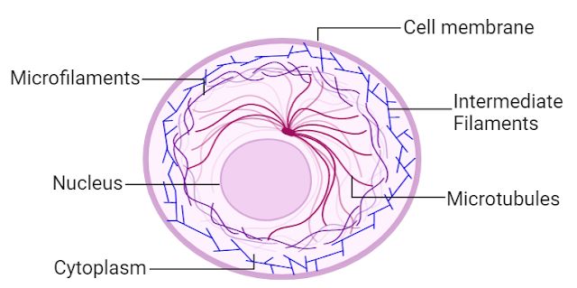
Structure of Cytoskeleton
- Comprising an elaborate network of protein filaments, the cytoskeleton supports mechanical movement, preserves cell form, and arranges interior cell structure.
- Microtubules, actin filaments (microfilaments), and intermediate filaments make up three primary varieties of filaments used in this work.
- Crucially important in mitosis and cell division, microtubules are hollow cylinders formed from alpha- and beta-tubulin dimers that act as pathways for intracellular movement.
- Crucially for cell mobility, muscle contraction, and the development of the cell cortex are helical polymers of actin filaments.
- Diverse proteins including keratins, vimentin, and laminates make up intermediate filaments; they strengthen tensile forces and improve cell general stability.
- Microtubule-associated proteins (MAPs) and actin-binding proteins among other regulatory proteins influence the construction, disassembly, and cross-linking of cytoskeletal components to vary cell dynamics.
- Highly dynamic and responsive to mechanical stressors and outside cues, the cytoskeleton remodels to enable cell migration, division, and intracellular transport.
- Underlining its vital importance in cell biology, changes in cytoskeletal structure or control have been connected to many illnesses, including cancer, neurological diseases, and developmental problems.
Functions of Cytoskeleton
- The cytoskeleton provides structural support by maintaining cell shape and resisting mechanical stress
- It organizes intracellular architecture by positioning organelles and establishing spatial order within the cell
- It facilitates intracellular transport by serving as tracks for motor proteins such as kinesin, dynein, and myosin, which move vesicles and organelles
- It plays a crucial role in cell division, contributing to the formation of the mitotic spindle that ensures accurate chromosome segregation
- It drives cell motility through the dynamic assembly and disassembly of actin filaments, enabling cell movement and shape changes
- It participates in cellular signaling and mechanotransduction, linking external stimuli to intracellular responses
- It supports cell adhesion by anchoring membrane proteins and connecting the cell to the extracellular matrix, thereby contributing to tissue integrity
Functions of Human Cell
- Cells generate energy using mitochondria
- They synthesize proteins via ribosomes
- They store and replicate genetic material in the nucleus
- They regulate growth and control cell division
- They facilitate intracellular transport and remove waste
- They communicate with other cells through signaling pathways
- They maintain structural integrity and cell shape
- https://mugberiagangadharmahavidyalaya.ac.in/images/ques_answer/1585930984BD%20Cell%20Unit%20-%201.2.pdf
- https://byjus.com/biology/cells/#:~:text=The%20cell%20structure%20comprises%20individual,%2C%20nucleus%2C%20and%20cell%20organelles.
- https://training.seer.cancer.gov/anatomy/cells_tissues_membranes/cells/structure.html
- https://www.sc.chula.ac.th/courseware/2303101j/VIII-Cell.pdf
- https://louis.pressbooks.pub/humananatomyandphysiology1/chapter/6-cell-structure-function/
- https://www.biologyonline.com/tutorials/cell-structure
- Text Highlighting: Select any text in the post content to highlight it
- Text Annotation: Select text and add comments with annotations
- Comment Management: Edit or delete your own comments
- Highlight Management: Remove your own highlights
How to use: Simply select any text in the post content above, and you'll see annotation options. Login here or create an account to get started.