What is Gnetum?
Gnetum represents a unique genus within the Gnetaceae family and the Gnetales order, characterized as a group of gymnosperms. The Gnetales order consists of three distinct extant genera: Ephedra, Gnetum, and Welwitschia, each exhibiting remarkable morphological differences. Historically, the phylogenetic placement of Gnetum has been contentious. While earlier classifications positioned it closer to angiosperms, contemporary studies suggest a closer relationship with conifers.
The Gnetum genus includes approximately 40 species, primarily distributed in tropical and humid environments. These plants manifest diverse forms such as trees, shrubs, lianas, and climbers, exhibiting prolific branching patterns and a distinctive phyllotaxy, which can be either decussate or whorled. Unlike many other gymnosperms, Gnetum species possess vessel elements within their xylem, a feature that enhances their efficiency in water transport. This trait, along with evidence suggesting that some Gnetum species were among the first to engage in insect pollination—based on fossil associations with extinct pollinating scorpionflies—underscores their evolutionary significance.
Gnetum species predominantly thrive at elevations below 1,500 meters, except for Gnetum microcarpum. Notably, five species are documented in India: Gnetum contractum, Gnetum gnemon, Gnetum montanum, Gnetum ula, and Gnetum latifolium. Among these, Gnetum ula is the most prevalent.
Species of Gnetum in India
- Gnetum ula: This species is a woody climber, characterized by branches featuring swollen nodes. It is found in various locations including the Western Ghats near Khandala, as well as forests in Kerala, the Nilgiris, the Godavari district of Andhra Pradesh, and Orissa.
- Gnetum contractum: A scandent shrub, Gnetum contractum grows primarily in Kerala, the Nilgiri Hills, and Coonoor in Tamil Nadu.
- Gnetum gnemon: Recognized as a shrubby plant, Gnetum gnemon is native to Assam, specifically in the Naga Hills, Golaghat, and Sibsagar regions.
- Gnetum montanum: This species is distinguished by its smooth, slender branches, which are swollen at the nodes. It is located in Assam, Sikkim, and parts of Orissa.
- Gnetum latifolium: A climber, Gnetum latifolium is found in the Andaman and Nicobar Islands.
Distribution of Gnetum
The distribution of Gnetum is primarily limited to tropical and humid regions, encompassing approximately 40 species within its genus. The following points elaborate on the geographical and ecological characteristics associated with Gnetum species:
- Global Distribution: Gnetum species thrive in tropical and humid climates, where environmental conditions favor their growth. The genus is predominantly found in areas characterized by abundant rainfall and warm temperatures.
- Altitude Preference: Most Gnetum species inhabit lowland areas, occurring at elevations below 1,500 meters. The only exception is Gnetum microcarpum, which is adapted to different altitudinal conditions.
- Species in India: Five specific species of Gnetum have been documented in India, highlighting the country’s ecological diversity. These include:
- Gnetum contractum: Found in Kerala and the Nilgiri Hills.
- Gnetum gnemon: Native to Assam, particularly in the Naga Hills, Golaghat, and Sibsagar regions.
- Gnetum montanum: Located in Assam, Sikkim, and parts of Orissa, characterized by its slender branches.
- Gnetum ula: The most prevalent species in India, discovered in various regions such as the Western Ghats, Kerala, Nilgiris, Godavari district of Andhra Pradesh, and Orissa.
- Gnetum latifolium: A climber found in the Andaman and Nicobar Islands.
- Ecological Importance: The distribution of Gnetum species in these tropical regions plays a crucial role in maintaining local ecosystems. Their presence contributes to biodiversity and provides habitat for various organisms, including pollinators.
Habit of Gnetum
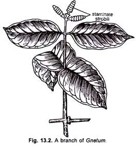
The habit of Gnetum is characterized by a diverse range of growth forms, primarily featuring climbing species, with some exceptions that manifest as shrubs or trees. This unique growth pattern contributes to the ecological versatility and adaptability of the genus. The following points outline the distinctive habits and structural characteristics of Gnetum:
- Growth Forms: The majority of Gnetum species are climbers, utilizing various strategies to ascend towards light. However, there are also species classified as shrubs and trees, expanding the morphological diversity within the genus. For example, Gnetum trinerve has been noted for its apparently parasitic nature, which is an unusual adaptation among Gnetum species.
- Branch Structure: Gnetum plants exhibit two distinct types of branches on their main stem: branches of limited growth and branches of unlimited growth. Each branch is composed of nodes and internodes, which are crucial for the plant’s overall architecture. The articulation of stems in several Gnetum species enhances their flexibility and adaptability, facilitating climbing.
- Short Shoots: In climbing species, branches of limited growth, or short shoots, tend to be unbranched and primarily serve to bear foliage leaves. This adaptation allows for efficient light capture, which is essential for growth in shaded environments.
- Leaf Arrangement: The leaves of Gnetum, typically numbering between nine and ten, are arranged in decussate pairs along the branches. This specific arrangement often results in the leaves lying in a single plane, creating the appearance of a pinnate leaf structure. This morphological characteristic is advantageous for maximizing light exposure and optimizing photosynthesis.
- Leaf Morphology: The leaves of Gnetum are generally large and oval-shaped, featuring entire margins and reticulate venation, a trait also found in dicotyledons. This reticulate pattern aids in structural strength and may enhance the plant’s ability to transport nutrients and water efficiently. Additionally, some species possess scaly leaves, which may serve protective functions or assist in moisture retention.

Characteristics of Gnetum
- Diversity and Growth Form:
- Gnetum species are predominantly dioecious, meaning individual plants are either male or female. They are characterized as evergreen and mostly consist of woody vines; however, some species can take the form of shrubs or trees. For instance, Gnetum gnemon is known as a tree, while others may appear as lianes or even stumpy, turnip-like forms, such as Welwitschia mirabilis.
- Stem Structure:
- The stems of Gnetum exhibit swollen nodes, which contribute to their unique structural characteristics.
- Leaf Morphology:
- Leaves are arranged oppositely on the stem and are petiolate, lacking stipules. They are simple in structure, typically elliptic, featuring pinnate venation and entire margins. Many leaves also have drip tips, which facilitate the efficient shedding of water.
- Vascular System:
- The secondary wood of Gnetum contains vessels, contributing to its structural integrity and fluid transport capabilities.
- Floral Characteristics:
- The reproductive structures, often referred to as “flowers,” are unisexual. Gnetum is predominantly dioecious, though some species may be monoecious.
- These flowers are arranged in compound strobili or inflorescences, with both male and female megastrobili appearing terminally or laterally. Occasionally, they cluster densely in cauliflorous arrangements on older stems.
- Structure of Megastrobili:
- Each megastrobilus features a straight axis topped with a basal pair of opposite, connate bracts. This axis typically bears three to six superposed cupules, which contain several to many male or female strobili.
- Male strobili comprise a stamen and perianth, while female strobili consist of an ovule encased in two integuments and a perianth.
- Ovule Characteristics:
- The nucellus of each ovule is enveloped by two or three layers of integuments, and the micropyle remains elongated, projecting outward as a bristle-like tube.
- Fertilization:
- During fertilization, the pollen tube contains two male nuclei, which facilitate successful reproduction.
- Embryonic Structure:
- Within the embryo, a unicellular primary suspensor is present, supporting the developing structure.
- Seed Features:
- Seeds are drupelike and encased in a fleshy false seed coat that can be red, orange, or yellow; in rare instances, this coat may be corky.
- The female gametophyte tissue surrounding the seeds is copious and succulent, providing nourishment for the developing embryo.
- Cotyledons and Germination:
- Each Gnetum seed typically contains two cotyledons. The germination process is epigeal, with cotyledons emerging above the soil surface.
- Wood Anatomy:
- The wood of Gnetum consists of tracheids typical of gymnosperms, but it also contains vessels, a trait associated with angiosperms. This vascular feature is believed to have evolved independently, lacking phylogenetic significance.
Anatomy of Gnetum
The anatomy of Gnetum reflects a complex and specialized structure that supports its diverse growth forms and ecological adaptations. Understanding the anatomical features of Gnetum provides insight into its physiological functions and ecological roles. The following points delineate the anatomical components of Gnetum, focusing on roots, stems, and leaves.

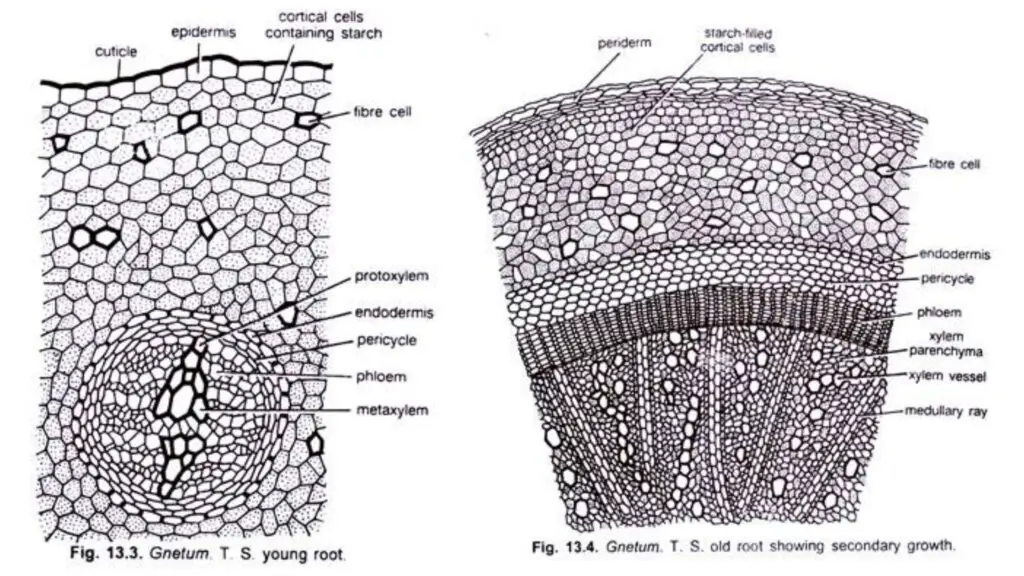
- Root Anatomy: The young root of Gnetum exhibits several layers of starch-filled parenchymatous cortex. These cells are large and polygonal in shape, facilitating storage and metabolic functions. A distinguishable endodermal layer is present, characterized by the presence of Casparian strips, which regulate water and nutrient uptake. Beneath the endodermis, a 4-6 layered pericycle can be observed. The roots are classified as diarch and exarch, with a small amount of primary xylem visible in the young roots, becoming less distinct following secondary growth. The secondary growth is typical, leading to a continuous zone of wood in older roots, comprising tracheids, vessels, and xylem parenchyma. The tracheids feature uniseriate bordered pits alongside the bars of Sanio, while the vessels contain simple or small multiseriate bordered pits. Some xylem elements contain starch grains, and the phloem consists of sieve cells and phloem parenchyma.
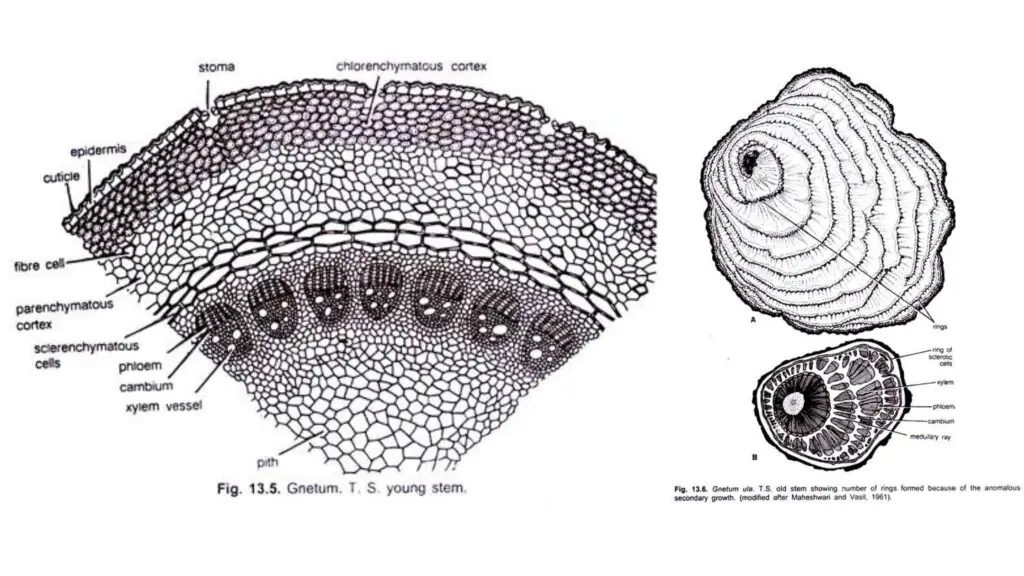
- Young Stem Anatomy: The young stem of Gnetum, upon transverse section, displays a roughly circular outline similar to that of typical dicotyledonous stems. It is encased by a single-layered epidermis that is thick and consists of rectangular cells, with some epidermal cells exhibiting papillate outgrowths and sunken stomata. The cortex comprises three distinct regions: an outer chlorenchymatous region consisting of 5-7 cells, a middle parenchymatous region of few cells, and an inner sclerenchymatous region that is 2-4 cells thick. The endodermis and pericycle regions are not sharply defined. Several conjoint, collateral, open, and endarch vascular bundles are arranged in a ring within the young stem. The xylem includes tracheids and vessels, indicative of angiospermic characteristics. The protoxylem elements are spiral or annular, while the metaxylem showcases bordered pits with circular outlines. The phloem consists of sieve cells and phloem parenchyma. A substantial pith, formed of polygonal parenchymatous cells, occupies the center of the young stem.
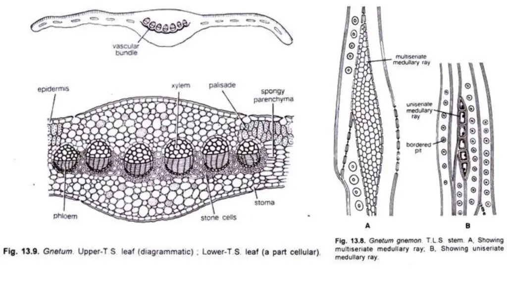
- Old Stem Anatomy: In older stems, Gnetum exhibits secondary growth, typically following the patterns seen in dicotyledons. In species such as G. gnemon, this secondary growth is normal; however, many species (e.g., G. ula and G. africanum) demonstrate anomalous secondary growth. The primary cambium is ephemeral, leading to the development of secondary cambium in various parts of the cortex in successive rings. Each cambium produces secondary xylem towards the interior and secondary phloem towards the exterior, ceasing function after a time. This process results in eccentric rings of xylem and phloem formation, characteristic of angiospermic lianes. The periderm develops from the outer cortex and includes lenticels. The cortex comprises chlorenchymatous and parenchymatous tissues, along with numerous sclereids. The secondary wood consists of tracheids and vessels, with tracheids possessing bordered pits on their radial walls and vessels containing simple pits. Transitional stages in the vessels often present multiple perforations at their terminal ends. In tangential longitudinal sections of the stem, both wood xylem and medullary rays are visible, with bordered pits on both radial and tangential walls. Medullary rays are either uniseriate or multiseriate, composed of polygonal parenchymatous cells.
- Leaf Anatomy: Internally, the leaves of Gnetum resemble those of dicotyledons, featuring a layer of thickly circularized epidermis on both surfaces. Stomata are predominantly located on the lower surface, except along the veins. The mesophyll is typically differentiated into a single-layered palisade parenchyma and a well-developed spongy parenchyma, which consists of loosely packed cells. Numerous stellate branched sclereids are present near the lower epidermis within the spongy parenchyma. The midrib region of the leaf showcases several vascular bundles organized in an arch or curve. A ring of thick-walled stone cells encircles the phloem, with each vascular bundle being conjoint and collateral. The xylem within each bundle is oriented toward the upper surface, while the phloem faces the lower surface. The xylem comprises tracheids, vessels, and xylem parenchyma, whereas the phloem consists of sieve cells and phloem parenchyma.


Reproduction of Gnetum
Gnetum, a dioecious genus of gymnosperms, exhibits a unique reproductive structure characterized by well-developed cones or strobili. These cones form organized inflorescences, typically of the panicle type, and can sometimes be terminally positioned.
- Structure of Cones:
- Each cone comprises a cone axis that features nodes and internodes, with two opposite connate bracts at its base.
- Whorls of circular bracts arise at the nodes, stacked to create collars or cupulas, which house the flowers. In G. gnemon, the upper collars may be reduced and sterile.
- Male Cones and Flowers:
- Male flowers are arranged in distinct rings above each collar on the male cone axis, typically numbering between three to six. The flowers in these rings alternate in arrangement, with a ring of abortive ovules or imperfect female flowers located above the male flowers.
- Each male flower consists of two coherent bracts forming the perianth, and features two unilocular anthers attached to a short stalk enclosed within the perianth.
- As the anthers mature, the stalk elongates, allowing the anthers to emerge from the perianth sheath. In G. gnemon, a few male flowers may occasionally fuse.
- Development of Male Flowers:
- In very young cones, specific cells beneath each collar become meristematic, dividing repeatedly to form a small outgrowth.
- The upper side of this outgrowth differentiates into ovule initials, leading to the formation of abortive ovules, while the lower side develops into the primordium of the male flower.
- The central cushion of cells formed in the male flower primordium becomes surrounded by a circular sheath called the perianth, which encloses a depression that later differentiates into two anther lobes.
- The basal portion of this central mass differentiates into a stalk, pushing the anther lobes outward, while each lobe remains enveloped by wall layers and tapetum, the innermost layer surrounding the sporogenous tissue.
- The sporogenous cells develop into microspore mother cells through meiosis, resulting in the formation of spore tetrads. The radial thickenings in the epidermal cells facilitate microsporangium dehiscence.
- Female Cones and Flowers:
- The female cones share similarities with male cones, featuring a single ring of four to ten ovules located above each collar. Only a subset of these ovules matures into seeds.
- Young female and male cones appear nearly identical; however, as they develop, certain ovules enlarge and mature into seeds while others remain undeveloped.
- Ovule Structure:
- Each ovule comprises a nucellus surrounded by three envelopes. The nucellus contains the female gametophyte and lacks a nucellar beak.
- The inner envelope extends to form the micropylar tube, with stomata, sclereids, and laticiferous cells present in the outer envelopes.
- Abnormal Cone Characteristics:
- Abnormalities such as multiple rings of ovules in male cones and spirally arranged collars in female cones have been observed. Occasionally, male cones may possess one or two fertile ovules alongside normal male flowers.
- Morphological Nature of Envelopes:
- The three envelopes surrounding the nucellus have been interpreted differently by researchers. Some suggest all three are integuments, while others propose that the outer envelope functions as a perianth and the inner envelopes as integuments.
- Mega-Sporangium and Female Gametophyte Development:
- In the female cone, four to ten ovular primordia develop similarly to the male cone’s structure. The ovular primordium undergoes multiple divisions to create a mass of cells, with all three envelopes forming around it.
- Archesporial cells develop and divide, forming a massive nucellus from the outer epidermal layer and primary parietal cells. The sporogenous cells eventually give rise to megaspore mother cells, with most degenerating, except for one, which can form a tetrasporic embryo sac.
- The female gametophyte is unique in its partially cellular and partially nuclear composition, lacking archegonia.
- Microsporangium and Micro-Sporogenesis:
- The development of the microsporangium begins in young anthers with archesporial cells dividing to form sporogenous cells.
- The microspore mother cells divide reductionally, resulting in haploid microspores, which may arrange in various configurations.
- Male Gametophyte Development:
- Pollen grains are spherical, uninucleate, and surrounded by a thick, spiny exine and a thin intine. Mature pollen grains are released at a three-nucleate stage, consisting of a prothallial nucleus, a tube nucleus, and a generative nucleus.
- The pollen tube forms as the intine ruptures the exine, with the tube nucleus and generative nucleus migrating into the pollen tube, where the latter divides to produce male gametes.
- Pollination:
- Wind facilitates the transfer of pollen grains to the micropylar tube of the ovule, where a secreted fluid entraps pollen grains that reach the pollen chamber.
- Fertilization:
- During fertilization, the pollen tube pierces the female gametophyte’s membrane, releasing male cells near cytoplasmic cells. One male cell enters the egg cell, leading to the formation of the zygote, characterized by a spherical shape and dense cytoplasm.
- Endosperm Formation:
- Unlike most gymnosperms, Gnetum exhibits free-nuclear divisions before fertilization. After fertilization, wall formation within the female gametophyte occurs, dividing the cytoplasm into compartments, each potentially containing multiple nuclei. This cellular development is variable across Gnetum species.
- Embryo Development:
- The initial division of the zygote reflects characteristics of both gymnosperms and angiosperms, showcasing both free-nuclear and cell divisions. A two-celled pro-embryo is established, with one nucleus undergoing free-nuclear divisions while the other remains unchanged.
- In Gnetum, zygote development can yield one functional zygote from several formed, with subsequent outgrowths developing into the primary suspensor, which assists in pushing the embryo into the endosperm.
- Seed Structure:
- Gnetum seeds are oval to elongated, varying in color from green to red, surrounded by a three-layered envelope enclosing the embryo and endosperm. The outer envelope is fleshy, while the middle envelope is hard and protective, consisting of three layers. The inner envelope is composed of parenchyma.
- Seed Germination:
- Germination occurs through an epigeal process, where the cotyledons emerge from the seed as the hypocotyl elongates. The cotyledons serve as the first green leaves, while the plumule develops into the first pair of foliage leaves. A persistent feeder remains present throughout germination.
Vegetative Structure of Gnetum
Gnetum, a genus known for its unique morphology and evolutionary significance, possesses several vegetative structures that contribute to its adaptability and functionality. Understanding these structures is crucial for comprehending the overall biology of Gnetum and its ecological roles.
- Root Structure:
- Gnetum exhibits a tap root system, complemented by well-developed lateral roots.
- The outermost layer is the epidermis, which is followed by a multilayered parenchymatous cortex filled with starch.
- Thick-walled fiber cells are prevalent in the cortex, enhancing structural support.
- The endodermis surrounds a multilayered pericycle, providing a barrier for selective absorption.
- Primary xylem is characterized as diarch, with tracheids featuring uniseriate bordered pits, allowing for efficient water transport.
- The phloem consists of uniform cells, facilitating nutrient distribution.
- Secondary growth in Gnetum occurs in a typical manner, contributing to the plant’s overall growth and stability.
- Stem Structure:
- The stem of Gnetum displays two types of branches: those with limited growth and those with unlimited growth. Notably, this distinction is absent in shrub and tree species such as G. gnemon.
- Some species possess articulate stems, where joints consist of two parts: one above and one below the node, separated by an annular groove.
- A young stem showcases a single-layered epidermis composed of rectangular cells coated with a thick cuticle, featuring sunken stomata.
- The cortex comprises 12-16 layers of parenchymatous cells, with some inner cells becoming fibrous and exhibiting narrow lumens.
- In older stems, a ring of parenchymatous cells transforms into sclerenchymatous cells within the inner cortex, referred to as the ring of sclerous cells.
- The endodermis and pericycle are not distinct, simplifying the structural organization.
- The vascular bundles, numbering between 20-24, are collateral and endarch, arranged in a circular formation. The xylem primarily consists of tracheids and a few vessels, with medullary rays between the vascular bundles being broad and prominent.
- Laticiferous elements are present in both the pith and cortex, contributing to the plant’s defense mechanisms.
- In tree species like G. gnemon, secondary growth occurs normally. Conversely, climbing species such as G. ula and G. africanum initially exhibit normal secondary growth but later develop new cambium, resulting in several rings of xylem and phloem, often leading to wedge-shaped bundles due to medullary rays.
- The secondary phloem consists of sieve cells and parenchyma, showing regular arrangement patterns; sieve cells are arranged in uniform rows while parenchyma occupies the angles between them.
- The wood of Gnetum is noteworthy for having vessels with a single pore on their end walls, with tracheids and xylem parenchyma also present. The tracheids are elongated, showcasing uniseriate bordered pits on both radial and tangential walls, while xylem parenchyma features simple pits.
- Stem Apex:
- The shoot apex of Gnetum displays characteristics akin to angiosperms, characterized by an atypical tunica-corpus organization.
- The tunica comprises the outermost layer of cells extending from the first pair of leaf primordia over the shoot apex.
- The corpus consists of subapical initials, a central mother cell zone, flanking layers, and a pith rib meristem, reflecting a complex developmental pattern.
- Leaf Structure:
- Gnetum’s leaves resemble those of dicotyledons, characterized by large, oval shapes with reticulate venation and entire margins.
- The short shoots typically bear 9 or 10 decussately arranged leaves on each branch.
- The leaf epidermis features undulating walls with a thick cuticle, enhancing protection and reducing water loss.
- The mesophyll is differentiated into palisade and spongy parenchyma, where the palisade consists of a single layer of compact cells, containing stellately branched sclereids near the lower epidermis.
- Fibers and latex tubes are abundant, particularly in the midrib region, contributing to the leaf’s structural integrity and chemical defense.
- Stomata are present only on the lower surface, oriented irregularly, and exhibit haplocheilic developmental patterns.
- Vascular bundles in the leaves are arranged in a curve, with xylem composed of vessels, tracheids, and parenchyma, while the phloem is organized in regular rows just beneath the xylem. Thick-walled pitted cells form a patch outside the phloem, enhancing the leaf’s structural support.
Reproductive structures of gnetum
The reproductive structures of Gnetum exhibit a fascinating complexity, reflecting its dioecious nature, where male and female strobili are produced on separate plants. This unique characteristic not only facilitates genetic diversity through cross-pollination but also highlights the distinct morphological differences between the male and female reproductive systems.
- Male Strobilus:
- The male strobilus is characterized by a solitary or fascicled panicle that develops axillary on a short shoot, remaining unbranched.
- It comprises microsporangia along with a whorl of sterile ovules. Each strobilus features an axis with two connate opposite bracts at the base, accompanied by a series of circular bracts, often referred to as cupules or collars, which are superposed.
- In its early stages, the strobilus appears compact due to a reduced axis with short internodes, causing the collars to seem continuous. However, as the axis elongates, these collars become separated.
- The upper collars in species like G. gnemon are typically reduced and sterile. Meristematic cells differentiate at the base of each collar, contributing to the formation of microsporangia.
- Each microsporangium originates from a tissue hump that becomes notched to form two anthers supported by a stalk. Two groups of hypodermal archesporial cells divide, forming a multicellular archesporium.
- The outer layer of archesporial cells divides, resulting in a primary parietal layer and sporogenous cells. The primary parietal layer subsequently gives rise to the wall layer and the tapetum. The tapetum, which is dense and binucleate, begins to degenerate post-meiosis, leaving remnants at the uninucleate stage of microspores.
- Pollen grains are shed at the 3-celled stage, with the microspore nucleus dividing to produce a small prothallial cell and a larger antheridial initial. The prothallial cell degenerates, while the antheridial initial further divides to form an antheridial cell, functioning directly as the spermatogenous cell. Occasionally, double pollen grains are observed, arising from the non-separation of cells within a tetrad. Notably, the pollen grains of Gnetum are sticky.
- Female Strobilus:
- The female strobilus initially resembles the male but becomes distinct as it matures. It features a ring of four to ten female flowers (ovules) positioned above each collar, with no male flowers present. Although all ovules appear similar initially, only a few mature.
- Female flowers arise from an annular meristem located below each collar. The ovular primordium sits on a cushion, with three envelopes (outer, inner, and integument) developing centripetally.
- The outer envelope, referred to as the perianth, is the first to differentiate, thickening and becoming succulent at maturity. Stomata develop on its outer epidermis.
- The inner envelope, also called the outer integument, follows, characterized by a tapering rim that develops stomata and laticifers during pollination. The integument, the last envelope to form, fuses with the nucellus at the lower part and elongates into a micropylar tube or style that projects beyond the outer envelope’s apical cleft.
- The nucellus is well-developed, with its epidermis dividing to form a cap. Below the megaspore mother cells, some nucellar cells divide, creating a nutritive tissue known as “pavement tissue,” which is absorbed as the female gametophyte grows.
- Megasporogenesis and Female Gametophyte:
- In the developing nucellus, two to four hypodermal archesporial cells differentiate, producing primary parietal and sporogenous cells. The sporogenous cells undergo division, forming a linearly arranged cluster that acts as megaspore mother cells.
- Following meiosis, a multinucleate coenomegaspore is generated, with the four nuclei moving from the center to the periphery. The development of the female gametophyte is tetrasporic.
- Although multiple coenomegaspore structures can develop, generally only two or three progress beyond the 16-nucleate stage. As the female gametophyte matures, a large vacuole appears centrally, and peripheral nuclei continue to divide.
- The gametophyte elongates, eventually resembling an inverted flask. Notably, Gnetum exhibits the absence of archegonia, a characteristic it shares with Welwitschia. Upon contact with the pollen tube, nuclei in the equatorial region of the gametophyte can be identified as egg nuclei, typically numbering two, though variations occur.
- The differentiation of eggs is stimulated by the presence of the pollen tube, and this process does not occur simultaneously across all eggs. Thus, egg differentiation can continue even post-fertilization of the first egg, with the upper part of the gametophyte transitioning from a free nuclear to a cellular state shortly after fertilization.
Pollination And Fertilization Of Gnetum
The processes of pollination and fertilization in Gnetum reveal a remarkable adaptation to ensure successful reproduction in these dioecious plants. This process not only highlights the intricate relationship between the male and female reproductive structures but also demonstrates how environmental factors influence these mechanisms.
- Pollination Process:
- Pollination occurs when the female gametophyte contains 250 nuclei in G. ula and either 32 or 64 nuclei in G. gnemon.
- Pollination can be facilitated by both wind and insects. A significant feature of the pollination process is the flaring and laceration of the micropylar tube’s tip.
- A pollination drop, rich in sugars, is exuded from the tip and serves to collect pollen. As this fluid dries, it creates a vacuum that draws pollen grains into the micropylar canal, ultimately lodging them in the pollen chamber.
- Ants have been observed visiting the pollination drop, which is formed from the degenerated cells of the nucellar tip.
- In the dioecious shrub species G. gnemon, it has been documented that both male and female strobili emit a strong, putrid odor during the evening, accompanied by the secretion of pollination droplets from the ovules.
- The slow evaporation of the exposed pollen droplet during the night in tropical rainforests enhances nocturnal pollination, achieved through the replacement of showy petals with a strong odor.
- Pollen Germination:
- Upon germination, the exine of the pollen grain is cast off, and the pollen grains lack germ pores.
- The pollen grain develops a tube that grows through intercellular spaces within the nucellus.
- As the pollen tube progresses, the spermatogenous cell moves into the tube, while the prothallial cell degenerates in situ.
- The spermatogenous cell undergoes division to produce two male gametes, which move towards the tip of the pollen tube, ahead of the tube nucleus.
- Pollen Chamber Development:
- A pollen chamber forms at the apical portion of the nucellus. Once megasporogenesis is complete, cells in the upper region begin to degenerate.
- This degeneration involves the breakdown of cytoplasm, nuclei, and cell walls, ultimately leading to the formation of a shallow pollen chamber where pollen grains are lodged.
- The cells between the pollen chamber and the female gametophyte accumulate starch grains, contributing to the nutrition of the developing endosperm, which nearly consumes the nucellus.
- In mature seeds, the apical cells become cutinized and persist.
- Post-Pollination Changes:
- Following pollination, a ring-shaped swelling appears just above the insertion of the inner envelope, accompanied by another proliferation in the apical region below the pollen chamber.
- A circular rim, or umbrella-shaped structure known as the “flange,” develops from the integument during this time; its precise function remains unclear.
- Micropylar Closing Tissue:
- A specific tissue called the closing tissue forms from the proliferation of the inner epidermis of the integument at the flange level, resulting in the closure of the micropylar canal.
- This closure is facilitated by the elongation and interlocking of the inner epidermal cells.
- The micropylar closing tissue may extend downward to fill the pollen chamber, acting as a plug referred to as the “obturator.”
Embryogeny and seed development
The processes of embryogeny and seed development in Gnetum illustrate the complex and adaptive strategies that these gymnosperms utilize for successful reproduction. This narrative synthesizes various aspects of these processes to provide a clearer understanding for students and educators.
- Zygote Development:
- Upon fertilization, the zygote may develop a small protuberance into which the nucleus migrates.
- This zygote can also divide into two cells, and either or both of these cells may produce a tube known as the primary suspensor tube. This designation helps distinguish it from the later-developed secondary suspensor.
- These primary suspensor tubes are characterized by their elongation, coiling, and septate nature, allowing them to penetrate into the female gametophyte or endosperm.
- Embryo Formation:
- The primary suspensor tubes consistently grow downwards toward the chalazal end, marking the direction of development.
- Embryo development initiates at the tips of some primary suspensor tubes. The nucleus at the tip divides into two unequal nuclei, with the smaller nucleus rounding up and dividing twice, resulting in four cells.
- Subsequent cell divisions occur irregularly, leading to a mass of cells where the upper cells differentiate into the secondary suspensor, also referred to as embryonal tubes.
- Cells at the apex of the secondary suspensor become compact, forming the embryo proper. The secondary suspensor then develops into a massive structure that resembles a folded plate of cells, with some cells elongating to form long tubes alongside the primary suspensor, which ceases to function.
- Embryo Development:
- Among the numerous primary suspensor tubes formed, only a few will continue to develop, while the rest, along with their associated embryos, degenerate.
- The growing embryo is observed as a conical mass of cells, with the shoot apex located at the cone’s tip.
- Active cell division at the sides of the embryonal mass leads to the formation of two cotyledons that cover the central zone of the shoot apex, while the root tip is differentiated at the opposite end.
- The root cap cells merge with the secondary suspensor, indicating their connection.
- As development progresses, a protrusion appears between the two apices, known as the “feeder.” This structure consists of epidermis, cortex, vascular bundles, and pith. In mature embryos, the feeder is notably prominent and typically exceeds the length of the hypocotyl.
- Polyembryony:
- Gnetum frequently exhibits polyembryony, which can be categorized into simple and cleavage polyembryony. Cleavage polyembryony may arise from the embryonal mass of either primary or secondary suspensor tubes.
- Occasionally, cells from the secondary suspensor may become meristematic, leading to the production of multiple embryos at the tip.
- Endosperm Development:
- In contrast to other gymnosperms, where cellular endosperm formation occurs before fertilization, Gnetum initiates cellularization prior to fertilization while retaining free nuclei within a portion of the gametophyte.
- This process results in multinucleate compartments where nuclei eventually fuse to form a single polyploid nucleus. As the endosperm develops, its shape evolves, with the lower part broadening and overgrowing the chalazal region.
- Transverse divisions of these cells generate a distinct area termed the axial tissue. Meanwhile, the upper section of the endosperm is progressively consumed by the growing suspensors, becoming compressed and crushed.
- Rich in starch, the endosperm provides essential nutrients for the undifferentiated embryo, which continues its growth even after the seed has fallen.
- Seed Characteristics:
- The seeds of most Gnetum species are oval, with colors ranging from green to red. They are shed before the embryo is fully developed, and the nucellus forms a thin strip at the apex, encased in a massive endosperm and a three-layered seed coat.
- There is typically a delay between seed shedding and germination. For example, in G. gnemon, seeds are shed in April and germinate by September, while G. ula seeds may take a full year to germinate.
- The germination process is epigeal, meaning the cotyledons emerge above the soil surface.
Relationships of Gnetum
Gnetum, a genus within the Gnetales, exhibits a unique blend of characteristics that connect it both to gymnosperms and angiosperms. This intricate relationship provides valuable insight into plant evolution and diversification.
- Resemblances with Gymnosperms:
- Wood Structure: Gnetum possesses wood with tracheids featuring bordered pits, a characteristic shared with gymnosperms.
- Absence of Specialized Cells: There are no sieve tubes or companion cells present in Gnetum, aligning it with gymnospermic features.
- Naked Ovules: Like other gymnosperms, Gnetum has naked ovules, a fundamental reproductive trait.
- No Fruit Formation: The absence of ovary in Gnetum leads to no fruit development, which is typical for gymnosperms.
- Pollination: Gnetum employs anemophilous pollination, a common trait among gymnosperms.
- Prothallial Cell Development: The development of prothallial cells indicates a reproductive similarity with gymnosperms.
- Cleavage Polyembryony: This phenomenon, where multiple embryos can develop from a single fertilized ovule, is also observed in Gnetum.
- Vascular Supply: The vascular supply in the peduncle of the cone of Cycadeoidea wielandii resembles that of a single flower of Gnetum, furthering the relationship.
- Basal Ovule Structure: The basal part of the ovule in Gnetum shows similarities with that in Bennettites, highlighting its gymnospermic affinity.
- Connections to Angiosperms:
- Evolutionary Perspective: Gnetum is regarded as a crucial link in discussions surrounding the origin of angiosperms, tracing back to a common ancestor known as “Hemi-angiosperm”.
- Close Relatives: Thompson (1916) suggested that the ancestors of Gnetum and angiosperms were closely related. Some researchers even propose that Gnetum should be classified within angiosperms.
- Relationship with Piperaceae: Hagerup (1934) identified a close relationship between Gnetales and the Piperaceae family.
- Phylogenetic Puzzle: Maheshwari and Vasil (1961) characterized Gnetum as largely a phylogenetic puzzle, exhibiting traits of both gymnosperms and angiosperms.
- Common Features with Angiosperms:
- Sporophyte Habit: The overall growth habit of many Gnetum species resembles that of angiosperms.
- Leaf Venation: Gnetum exhibits reticulate venation in its leaves, a distinctive feature of angiosperms.
- Xylem Structure: The presence of vessels in the xylem is a significant angiospermic characteristic.
- Shoot Apex Configuration: Gnetum features a clear tunica and corpus arrangement at the shoot apices, similar to angiosperms.
- Strobili Structure: The strobili of Gnetum bear a closer resemblance to those of angiosperms than to any other gymnosperms.
- Micropylar Tube Functionality: The micropylar tube in Gnetales functions similarly to the style in angiosperms, facilitating similar roles in reproduction.
- Female Gametophyte Development: Tetrasporic development of the female gametophyte aligns Gnetum more closely with angiosperms.
- Lack of Archegonia: The absence of archegonia in Gnetum further narrows the gap between it and angiosperms.
- Embryonic Characteristics: The dicotyledonous nature of the Gnetum embryo places it in closer relation to dicotyledonous angiosperms.
- Shared Traits Among Gnetales:
- Leaf Arrangement: Gnetum, Ephedra, and Welwitschia all feature opposite leaves.
- Wood Vessels: Each genus possesses vessels in their secondary wood.
- Perforation Plate Development: The structure and development of perforation plates in their vessels are strikingly similar.
- Vessel Development: They exhibit a Gnetalean mode of vessel development through the dissolution of the torus and middle lamella of bordered pits.
- Phloem Structure: The structure of sieve cells and phloem parenchyma is akin across these genera.
- Protoxylem Elements: Spiral or annular elements are present in their protoxylem.
- Flower Arrangement: The arrangement of flowers in compound strobili is consistent among Gnetales.
- Flower Structure: Gnetales feature unisexual flowers, with dioecious plant structures.
- Male Flower Composition: Stalked male flowers possess synangia, made of 1-6 or more sporangia.
- Microsporangia: The wall structure of microsporangia is consistently similar.
- Pollen Characteristics: Gnetales exhibit wingless pollen grains.
- Ovule Characteristics: Orthotropous ovules are common among these genera.
- Surrounding Envelopes: Ovules are encased in several envelopes, interpreted variously as integuments or perianth.
- Micropylar Tube: The elongated micropylar tube is a notable feature.
- Primary Suspensors: Formation of unicellular primary suspensors occurs.
- Embryo Characteristics: All exhibit a dicotyledonous embryo.
- Polyembryony: They share a simple type of polyembryony.
Difference Between Gnetum, Ephedra, and Welwitschia
Gnetum, Ephedra, and Welwitschia are three distinct genera within the Gnetales, each displaying unique characteristics and adaptations that reflect their respective environments. Understanding these differences can enhance comprehension of plant diversity and evolutionary biology.
- Gnetum:
- Distribution: Gnetum is primarily found in tropical or subtropical and humid regions across several countries worldwide.
- Habit: Most species of Gnetum are lianes or trees, with only a few being shrubs.
- Leaf Characteristics: Gnetum features foliaceous, green leaves that resemble those of angiosperms.
- Venation: The venation in Gnetum leaves is reticulate and unicostate.
- Stomatal Development: Gnetum exhibits syndetocheilic stomatal development.
- Shoot Apex: The shoot apex follows an open system of growth and has a definable tunica layer.
- Strobili Structure: Gnetum’s compound strobili are predominantly cauline and only rarely terminal.
- Male Gametophyte:
- Contains a single prothallial cell.
- Lacks sterile cells and spermatogenous cells in the young male gametophyte.
- Female Gametophyte:
- Normal archegonia are absent; a free nucleus organizes cytoplasmic material to function as an egg.
- The upper portion of the female gametophyte remains free-nuclear at fertilization, while the lower portion becomes cellular.
- Endosperm Formation: Similar to angiosperms, the endosperm in Gnetum forms after fertilization.
- Fertilization: Occurs within an embryo sac.
- Embryogenesis: There is no free-nuclear division phase in embryogenesis.
- Feeder Presence: The feeder is present in the embryo.
- Haploid Chromosome Number: Gnetum has a haploid chromosome number of 22.
- Ephedra:
- Distribution: Ephedra grows in dry and arid regions of North and South America, as well as parts of the Old World, including India and China.
- Habit: Most species are bushy or shrubby, with only a few being lianes.
- Leaf Characteristics: The leaves of Ephedra are small and scale-like.
- Venation: The leaves exhibit parallel venation.
- Stomatal Development: Ephedra displays haplocheilic stomatal development.
- Shoot Apex: The shoot apex structure is similar to that of Gnetum.
- Strobili Structure: In Ephedra, compound strobili are always terminal and never cauline.
- Male Gametophyte:
- Contains a pair of prothallial cells.
- Includes both a sterile cell and a spermatogenous cell.
- Female Gametophyte:
- Normal archegonia are present.
- The entire female gametophyte becomes cellular before fertilization.
- Endosperm Formation: No endosperm is formed in Ephedra.
- Fertilization: Occurs in an archegonium.
- Embryogenesis: Free-nuclear divisions occur in the zygote until about eight nuclei are formed.
- Feeder Presence: The feeder is absent.
- Haploid Chromosome Number: Ephedra has a haploid chromosome number of 7 or 14.
- Welwitschia:
- Distribution: Welwitschia is found exclusively along a narrow coastal belt of approximately 1000 km in southwest Africa.
- Habit: This genus is represented by only one known species, which resembles a gigantic turnip.
- Leaf Characteristics: Welwitschia has only two large, thick, foliaceous, ribbon-like leaves that can reach lengths of about two meters.
- Venation: The leaves exhibit parallel venation, with veins joined by transverse or interwoven veinlets.
- Stomatal Development: Similar to Gnetum, Welwitschia has syndetocheilic stomatal development.
- Shoot Apex: The shoot apex follows a closed system of growth, ceasing activity after initiating a pair of foliage-leaf primordia.
- Strobili Structure: The compound strobili are cauline and rarely terminal.
- Male Gametophyte:
- Contains a single prothallial cell.
- Lacks sterile and spermatogenous cells in the young male gametophyte.
- Female Gametophyte:
- Only archegonial initials are present, functioning directly as eggs.
- The entire female gametophyte becomes cellular before fertilization.
- Endosperm Formation: No endosperm is present in Welwitschia.
- Fertilization: Takes place within the pollen tube, a unique feature not observed in other gymnosperms or angiosperms.
- Embryogenesis: There is no free-nuclear division phase in embryogenesis.
- Feeder Presence: The feeder is present.
- Haploid Chromosome Number: Welwitschia has a haploid chromosome number of 21.
- https://www.biologydiscussion.com/gymnosperm/gnetum-distribution-habitat-and-relationships-gnetales/22583
- https://ramsadaycollege.com/upload/eclassroom/Botany/GDM_Botany_Gnetum_sem2_03.pdf
- https://egyankosh.ac.in/bitstream/123456789/16723/1/SubUnit-4B.pdf
- https://www.biologydiscussion.com/essay/gymnosperms/essay-on-the-life-cycle-of-gnetum-class-gnetopsida-gymnosperms-botany/76855
- https://hmmcollege.ac.in/uploads/dept_teaching_plan/Unit_6_Gymnosperm_morphology.pdf
- http://ndl.iitkgp.ac.in/he_document/cec/nYzKXLFBx48_PLNsppmbLKJ8L4M0x5ioi4TA6DQFSLGb-W
- https://academichunt.blogspot.com/2021/04/Gnetum-gnetales-morphology-of-gnetum-anatomy-of-gnetum-reproduction-of-gnetum-gauhati-university-tdc-botany-gauhatiuniversity.html
- https://www.biologydiscussion.com/gymnosperm/difference-among-gnetum-ephedra-and-welwitschia-gnetales/22471
- https://www.bergianska.se/polopoly_fs/1.521706.1602513117!/menu/standard/file/Jorgensen_Rydin_2015.pdf