What are Fish Fins and Swim bladder?
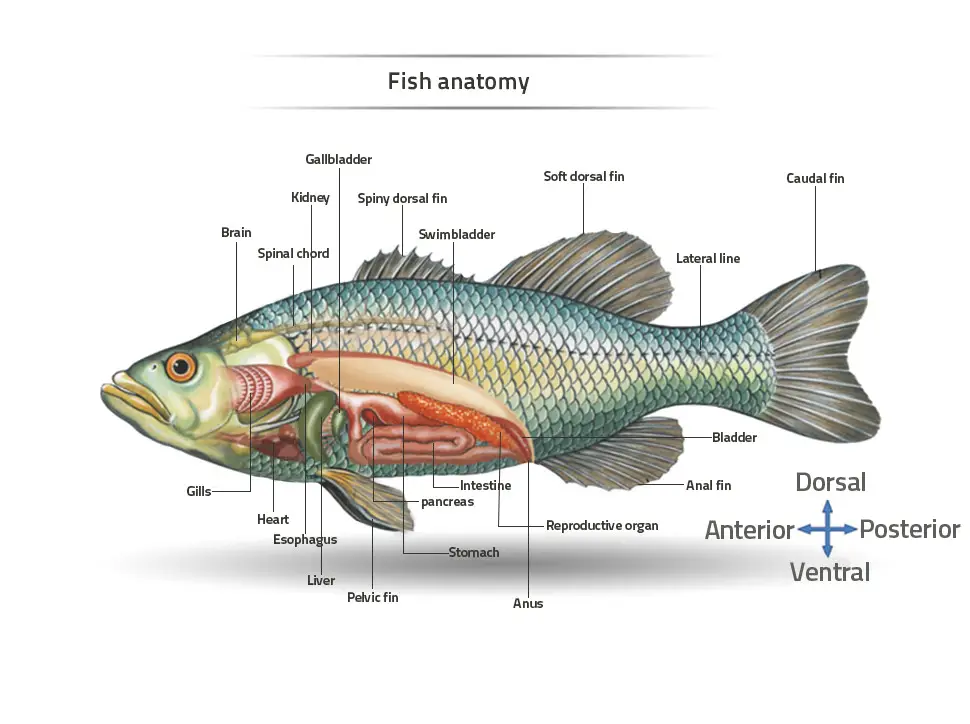
- Fish possess specialized structures known as fins, which are essential for locomotion. Fins can be categorized into two primary types: unpaired (or median) fins and paired fins. The median fins consist of three main components: the dorsal fin located on the back, the anal fin situated on the ventral side, and the caudal fin at the tail’s end. Paired fins include the pectoral fins, which resemble the forelimbs of terrestrial animals, and the pelvic fins, analogous to hind limbs. These fins are supported by skeletal structures known as radials and dermal fin rays. In teleosts, the fin rays are bony, branching structures termed lepidotrichia.
- Fins serve a critical role in the movement and stability of fish in water. Each fin’s location and structure cater to specific functions, including propulsion, maneuvering, stabilization, and braking. For instance, the caudal fin primarily propels the fish forward, while the pectoral fins assist in steering and balancing. Some species exhibit unique adaptations of their fins; for example, flying fish utilize their pectoral fins to glide above the water’s surface, while frogfish use their fins for crawling along the ocean floor. Additionally, fins can serve reproductive functions, as seen in certain shark species, where modified fins assist in the transfer of sperm.
- The composition of fins includes bony spines or rays covered by skin, arranged in a webbed structure in most bony fish. This design allows for flexible movement while providing the necessary support without direct attachment to the vertebral column, relying instead on muscular connections. Furthermore, certain fish have evolved specialized fins for defense or predation, such as the venomous spines in reef stonefish dorsal fins or the anglerfish’s use of a dorsal spine to lure prey.
- In addition to fins, many bony fish possess a swim bladder, an internal gas-filled organ that aids in buoyancy regulation. Positioned between the alimentary canal and the kidneys, the swim bladder allows fish to maintain depth in the water column without expending energy on constant swimming. This organ typically contains gases such as oxygen, enabling the fish to adjust its buoyancy effectively. In some primitive fish, the swim bladder also functions as a lung, providing respiratory support. However, it is essential to note that elasmobranchs, including sharks and rays, lack a swim bladder, relying instead on their body composition and movement to regulate buoyancy.
- The evolutionary significance of the swim bladder is evident in its origin; it develops from the dorsal wall of the gut and receives blood supply from the dorsal aorta. This is in contrast to the lungs of higher vertebrates, which originate from the pharynx and receive blood from the sixth aortic arch. The swim bladder is homologous to lungs, albeit differing in blood supply and developmental origin.
Fish fins Origin and evolution
The origin and evolution of fish fins represent a complex interplay of anatomical adaptations and evolutionary processes. Fins are integral to fish locomotion and have developed into two main types: unpaired (median) fins and paired fins. Median fins include the dorsal fin on the back, the anal fin along the belly, and the caudal fin at the tail’s end. Paired fins consist of the pectoral and pelvic fins. Notably, these structures were absent in ancestral fish forms, indicating a significant evolutionary transition.

The emergence of paired appendages in vertebrates is widely accepted to have occurred independently from invertebrate structures. This divergence can be traced back to ancient ostracoderms, which exhibit primitive appendages. Four primary theories have been proposed to elucidate the origins of paired fins:
- Gill Arch Theory
- Proposes that paired fins evolved from gill arches, suggesting that the pectoral and pelvic girdles formed from gill arches, and their skeletons derived from gill rays.
- This theory posits that early gnathostomes had extended gill arches, which migrated backward in development.
- However, this theory is challenged by several observations:
- It does not explain the involvement of multiple muscle segments in fin formation.
- Paired fins do not appear as a dorsoventral fold during development, but as longitudinal ridges.
- There is insufficient fossil evidence to support the backward migration of pelvic fins.
- The proposed muscle and nerve arrangements do not align with the expected anatomical structures derived from gill arches.
- Overall, this theory faces significant criticism and lacks supporting evidence.
- External Gill Theory
- Suggests that paired fins originated from external gills found in certain early vertebrates, such as crossopterygians and lungfish.
- This theory indicates that gill arches served as supports for these external structures, which were initially respiratory organs.
- Despite superficial similarities, external gills fundamentally differ from the structure and function of paired fins.
- Consequently, this theory has not gained widespread acceptance.
- Lateral Fin Fold Theory
- Posits that paired fins evolved from longitudinal lateral folds in the body.
- According to this hypothesis, the paired fins and their girdles arose from the hardening of these folds, which share a developmental lineage with unpaired fins.
- Evidence supporting this theory includes the presence of extensive muscle bands and nerve networks connecting the pectoral and pelvic fins.
- This theory is bolstered by findings from paleontology and embryology, indicating that paired fins exhibit continuity with the median fins.
- Ostracoderm Theory
- Suggests that the origin of paired appendages can be traced back to ostracoderms, which possessed lateral lobes that may have evolved into pectoral fins.
- The theory postulates that these lobes gave rise to the pectoral girdle, with dermal bones forming from the bony plates of thoracic shields.
- Additionally, some ostracoderms had ventral spines that facilitated movement in swift waters, which could have contributed to the evolution of paired appendages.
- Anatomical and embryological evidence challenges the fin fold theory and emphasizes the significance of ostracoderm features in understanding fin evolution.
Further complexity arises from the positional changes of paired fins throughout evolutionary history. These shifts are not merely the result of migration but may reflect three additional concepts:
- Theory of Intercalation
- Suggests that segments may be added or removed, causing shifts in fin position.
- However, this theory struggles to explain the observed independence of the median and paired fins.
- Theory of Redivision
- Proposes that variations in the number of segments among fins arise from subdivision rather than addition or subtraction of segments.
- This theory lacks clear evidence and fails to provide a compelling explanation for the observed anatomical patterns.
- Theory of Progressive Migration and Transposition
- Argues that the gradual growth of one fin area, combined with a corresponding reduction in another, can lead to positional changes.
- While it aligns more closely with comparative anatomy, it remains an incomplete explanation for the observed fin morphologies.

Types of fins
Primarily, fish fins are classified into two main types: median (unpaired) fins and paired fins. Each type possesses distinct anatomical and functional characteristics, contributing to the overall fitness of fish species.
- Median (Unpaired) Fins
- Median fins include the dorsal, anal, and caudal fins, which are positioned along the midline of the fish’s body.
- These fins develop from a continuous embryonic fin fold that runs dorsally along the fish’s body from the tail to the cloaca.
- In primitive forms, such as lampreys, this embryonic fold retains a more primordial structure, while in advanced bony fishes, it differentiates into distinct fins.
- Formation Process:
- Initially, a tissue fold emerges during embryonic development, forming a continuous structure.
- A series of cartilaginous rods strengthens this fold, leading to the formation of differentiated fins in higher fish species.
- Distinct dorsal, anal, and caudal fins arise from localized concentrations of skeletal elements, specifically radials, while degeneration of the fold occurs in the interstitial spaces.
- Evolutionary Context:
- The middle fins of sturgeons represent a primitive example among modern bony fish, showcasing a fleshy lobe at their bases, encasing the radial and muscular structures.
- Over evolutionary time, this fleshy lobe has diminished in most advanced bony fish, leading to radials that are often reduced to nodules of bone or cartilage.
- Types of Caudal Fins:
- Caudal fins can be classified into three types based on their structural arrangement:
- Protocercal: A primitive type where the tail fin extends symmetrically on both sides.
- Heterocercal: An asymmetrical configuration, with one lobe typically larger than the other, providing lift and thrust.
- Homocercal: A symmetrical fin structure, common in advanced bony fish, facilitating streamlined movement.
- Caudal fins can be classified into three types based on their structural arrangement:
- Paired Fins
- Unlike median fins, paired fins are a more recent evolutionary development, absent in the early ancestors of vertebrates.
- These fins consist of the pectoral and pelvic fins, which play a significant role in steering, balance, and propulsion during swimming.
- Anatomical Structure:
- The original state of paired fins is referred to as the “archipterygium,” characterized by a jointed axis with anterior (preaxial) and posterior (post-axial) sets of radials.
- The radials decrease in size toward the fin tip, arranged on either side of the central axis in a biserial pattern.
- Evolutionary Development:
- The “biserial archipterygium” is primarily observed in the genus Ceratodus from the Devonian period, showcasing an acutely lobate fin shape.
- Over time, the paired fins of modern teleosts evolved into a “pleuroarchic” or uniserial skeletal structure. This transition involved a shortening of the median axis and a reduction of post-axial radials, which eventually disappeared altogether.
- Functional Importance:
- Paired fins are vital for executing precise movements, maintaining stability, and aiding in locomotion, allowing fish to navigate diverse aquatic environments efficiently.
Structure of fins
The structure of fish fins exemplifies a remarkable adaptation that facilitates locomotion, stability, and maneuverability in aquatic environments. These appendages vary significantly in morphology and function, reflecting the diverse evolutionary paths taken by different fish species. Fins can be broadly categorized into two main types: median (unpaired) fins and paired fins. Each category encompasses distinct anatomical features that contribute to the fish’s overall capabilities in its habitat.
- Median (Unpaired) Fins
- Median fins are singular structures positioned along the midline of the fish’s body. These include the dorsal, anal, and caudal fins, each playing critical roles in swimming and stabilization.
- Dorsal Fin:
- Located on the back of the fish, the dorsal fin aids in maintaining balance and preventing rolling during swimming.
- It consists of three segments: proximal, central (or middle), and distal dorsal fins.
- Some species, such as certain boxfish and pufferfish, utilize their dorsal fins in conjunction with their anal fins for propulsion, while Gymnarchus relies solely on its dorsal fin for movement.
- Anal Fin:
- Found on the ventral side, just behind the anus, the anal fin provides stability during swimming and assists in controlling undulatory movements.
- It acts as a counterbalance to the dorsal fin, helping to maintain directional control.
- Caudal Fin (Tail Fin):
- The caudal fin is critical for propulsion and is often referred to as the tail fin. It is typically located at the caudal end of the body, supported by the caudal peduncle, which contains strong swimming muscles.
- This fin can be classified into three distinct types based on its structural arrangement:
- Protocercal Fin: Characterized by an equal division of the caudal fin into upper (epichordal) and lower (hypochordal) lobes, connected to the notochord. This primitive fin type is seen in species such as Amphioxus and Cyclostomata.
- Heterocercal Fin: Found in Chondrichthyes (sharks and rays) and some early bony fish, the heterocercal fin features an asymmetrical shape, with the ventral lobe significantly larger than the dorsal lobe. The notochord extends into the larger lobe, providing enhanced lift and thrust.
- Homocercal Fin: Common in advanced bony fishes, this fin type exhibits symmetrical upper and lower lobes. While the external appearance may suggest equality, the internal structure is asymmetrical, with the vertebral column not extending to the fin’s tip. This design enables efficient forward propulsion and speed.
- Paired Fins
- Paired fins consist of pectoral and pelvic fins, which are critical for maneuverability and stabilization during swimming.
- Pectoral Fin:
- Located on both sides of the fish, typically just behind the operculum, the pectoral fin is homologous to the forelimbs of tetrapods.
- It aids in swimming, providing lift and enabling the fish to execute turns. The pectoral fin’s movement can create dynamic forces that enhance propulsion and stability.
- Pelvic Fin:
- Situated ventrally beneath and slightly behind the pectoral fins, pelvic fins are integral for maintaining position in the water column.
- In some species, such as members of the cod family, pelvic fins may even be located anterior to the pectoral fins. They help in controlling the fish’s vertical movement and can aid in slowing down.
- Adipose Fin:
- Located between the dorsal and caudal fins, the adipose fin is soft and fleshy. It is primarily found in catfishes and serves to stabilize the fish in turbulent waters.
Modifications of fins
Below are some of the specific adaptations observed in caudal fins:
- Leptocercal or Isocercal Fin:
- Characterized by a tapering structure, the isocercal caudal fin possesses a long, straight, rod-like spinal component.
- This fin type provides a balanced shape, aiding in streamlined swimming.
- Species exhibiting isocercal fins include rat tails (family Macruidae), blennies (family Blennidae), eels (order Anguilliformes), featherbacks (family Notopteridae), and gymnarchids (family Gymnarchidae).
- Internally Symmetrical Caudal Fin:
- In this fin type, several fin components are fused, resulting in a more compact structure.
- Internally symmetrical caudal fins are commonly observed in cods (order Gadiformes).
- This fusion can enhance the fin’s efficiency in producing thrust and stability during swimming.
- Pseudo-cercal Caudal Fin:
- Found primarily in Dipnoi (lungfish), the pseudocercal fin is formed when the dorsal and ventral elements develop ventrally before transitioning into fin structures.
- This adaptation reflects a unique evolutionary strategy for navigating complex environments, particularly in shallow waters.
- Hypocercal Caudal Fin:
- The hypocercal fin features a significantly larger dorsal lobe compared to its ventral counterpart, resembling an inverted heterocercal fin.
- This fin type is often found in early Agnathans (jawless fish), where the vertebral axis bends downward, resulting in the dorsal lobe extending further from the body.
- Such a structure can enhance lift and maneuverability in specific swimming scenarios.
- Gephyrocercal Caudal Fin:
- The gephyrocercal fin, often referred to as a bridge caudal fin, presents a unique structure that frequently resembles an isocercal fin.
- However, in this case, the caudal lobe is diminished due to the absence of hypurals in the spinal column.
- Species like pearlfishes (Carpus), Flerasfer, and Orthagoriscus exhibit gephyrocercal fins, which are remnants of more developed structures.
Functions of fins
Below are some of the key functions of fins in fish:
- Pectoral Fins:
- Primarily involved in turning, pectoral fins enable fish to maneuver efficiently in water.
- Certain species, such as Cirrhitichthys, utilize their pectoral fins to stabilize themselves while resting on the seafloor or in reef habitats.
- Flying fish (family Exocoetidae) are noted for their large pectoral fins, which they use to glide above the water’s surface, aiding in evasion from predators.
- Bottom-dwelling fish, including threadfins (family Polynemidae), possess taste buds and touch receptors on their pectoral fins, facilitating the detection of food in their environment.
- Pelvic Fins:
- These fins contribute significantly to a fish’s buoyancy, helping them maintain a stable position in the water column.
- Some species, such as clingfish (family Gobiesocidae), have evolved pelvic fins that function as sucking appendages, enabling them to cling to stationary surfaces on the ocean floor.
- Additionally, fish like the Freshwater Butterflyfish (Pantodon buchholzi) utilize their pelvic fins for gliding, enhancing their movement efficiency in the water.
- The sea robin is another example of a fish that employs its pelvic fins for locomotion along the substrate.
- Dorsal Fins:
- The dorsal fin plays a critical role in maintaining stability and orientation during swimming. It acts as a keel, anchoring the fish in the water.
- Many bony fishes utilize their dorsal fins for quick changes in direction, enhancing their agility in aquatic environments.
- Certain species, such as angelfishes in the phylum Lophiiformes, use their dorsal fins as lures to attract prey, showcasing an evolutionary adaptation for hunting.
- The African knife fish (Gymnarchus niloticus) employs its dorsal fin to create undulations, allowing it to swim both forward and backward.
- In some cases, fish from the Echeneidae family have modified their dorsal fins into sucking discs, enabling them to attach to larger marine animals for transportation.
- Anal Fins:
- The anal fin contributes to the overall stability of the fish and aids in reproductive activities in certain bony fishes.
- By maintaining balance during swimming, the anal fin enhances maneuverability and control.
- Caudal Fins:
- Serving as the primary propulsive appendage, the caudal fin is essential for locomotion in most fish.
- Fast swimmers, such as tunas, possess lunate caudal fins, which enable them to sustain high speeds over extended periods.
- The design and structure of the caudal fin can vary among species, affecting their swimming styles and ecological roles.
Swim bladder species
The swim bladder, also known as the gas bladder, is a vital anatomical feature in many bony fish, facilitating buoyancy and aiding in locomotion. Its structural variations among different species reflect adaptations to diverse ecological niches. Below are detailed observations regarding the swim bladder across various groups of fish:
- Chondrostei:
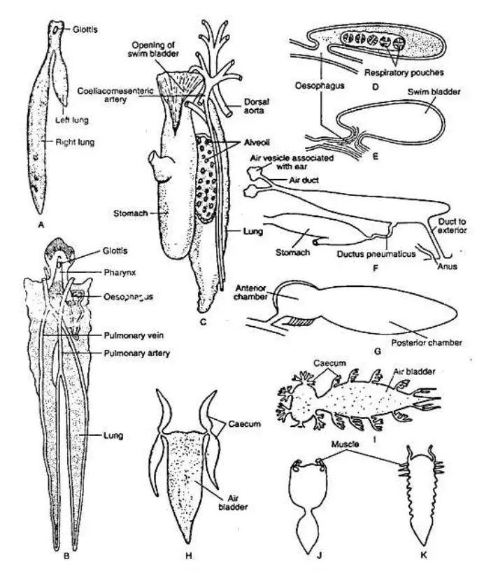
- In the species Polypterus, the swim bladder is characterized by two lobes: a short, oval-shaped left lobe and a longer, tubular right lobe. These lobes converge at the anterior end, forming a single chamber that opens into the esophagus on the ventral side. Notably, this organ lacks internal sacculations, and its walls are smooth.
- The Acipenser species features a similar bladder that is oval-shaped with a smooth wall, accompanied by a prominent glottis, which serves as the entry point to the esophagus.
- Holostei:
- In Lepidosteus, the swim bladder presents as an unpaired, elongated sac with a glottic opening into the esophagus. Its structure includes fibrous bands that form alveoli, which are further divided into smaller sacculi.
- Amia, in contrast, has a significantly larger air bladder than Lepidosteus, characterized by a greater number of smaller alveoli.
- The ductus pneumaticus is notably short in these species, allowing the bladder to attach directly to the esophagus. Unlike other groups, these fish do not possess red bodies or red glands, which are often associated with gas exchange.
- Dipnoi:
- In lungfish such as Neoceratodus, the ventral side exhibits a muscular vestibule leading to the esophagus, while species like Lepidosiren and Protopterus feature double vestibules.
- The swim bladder walls are vascularized, enhancing gas exchange, although visible red bodies or glands are absent. Protopterus also has alveoli that may connect to caecal sacculi, which play a role in respiratory functions.
- Teleostei:
- While the swim bladder is prevalent in many teleost fish, certain groups, including flatfishes (Pleuronectiformes), Saccopharyngiformes, and Echeneiformes, lack this structure entirely.
- The swim bladder can exhibit various shapes—tubular, oval, heart-shaped, and more—depending on the species. For instance, members of the Cyprinidae family have their bladders divided into two intercommunicating chambers.
- In some species, such as those from the Notopteridae, Sparidae, and Carangidae families, the swim bladder extends into the tail region, manifesting as paired caeca.
- Certain highland fish like Psillorhynchus and Nemacheilus have adapted their swim bladders to contain only a small anterior chamber, enclosed in bone, while the posterior portion may be absent.
- Air-breathing fish, such as Heteropneustes fossilis and Clarias batrachus, possess smaller, bony swim bladders that facilitate aerial respiration.
- Caecal Outgrowths:
- Many teleosts exhibit caecal outgrowths resembling fingers sprouting from the swim bladder, particularly in vocal species. For instance, the air bladder of Gadus features two caecal extensions that reach into the cranial region.
- In Otolithus, small tubular outgrowths extend forward and backward from the bladder’s anterolateral wall, while Carvina lobata from the Scianidae family showcases a series of tubular caeca that emerge from its lateral walls.
- Chambered Structure:
- The swim bladder frequently contains two or three chambers, separated by internal septa or partitions. For example, in many fish, the cavity is split into two communicating compartments by a transverse diaphragm, which is regulated by sphincter muscles.
- In the case of Notopterus, a longitudinal wall divides the bladder into two lateral chambers, while a T-shaped septum is present in Mystus seenhala.
- Physostomous and Physoclistous Development:
- Initially, all teleosts are classified as physostomous, possessing a pneumatic duct that opens along the dorsal line. However, many species undergo a transition to physoclistous as they mature, where the duct becomes non-functional.
- In some clupeid species, such as Caranx, Clupea, and Sardinella, the swim bladder opens to the exterior at the hind end, although species like Hilsa and Gadusia lack this feature.

Lepisosteus (E) Acipenser (F) Clupea harengus (G) Essox (H)Gadus (I) Otolithus (J) Corvina lobata (K) Pangassius
Structure of swim bladder
The following points outline the fundamental aspects of swim bladder structure, focusing on its anatomical features and physiological functions:
- General Structure:
- The swim bladder can be classified into two main regions: the anterior and posterior sections. The anterior portion is specialized for gas secretion, while the posterior part facilitates gas absorption into the circulatory system. This dual functionality is essential for regulating buoyancy.
- Specializations in Physoclistous Forms:
- In more specialized physoclistous fish, such as Mugil, Balistes, and Gadus, the posterior section is adapted into an oval shape. This modification is equipped with a sphincter and dilator muscles, which help regulate the opening for gas exchange.
- The red body or red gland, located in the anterior region, serves a crucial role in gas secretion. This gland is particularly developed in certain species of the family Syngnathidae.
- Chamber Division in Specific Families:
- In fish families such as Gadidae, Labridae, and Triglidae, the swim bladder is enclosed and divided into two distinct chambers. The anterior chamber contains the gas gland responsible for gas secretion, while the posterior chamber features thinner walls designed for efficient gas diffusion.
- In the Cyprinidae, the swim bladder is also divided into two chambers but possesses a pneumatic duct connecting them. The anterior chamber primarily serves an auditory function, while the posterior chamber aids in hydrostatic regulation.
- Histological Features:
- The anterior chamber of a cyprinid swim bladder exhibits a multilayered structure:
- Tunica Externa: Composed of dense, fibrous collagen, providing structural support.
- Submucosa: Consisting of loose connective tissue, contributing to flexibility.
- Muscularis Mucosa: A layer of dense smooth muscle fibers that aid in bladder function.
- Lamina Propria: A thin layer of connective tissue supporting the epithelium.
- Epithelial Layer: The innermost layer, consisting of epithelial cells that line the chamber.
- The anterior chamber of a cyprinid swim bladder exhibits a multilayered structure:
- Posterior Chamber Composition:
- The internal epithelium of the posterior chamber varies in height throughout its extent, adapting to specific physiological requirements. A glandular layer exists outside the smooth muscle, featuring large, atypical cells with coarsely granulated cytoplasm.
- Blood capillaries from the rete mirabile are abundant in this glandular layer, providing rich blood supply essential for gas exchange processes.
- Connecting Structures:
- The ductus communicans serves as a muscular connection between the two chambers, reinforced with dense muscle layers supplied by nerve fibers. This muscular structure likely functions as a sphincter, regulating the inter-chamber flow of gases.
- The ductus pneumaticus connects the swim bladder to the esophagus in lower teleosts, with its length and width varying among species. This duct plays a vital role in the transport of gases, primarily oxygen, along with smaller quantities of carbon dioxide and nitrogen.
- Gas Composition:
- The gas secreted by the swim bladder is predominantly oxygen, essential for buoyancy regulation. The ability to adjust gas levels enables fish to maintain their position within the water column, optimizing energy expenditure during swimming.
Types of swim bladder
The swim bladder in fish is an essential organ that varies significantly across species, primarily categorized into two major types: Physostomous and Physoclistous. These classifications are based on the anatomical features and functional mechanisms associated with the connection between the swim bladder and the esophagus. Understanding these types is critical for comprehending how fish regulate buoyancy and gas exchange in their aquatic environments. Below is a detailed overview of the different types of swim bladders:
- Physostomous Condition:
- The physostomous swim bladder is characterized by the presence of a ductus pneumaticus, which connects the swim bladder to the esophagus.
- This connection allows fish to gulp air directly from the water’s surface or release gas into the surrounding environment.
- Blood supply to the swim bladder originates from the coeliacomesenteric artery, which provides oxygen-rich blood that contributes to the organ’s function.
- The blood from the swim bladder is returned to the heart via a vein that joins the hepatic portal vein, facilitating gas exchange.
- Species exhibiting this condition include various Dipnoans (lungfish), soft-rayed teleosts, and several bony fishes.
- The presence of the duct allows for rapid adjustments in buoyancy, making it advantageous in environments where vertical positioning is necessary.
- Physoclistous Condition:
- In contrast, the physoclistous swim bladder lacks an active connection to the esophagus as the ductus pneumaticus is either occluded or significantly reduced.
- This type is typical among spiny-rayed fishes, which rely on a more complex internal gas exchange mechanism.
- The swim bladder features two significant regions:
- Ovale: Located in the posterodorsal area, this region is specialized for gas absorption into the bloodstream.
- Gas Gland: Positioned in the anteroventral section, this gland is responsible for secreting gas into the swim bladder.
- The gas gland receives blood from the coeliacomesenteric artery and branches from the dorsal aorta, which supply oxygen-rich blood to facilitate gas secretion.
- The rete mirabile, a network of blood vessels associated with the gas gland, plays a crucial role in maintaining the gas levels within the swim bladder, ensuring proper buoyancy.
- Blood from the gas gland returns to the heart via the hepatic portal vein, while blood from the remaining parts of the swim bladder is drained through the posterior cardinal veins.
- Innervation is also distinct, with sympathetic nerves innervating the ovale and lateral branches of the vagus nerve influencing the gas gland’s activity.
- Some dipnoans also display this condition, emphasizing the diversity in swim bladder structures across different fish species.
Functions of swim bladder
The following outlines the primary functions of the swim bladder:
- Hydrostatic Organ:
- The swim bladder acts as a hydrostatic organ, balancing the fish’s body weight with the water it displaces.
- By modifying the gas volume within the bladder, fish can adjust their buoyancy, aiding in maintaining their position in the water column.
- In physostomous fish, the presence of the ductus pneumaticus allows for direct expulsion of gas, while in physoclistous fish, excess gas is removed through diffusion, demonstrating different methods of buoyancy control.
- Adjustable Float:
- Serving as an adjustable float, the swim bladder enables fish to swim at varying depths with minimal energy expenditure.
- As a fish descends, the swim bladder can expand, reducing its specific gravity, thereby allowing it to ascend or maintain equilibrium at different depths.
- This mechanism of gas adjustment ensures that fish can navigate their environment efficiently.
- Maintenance of Centre of Gravity:
- The swim bladder aids in maintaining the fish’s proper center of gravity by redistributing gas within its chambers.
- This capability facilitates a range of movements, allowing fish to perform intricate swimming maneuvers and maintain stability in the water.
- Respiratory Function:
- The swim bladder plays a significant role in respiration, especially in environments where oxygen levels are low.
- In certain species, particularly among dipnoans (lungfish), the swim bladder can function as a lung, allowing the fish to intake atmospheric air.
- The oxygen stored in the swim bladder can serve as a supplementary oxygen source, contributing to the overall respiratory efficiency of the fish.
- Sound Resonance:
- The swim bladder also functions as a resonator, amplifying sound vibrations.
- These vibrations are transmitted to the inner ear through specialized structures known as Weberian ossicles, enhancing the fish’s ability to perceive sound.
- This function is particularly important for communication and navigation in the aquatic environment.
- Production of Sound:
- Many fish species utilize the swim bladder for sound generation.
- Fish such as Doras, Platystoma, and Trigla produce drumming, hissing, or grunting noises through vibrations of the incomplete septa located within the swim bladder.
- These sounds result from the movement of trapped air within the swim bladder, which can be enhanced by the contraction of intrinsic and extrinsic muscles.
- Notably, species like Polypterus and Protopterus can actively expel gas from the swim bladder to create sound, indicating a complex interaction between anatomy and behavior.
- https://biologyeducare.com/fish-fins-its-types-and-functions
- https://www.biologydiscussion.com/fisheries/fish/swim-bladder-development-structure-and-types-
fishes/40812