The morphology of insects encompasses their structural features, including body shape, size, and arrangement of various parts. Insects possess a three-part body plan consisting of the head, thorax, and abdomen.
- Head: This segment houses sensory organs, including compound eyes and antennae, as well as the mouthparts, which vary among species based on feeding habits (e.g., chewing, piercing, or sucking).
- Thorax: Comprised of three segments (prothorax, mesothorax, and metathorax), the thorax is primarily responsible for locomotion. Each segment typically bears a pair of legs (three pairs total) and may also support wings, which are crucial for flight in many species.
- Abdomen: The abdomen is segmented and contains vital organs for digestion, reproduction, and excretion. It often houses the reproductive structures, including the ovipositor in females.
Insects also exhibit a variety of appendages and surface modifications that serve specific functions, such as camouflage, locomotion, and sensory perception. Their exoskeleton, made of chitin, provides protection and structural support, while also facilitating mobility through a system of joints. Overall, insect morphology is highly diverse, reflecting adaptations to a wide range of ecological niches and behaviors.
General features of insects
Insects represent a highly diverse and adaptable group within the animal kingdom, primarily characterized by their segmented bodies and specialized structures. The study of insect morphology provides vital insights into their classification, development, and ecological roles. Below are the general features of insects, presented in a structured format for clarity and depth.
- Body Segmentation: Insects exhibit a tripartite body structure, divided into three distinct regions: the head, thorax, and abdomen. This segmentation is a defining characteristic of the phylum Arthropoda.
- Exoskeleton: The external body is covered by a rigid exoskeleton known as the cuticle, primarily composed of chitin. This exoskeleton serves multiple functions, including protection against physical damage and desiccation, as well as providing structural support.
- Jointed Appendages: A hallmark of the class Insecta (or Hexapoda) is the presence of three pairs of jointed legs. These legs enable insects to adapt to a variety of environments, facilitating movement and interaction with their surroundings.
- Wing Classification: Insects can be classified based on the presence or absence of wings.
- Pterygota: This group comprises insects that possess wings, facilitating flight and a wide range of ecological interactions.
- Apterygota: In contrast, this group includes primitive wingless insects, which rely on alternative survival strategies.
- Developmental Types: Insects exhibit three main types of developmental processes, each with distinct characteristics:
- Ametabolous Development: Insects undergoing this type show minimal changes from larvae to adults, maintaining a similar form throughout their life stages.
- Hemimetabolous Development: This involves a gradual transformation where immature forms, known as nymphs, resemble the adults but lack fully developed wings.
- Holometabolous Development: This process is marked by a complete metamorphosis, including a pupal stage where the organism undergoes significant structural changes, transforming from a larval stage to a winged adult.
- Morphological Structures:
- Head: The head region houses critical sensory organs, such as compound eyes and antennae, and is equipped with mouthparts adapted for various feeding strategies.
- Thorax: This segment is responsible for locomotion, containing three pairs of legs and, in winged insects, the wings themselves. The thorax is divided into three parts: the prothorax, mesothorax, and metathorax, each contributing to movement and support.
- Abdomen: Typically segmented, the abdomen contains vital organs for digestion, reproduction, and excretion. Some insects possess specialized structures, such as ovipositors for egg-laying.
- Tagmosis: The process of tagmosis refers to the specialization of body segments, resulting in functional regions that enhance the insect’s efficiency and adaptability. This evolutionary adaptation allows insects to thrive in diverse ecological niches.
- Ecological Roles: Insects play crucial roles in ecosystems as pollinators, decomposers, and prey for other animals. Their varied adaptations allow them to occupy different ecological niches, thus contributing to biodiversity and ecosystem stability.
External morphology of an insect
The external morphology of insects is a crucial aspect of their biological organization, reflecting a highly specialized design that facilitates various functions essential for survival. The body of an insect is generally segmented, featuring distinct regions that are adaptations to environmental and ecological demands. This segmentation is referred to as somite or metamere in more primitive arthropods. Over time, some of these segments have evolved to form cohesive groups known as tagmata, a process termed tagmosis. In insects, the body is divided into three primary regions: the head, thorax, and abdomen.
- Head:
- The head is the anterior region of the insect and plays a vital role in sensory perception and feeding.
- It houses a pair of compound eyes that provide a wide field of vision, along with simple eyes known as ocelli, which help detect light intensity.
- The mouthparts consist of several structures:
- Mandibles: These are hardened jaw-like structures used for biting and grinding food.
- Maxillae: Located behind the mandibles, these assist in manipulating food and may possess sensory functions.
- Labium: This is a lower lip that helps in holding food in place during consumption.
- A pair of antennae is also present, which are sensory appendages used for detecting chemical signals and environmental cues.
- Thorax:
- The thorax is the central region of the insect body and is divided into three segments: the prothorax (first segment), mesothorax (middle segment), and metathorax (last segment).
- Each thoracic segment is reinforced with hardened plates known as sclerites.
- The dorsal plates are referred to as nota or terga (singular: notum), while the lateral plates are termed pleura (singular: pleuron), and the ventral plates are called sterna (singular: sternum).
- The three pairs of legs arise from the thorax, each pair adapted for different functions, such as walking, jumping, or swimming.
- In addition, two pairs of wings may also originate from the thoracic region, providing insects with the ability to fly, which is crucial for escape from predators and seeking resources.
- Abdomen:
- The abdomen serves as the metabolic and reproductive center of the insect, where essential processes such as digestion, excretion, and reproduction occur.
- It generally consists of 11 segments, each playing a role in the insect’s physiology.
- The posterior abdominal segments are often modified for reproductive purposes, including mating and oviposition (egg-laying).
- In many insects, these modifications may include specialized structures such as ovipositors in females, which are adapted for laying eggs in specific substrates.
1. Tagmosis
Tagmosis refers to the evolutionary process through which the body segments of insects are organized into functional units known as tagmata. This structural specialization allows insects to adapt efficiently to various ecological niches. Understanding tagmosis provides insight into the anatomy and functionality of insects, which can be critical for both students and teachers in the biological sciences.
- Segmental Organization: The insect body consists of numerous segments that have undergone sclerotization. Each segment is defined by rigid plates called sclerites, which form distinct areas of the exoskeleton. The segments are interconnected by intersegmental membranes, which are unsclerotized, allowing for flexibility between the articulated sclerites.
- Formation of Tagmata: During the process of tagmosis, the originally segmented body is divided into three main regions:
- Head: Typically formed from six or seven embryologically detectable segments, the head houses vital sensory organs and mouthparts adapted for feeding.
- Thorax: This region is composed of three segments and is primarily responsible for locomotion. It supports three pairs of jointed legs and, in most adult insects, two pairs of wings.
- Abdomen: Generally consisting of nine to eleven segments plus a telson, the abdomen contains reproductive and digestive structures. It is notable for housing sensory organs, such as cerci, and external genitalia in some species.
- Sclerite Composition: Each body segment is comprised of four principal sclerites:
- Tergum: The dorsal plate, forming the upper surface of the segment.
- Sternum: The ventral plate, forming the lower surface.
- Pleura: Two lateral plates that separate the tergum from the sternum. The pleura serve as attachment sites for appendages, enhancing the structural integrity and functionality of the insect.
- Head Structure: The head is characterized by the presence of critical sensory structures:
- Compound Eyes: These provide a broad field of vision and are essential for navigation and predator avoidance.
- Ocelli: In many insects, up to three ocelli are present, contributing to light detection and spatial orientation.
- Antennae: Typically, there is a single pair of antennae, which function in sensory perception. Notably, the order Protura lacks antennae altogether.
- Thoracic Features: The thorax plays a vital role in mobility:
- Legs: It bears three pairs of legs, which are adapted for various forms of locomotion, including walking, jumping, or swimming.
- Wings: Most adult insects possess two pairs of wings, although some may be reduced or absent, reflecting adaptations to their specific environments.
- Abdominal Characteristics: The abdomen is primarily involved in reproductive and digestive functions:
- Segments: It typically consists of nine to eleven segments, providing flexibility and space for various organs.
- Sensory Structures: A terminal pair of cerci, which are sensory appendages, is often present, enhancing the insect’s ability to perceive environmental stimuli.
- Genital Structures: The abdomen may also contain external genitalia, facilitating reproduction.
- Functional Implications: The tagmatization of insects allows for specialization of function within each tagma, leading to greater efficiency in survival tasks. For example, the head’s sensory organs enable effective foraging, while the thorax’s legs and wings enhance mobility.
2. Body Wall (Integument)
The integument of insects, commonly referred to as the body wall, serves multiple essential functions, including protection, support, and prevention of water loss. Comprising two distinct layers—the epidermis and the cuticle—this structure plays a pivotal role in the survival and adaptability of insects across various environments.
- Layer Composition: The integument consists of two main layers:
- Epidermis: This is the inner layer, formed by a single layer of cells. It is responsible for secreting the components of the cuticle and contains ectodermal glands that contribute to various secretions.
- Cuticle: The outer layer, known as the cuticle, provides structural support and protection. It prevents desiccation and acts as a barrier against environmental hazards. The cuticle itself is further divided into two primary layers:
- Epicuticle: This is a thin, waxy, and waterproof outermost layer, which is essential for minimizing water loss.
- Procuticle: Situated beneath the epicuticle, the procuticle is thicker and composed of two sub-layers:
- Exocuticle: This outer layer is rigid and sclerotized, providing additional structural support.
- Endocuticle: This inner layer is tougher and more flexible, composed mainly of chitin and proteins.
- Chitin Structure: Chitin, a long-chain polymer of N-acetylglucosamine, forms the primary structural component of the procuticle. This biochemical composition provides the cuticle with its strength and resilience, enabling it to withstand various physical stresses.
- Glandular Secretions: The epidermis contains various ectodermal glands that secrete substances essential for the insect’s survival. These glands include:
- Wax Glands: Responsible for producing the wax layer that contributes to the waterproofing properties of the epicuticle.
- Lac Glands: Involved in the secretion of protective substances.
- Scent Glands: Used for communication and deterrence against predators.
- Poisonous Glands: Found in some species for defense mechanisms.
- Mucous Glands: Aid in moisture retention and locomotion.
- Pore Canals: The epidermal cells extend cytoplasmic processes into pore canals, which run through the epicuticle. These canals facilitate the transport of cuticular materials to the surface and anchor the cuticle to the underlying epidermis. This structural connection is vital for maintaining the integrity of the integument.
- Exuviae and Molting: As insects grow, the rigid nature of the cuticle necessitates periodic shedding in a process known as molting. The old cuticle, referred to as exuviae, is cast off to allow for the growth of a new, more spacious cuticle. This new cuticle is soft initially and expands before hardening.
- Waterproofing Mechanism: The waterproof properties of the epicuticle arise from its layered structure, which includes:
- Inner Cuticulin Layer: Provides structural support.
- Middle Wax Layer: Composed of long-chain hydrocarbons and fatty acid esters that orient themselves to prevent water penetration.
- Outer Cement Layer: A thin protective layer that safeguards the underlying wax.
- Functional Significance: The integument serves various critical functions beyond mere protection:
- Muscular Support: The cuticle provides an anchoring structure for muscle attachment, enabling movement.
- Environmental Barrier: It acts as a first line of defense against physical damage, pathogens, and desiccation.
- Sensory Functions: Certain modifications of the integument, such as bristles and setae, serve sensory functions, helping insects detect environmental changes.
3. Head
The insect head is a highly specialized structure that serves as the primary site for sensory perception, feeding, and communication. It is composed of a hard, sclerotized capsule formed through the fusion of six distinct segments. This design enables various functional adaptations that are critical for the insect’s survival.
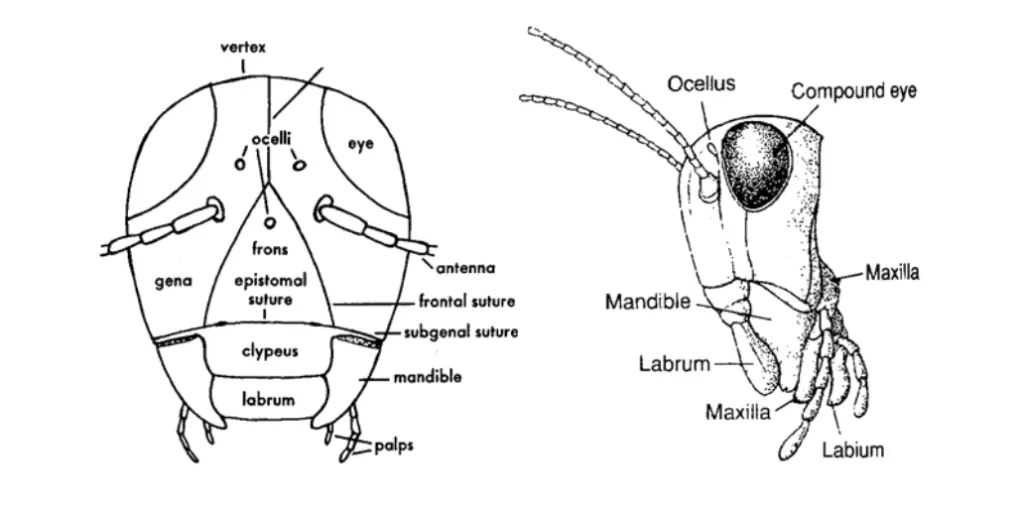
- Structure and Composition:
- The head capsule is predominantly formed by rigid sclerites, which are hardened segments that provide strength and support.
- Major components of the head include a pair of compound eyes, a pair of simple eyes known as ocelli, mouthparts consisting of mandibles, maxillae, and labium, and a pair of antennae.
- Orientation of the Head:
- The orientation of the head varies among insect species, classified into three primary types:
- Hypognathous:
- The mouthparts are oriented downward, aligning with the legs, which is characteristic of many herbivorous insects.
- This orientation is typical of species such as grasshoppers and cockroaches.
- Prognathous:
- The mouthparts are directed forward, facilitating active predation.
- This orientation is observed in carnivorous insects like beetles and ants, as well as in certain larvae that utilize their mandibles for burrowing.
- Opisthorhynchous:
- The elongated proboscis slopes backward between the front legs, common in insects that feed on liquids.
- This type is found in species such as bugs and mosquitoes.
- Hypognathous:
- The orientation of the head varies among insect species, classified into three primary types:
- Sclerites of the Insect Head Capsule:
- The head capsule comprises various hardened sclerites, each fulfilling specific functions:
- Vertex (Epicranium):
- Located on the dorsal side of the head, between the eyes, and is associated with sensory organs like ocelli and antennae.
- Frons:
- Positioned on the anterior face and contains the median ocellus. It is bounded ventrally by the frontoclypeal suture.
- Clypeus:
- A tip-like structure situated between the frontoclypeal suture and the labrum, connecting the two structures.
- Labrum:
- Known as the upper lip, it is a simple, fused sclerite that functions in food manipulation.
- Gena:
- The lower part of the head, located beneath the eyes and posterior to the frons. It is separated from the frons by a general suture.
- Post gena:
- These sclerites are located below the genae and above the mandibles, contributing to the structural integrity of the head.
- Occiput:
- This area comprises most of the head’s posterior region and is divided from the vertex and genae by the occipital suture.
- Post-occiput:
- This ring-like structure forms the margin of the occipital foramen, which connects the head to the body.
- Ocular sclerites:
- These ring-like structures surround the compound eyes, providing additional support and protection.
- Antennal sclerites:
- These sclerites form the base for the antennae and are notably well-developed in certain groups such as stoneflies.
- Vertex (Epicranium):
- The head capsule comprises various hardened sclerites, each fulfilling specific functions:
- Sutures of the Insect Head:
- The head capsule features several distinct sutures that separate the sclerites:
- Epicranial Suture (Ecdysial Suture):
- An inverted “Y” shaped suture that demarcates the vertex from the frons. This suture is critical during molting, as it is a line of weakness.
- Fronto-Clypeal Suture (Epistomal Suture):
- This line separates the frons from the clypeus.
- Clypeo-Labral Suture:
- A line that separates the clypeus from the labrum.
- Fronto-Genal Suture:
- Located on either side of the head, this suture distinguishes the facial region from the gena.
- Sub-Genal Suture:
- This line runs beneath the gena on both sides of the head.
- Occipital Suture:
- A “U”-shaped line that separates the occiput from the post-occiput.
- Post-Occipital Suture:
- The only true suture that divides the maxillary and labial segments, separating the head from the neck.
- Antennal Suture:
- A marginal depressed ring surrounding the antennal socket.
- Epicranial Suture (Ecdysial Suture):
- The head capsule features several distinct sutures that separate the sclerites:
a. Mouth parts
Insects exhibit a remarkable diversity in their mouthparts, which are specifically adapted to their feeding habits and ecological niches. The structure and function of these mouthparts are critical for the insect’s survival, as they play a central role in the uptake of food. Typically, the insect mouth consists of five main components, each serving distinct functions in the feeding process.
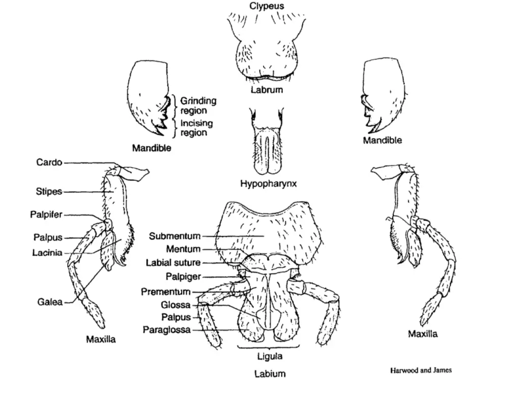
- Components of Insect Mouthparts:
- Upper Lip (Labrum):
- This structure is a simple fused sclerite that functions as the upper lip. It moves longitudinally and is hinged to the clypeus, assisting in the manipulation of food.
- Anterior Jaws (Mandibles):
- Highly sclerotized and paired, mandibles are robust structures that move laterally (at right angles to the body). Their primary function is to bite, chew, and sever food, making them essential for insects that consume solid materials.
- Accessory Jaws (Maxillae):
- The maxillae are paired structures that also move laterally and are equipped with segmented palps. These appendages aid in holding and positioning food for ingestion, thereby playing a supportive role in the feeding process.
- Lower Lip (Labium):
- The labium, often referred to as the lower lip, is another fused structure that moves longitudinally. It also features a pair of segmented palps, contributing to food manipulation and the ingestion process.
- Tongue-like Structure (Hypopharynx):
- The hypopharynx is a specialized structure that aids in the delivery of saliva to the food, facilitating digestion.
- Upper Lip (Labrum):
- Classification of Mouthparts:
- Insects can generally be classified into two main categories based on their mouthpart adaptations:
- Chewing and Biting Type (Mandibulate):
- This type is considered primitive and is characterized by mouthparts that are suitable for biting and chewing solid food. Examples include:
- Nymphs and adults of grasshoppers and cockroaches.
- Adult beetles and caterpillars.
- This type is considered primitive and is characterized by mouthparts that are suitable for biting and chewing solid food. Examples include:
- Sucking Type (Haustellate):
- This category includes insects that have evolved specialized mouthparts for sucking liquids. Examples include:
- Aphids and bugs.
- Mosquitoes and lice.
- This category includes insects that have evolved specialized mouthparts for sucking liquids. Examples include:
- Chewing and Biting Type (Mandibulate):
- Insects can generally be classified into two main categories based on their mouthpart adaptations:
- Variations in Mouthparts:
- The diversity in mouthpart structures leads to various modifications adapted to specific feeding strategies:
- Piercing and Sucking Type: Found in insects like aphids and mosquitoes, these mouthparts are designed to penetrate plant or animal tissues to extract fluids.
- Rasping and Sucking Type: Present in thrips, these mouthparts allow for scraping and sucking up plant juices.
- Sponging Type: Characteristic of house flies, this adaptation allows them to soak up liquid food through a sponging mechanism.
- Chewing and Lapping Type: Found in honey bees, these mouthparts are used for both chewing solid substances and lapping up liquids.
- Siphoning Type: Adapted for feeding on nectar, butterflies and moths possess elongated mouthparts that function like a straw.
- Mask Type: Present in the aquatic nymphs of dragonflies, this type allows for capturing prey efficiently.
- Degenerate Type: Exhibited by maggots, these mouthparts are reduced and adapted for a specific diet, often associated with decaying organic matter.
- The diversity in mouthpart structures leads to various modifications adapted to specific feeding strategies:
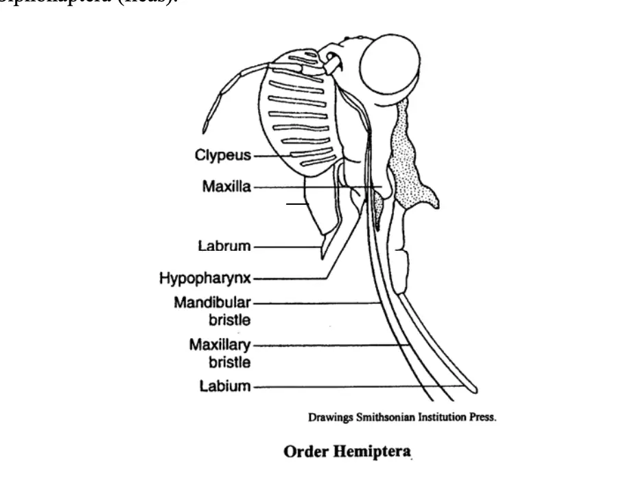
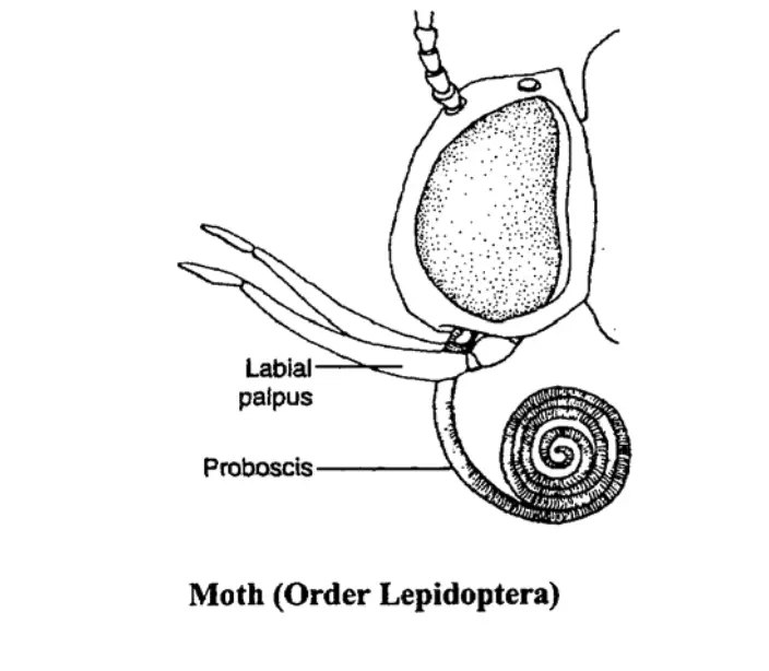
Mouth Part and its Modifications
The mouthparts of insects exhibit remarkable diversity, reflecting adaptations to various feeding strategies. Understanding these modifications provides insights into the ecological roles and feeding behaviors of different insect groups.
- Chewing-Lapping Type:
- Found in honey bees, these mouthparts are specialized for nectar and pollen collection, as well as wax manipulation.
- The mandibles are short and spoon-shaped, located on either side of the labrum, and are primarily used for shaping wax.
- The labium is modified with reduced paraglossae and well-developed glossae, forming an elongated tongue-like structure.
- A small structure called the labellum (or flabellum), resembling a honey spoon, is present at the tip of the glossae.
- The labial palps are elongated, and together with the galea, they form a tube around the glossae, enabling the bee to collect nectar efficiently by moving the glossae up and down within this tube.
- Biting and Chewing Type:
- Common in insects like cockroaches, this type features a rectangular labrum and paired mandibles equipped with inner teeth.
- The mandibles work in opposition, effectively masticating food.
- Each maxilla lies behind the mandibles, equipped with tactile maxillary palps that assist in food manipulation.
- The labium also plays a role in holding and pushing food toward the mouth, demonstrating a coordinated mechanism for processing solid food.
- Piercing and Sucking Type:
- Present in mosquitoes, this type is characterized by an elongated proboscis, which is a modified labium.
- The labial palps are transformed into labella at the proboscis tip, while the labrum is needle-like, covering the labial groove.
- The mandibles, maxillae, and hypopharynx are elongated and situated within the labial groove, forming a food channel.
- When feeding, the stylets separate to create a channel for blood to be drawn in. The hypopharynx includes a salivary channel that injects saliva into the host’s bloodstream, preventing coagulation.
- Bed bugs display similar adaptations, with elongated stylets derived from mandibles and maxillae, allowing them to pierce skin effectively and draw blood.
- Sucking Type:
- House flies possess mouthparts adapted for liquid feeding. They have a modified labium that forms a long, retractable proboscis.
- The proboscis consists of three parts: the rostrum, the haustellum, and the labella.
- The haustellum features a mid-dorsal oral groove with a blade-like hypopharynx located deep within this groove, equipped with a salivary channel.
- Labella at the proboscis’ distal end contain fine channels called pseudotracheae, allowing the house fly to suck up fluids efficiently.
- Siphoning Type:
- This type is found in butterflies and moths, adapted for feeding on nectar from flowers.
- The mouthparts include a small labrum and a coiled proboscis formed from modified galeae of the maxillae.
- Mandibles and labium are significantly reduced, and the hypopharynx is absent.
- The proboscis is elongated and can coil beneath the head when not in use. During feeding, it straightens to access nectar, employing capillary action to draw the liquid up through the food channel formed by the opposing galeae.
4. Antenna
Insects possess specialized sensory appendages known as antennae, which play a vital role in their interaction with the environment. These structures are primarily responsible for detecting chemical signals, vibrations, and other stimuli, thereby facilitating communication and navigation. The anatomy of the insect antenna consists of several distinct components that contribute to its functionality.
- Anatomy of the Antenna:
- Basal Components:
- The antenna is composed of three main parts: the scape, the pedicel, and the flagellum.
- Scape: This basal segment is attached to the head wall at a flexible joint called the antennifer, allowing for multi-directional movement.
- Pedicel: This segment connects the scape to the flagellum and functions as a transitional component.
- Flagellum: This elongated portion is made up of numerous segments, or annuli, that are interconnected by membranes, providing flexibility and range of motion.
- The antenna is composed of three main parts: the scape, the pedicel, and the flagellum.
- Muscle Control:
- The movement of the antennae is controlled by a complex system of muscles. Levator and depressor muscles arise from the anterior tentorial arms and attach to the scape, while flexor and extensor muscles originate in the scape and connect to the pedicel. This muscular arrangement facilitates precise and adaptable movements.
- Basal Components:
- Types of Insect Antennae:
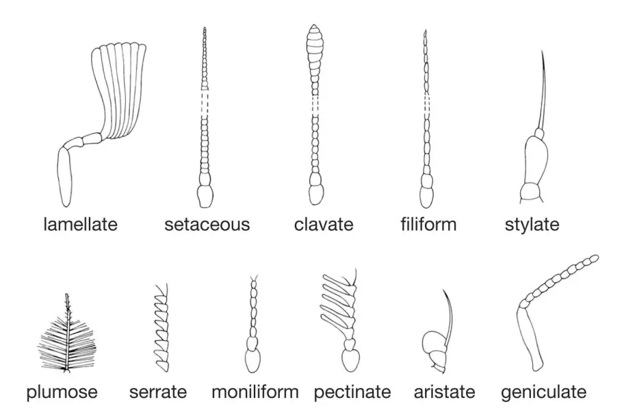
- The diversity of antennae among insects reflects their ecological niches and sensory requirements. Various types include:
- Setaceous: Characterized by a bristle-like appearance, these antennae decrease in size from base to apex and end in a bristle (e.g., leafhoppers, dragonflies).
- Filiform: Resembling a thread, these antennae consist of numerous cylindrical segments (e.g., orthopterans, moths).
- Moniliform: Beaded in appearance, these antennae have globular segments separated by prominent constrictions (e.g., termites).
- Clavate: These antennae gradually enlarge toward the tip, resembling a club (e.g., blister beetles).
- Capitate: The terminal segments are significantly enlarged, forming a knobbed structure (e.g., butterflies).
- Hooked: These feature a hooked end at the tip of the antenna (e.g., skippers, sphingids).
- Bipectinate: Exhibiting a double comb-like structure, these have long slender lateral processes on both sides (e.g., silkworm moths).
- Unipectinate: Similar to bipectinate but with processes only on one side (e.g., sawflies).
- Plumose: Known for their feathery appearance, these antennae have dense, long whorls of hairs (e.g., male mosquitoes).
- Pilose: Less feathery, these antennas possess fewer hairs at the junction of flagellomeres (e.g., female mosquitoes).
- Aristate: Comprising three segments, these antennae bear a terminal bristle known as an arista (e.g., house flies).
- Stylate: These have three segments with a terminal segment ending in a style-like process (e.g., robber flies, horse flies).
- Serrate: Characterized by saw-like segments with short triangular projections (e.g., longhorned beetles, jewel beetles).
- Lamellate: Small-sized with laterally expanded, flat plates at the tip (e.g., rhinoceros beetles, ground beetles).
- Geniculate: Elbowed in structure, where the basal scape is longer, and the remaining segments are angled (e.g., ants, honey bees).
- Flabellate: Very small antennae with side processes that create a fan-like arrangement (e.g., strepsipterans).
- The diversity of antennae among insects reflects their ecological niches and sensory requirements. Various types include:
Modifications in antennae
The modifications of antennae in insects reflect adaptations to various ecological niches and sensory requirements. Each type serves unique functions that facilitate environmental interaction, navigation, and communication. Understanding these variations is essential for comprehending insect behavior and ecology.
- Setaceous Antennae:
- Found in cockroaches, this type features segments that gradually narrow toward the apex.
- The tapered structure may enhance sensitivity to air movement or chemical signals.
- Filiform Antennae:
- Present in grasshoppers, these antennas have annuli of uniform width, resulting in a slender, rod-like appearance.
- This design likely aids in the detection of subtle environmental changes.
- Moniliform Antennae:
- Observed in termites, these antennas consist of annuli that resemble beads or ovoid shapes.
- The beaded structure may enhance tactile sensitivity, crucial for communication and navigation within colonies.
- Pectinate Antennae:
- Characteristic of sawflies, beetles, and moths, this type has flagellum annuli that feature projections on one side, resembling a comb.
- The comb-like structure may function to increase the surface area for sensory reception.
- Serrate Antennae:
- Found in pulse beetles, these antennae have tooth-like projections along the annuli.
- This serrated design might improve grip or tactile sensitivity during interaction with the environment.
- Clavate Antennae:
- Observed in butterflies and moths, clavate antennas are club-like, with the distal annuli gradually broadening.
- The club shape may enhance the ability to detect chemical signals, aiding in mate selection.
- Capitate Antennae:
- Seen in the Khapra beetle, these antennas feature an abruptly clubbed end.
- The capitate structure is likely adapted for specific sensory functions, possibly enhancing tactile or olfactory capabilities.
- Geniculate Antennae:
- Characteristic of ants, these antennas exhibit an elbow joint where the pedicel connects to the flagellum.
- This jointed structure allows for versatile movement, improving the insect’s ability to sense its surroundings.
- Lamellate Antennae:
- Found in bark beetles, these antennas have distal annuli that extend into leaf-like plates.
- The lamellate design likely increases the surface area for sensory receptors, aiding in environmental detection.
- Plumose Antennae:
- Seen in female mosquitoes, these antennas have thick, long whorls of hair at the base of the flagellum annuli.
- The plumose structure enhances the sensitivity to air currents, crucial for locating mates.
- Pilumose Antennae:
- Also found in female mosquitoes, these antennas feature short, thin whorls of hair at the base of the annuli.
- The pilumose design aids in detecting subtle changes in air movement, facilitating communication during mating.
- Flabellate Antennae:
- Observed in male stylops, these antennas have a basal annulus modified into a fan-like structure.
- The flabellate design may enhance chemical detection, critical for reproductive behavior.
- Bipectinate Antennae:
- Characteristic of silk moths, these antennas feature long, stiff projections on both sides of the annuli.
- The bipectinate structure may significantly enhance olfactory sensitivity.
- Aristate Antennae:
- Found in houseflies, these antennas consist of a single-segmented flagellum bearing a stiff hair known as an arista.
- The arista, with hairs on both sides, may improve tactile sensitivity, aiding in navigation.
5. Eyes
Insects exhibit a remarkable diversity of visual systems, primarily categorized into simple eyes (ocelli) and compound eyes. Each type plays a vital role in how insects perceive their environment, enabling them to adapt to various ecological niches.
- Simple Eyes (Ocelli):
- Found in many adult insects and some nymphs, simple eyes usually number three, arranged in a triangular formation between the compound eyes on the head.
- The transparent cuticular layer covering the ocellus functions as a lens, while the underlying epidermis remains transparent and colorless.
- Beneath the epidermis, nerve cells are organized into groups that form rhabdomeres, with pigment possibly present among these groups.
- Unlike compound eyes, ocelli do not form images; instead, they are sensitive to changes in light intensity.
- Lateral ocelli, known as stemmata, are present in the larvae of certain insect orders and serve solely as visual organs, enhancing their ability to detect light.
- Compound Eyes:
- Compound eyes serve as the primary visual organs in insects and crustaceans, characterized by numerous repeating units called ommatidia.
- The number of ommatidia can range from a few to thousands, each functioning as an independent photoreception unit with its optical system and light-sensitive cells.
- Ommatidia typically exhibit a hexagonal shape, contributing to the overall compound eye’s mosaic structure.
- Each ommatidium contains a focusing system, light-sensitive cells, and screening pigment to delineate the individual units.
- The transparent, biconvex cuticular end of the ommatidium, known as the lens (or cornea), is rigid and fixed, possessing a consistent focal length.
- Images formed by compound eyes create a mosaic effect, where each ommatidium captures light from a single point, and the brain integrates these signals to form a composite image.
- Types of Compound Eyes:
- Compound eyes can be further classified into two primary types based on their adaptation to light conditions: appositional and superpositional.
- Apposition Eyes:
- In bright light, screening pigments are maximally dispersed within the ommatidia, which prevents light from one ommatidium from affecting its neighbors.
- Each ommatidium responds to a single point of light, sending a corresponding signal to the brain that is perceived as a distinct point in the overall mosaic image.
- This type of eye is common in insects such as honeybees and locusts.
- Beneath the cornea, the crystalline cone acts as a second lens, and the retinular cells, containing microvilli, form the sensory complex of the ommatidia.
- Superposition Eyes:
- Adapted for low-light conditions, superposition eyes operate by allowing light to enter multiple ommatidia due to the contraction of pigment cells.
- This configuration permits the rhabdomes to capture light from various lenses, leading to the formation of a superposition image.
- This mechanism allows nocturnal insects to optimize their vision in dim lighting, facilitating their activity in such environments.
Difference between apposition and superposition eyes.
Understanding the differences between apposition and superposition eyes is essential for appreciating how various insects adapt their visual systems to their environments. Each type of eye has unique structural and functional characteristics that enable them to perform optimally under specific lighting conditions.
- Fundamental Differences:
- Light Reception:
- In apposition eyes, each rhabdom (the light-sensitive structure) in an ommatidium (the individual optical unit) receives light solely from its corresponding cornea, ensuring precise localization of light sources.
- Conversely, superposition eyes allow the rhabdom to receive light from multiple corneas, leading to a more generalized perception of light rather than distinct light sources.
- Presence of Cone Stalk:
- Apposition eyes lack a cone stalk, maintaining a direct connection between the crystalline cone and the rhabdom.
- In superposition eyes, a cone stalk is present, linking the crystalline cone and the rhabdom, which facilitates the integration of light from various ommatidia.
- Light Reception:
- Development of Screening Pigments:
- The screening pigments in apposition eyes are well developed, creating a curtain of pigment that separates adjacent ommatidia, thereby reducing crosstalk between them.
- In contrast, superposition eyes have concentrated screening pigments that do not form a barrier between neighboring ommatidia, which allows for a collective reception of light.
- Crystalline Cone Length:
- In apposition eyes, the length of the crystalline cone is approximately equal to its focal length, ensuring effective light focusing.
- Superposition eyes exhibit a crystalline cone that is about twice its focal length, which aids in capturing more light in dim conditions.
- Connection Between Structures:
- In apposition eyes, the lower end of the cone is contiguous with the upper end of the rhabdom, facilitating direct light transmission.
- Superposition eyes feature a discontiguous relationship between the cone and the rhabdom, connected instead by a cone stalk.
- Reticular Cell Structure:
- The reticular cells in apposition eyes are elongated, extending from the crystalline cone to the basement membrane, providing an efficient pathway for light transmission.
- In superposition eyes, these reticular cells are relatively short and remain confined to the bases of the ommatidia, limiting their extension.
- Image Formation:
- Apposition eyes create a complete image of the visual field, with each single point of light corresponding to a distinct point in the resulting mosaic image.
- Superposition eyes produce either a blurred image or no image at all; however, the brain can still register the presence of light in the environment, indicating a broader awareness rather than detailed visualization.
- Adaptation to Light Conditions:
- Apposition eyes are well-suited for bright light environments, making them typical in diurnal insects, such as honeybees.
- On the other hand, superposition eyes are adapted for weak or dim light, commonly found in nocturnal insects, like moths.
6. Thorax
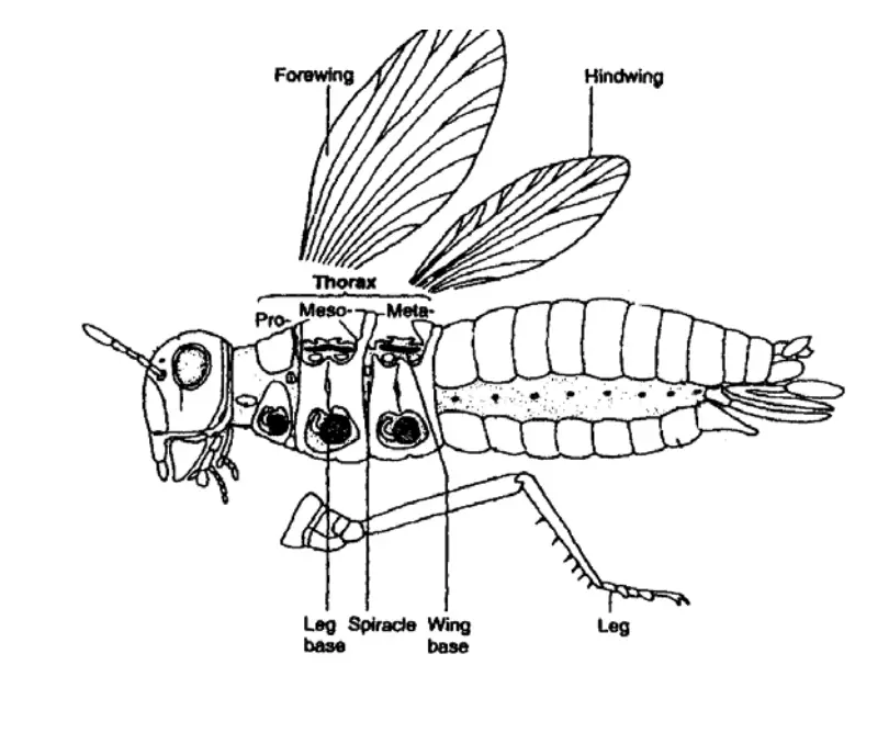
The thorax is a vital anatomical structure in insects, consisting of three distinct segments: the prothorax, mesothorax, and metathorax. This region plays a crucial role in locomotion and flight, contributing significantly to the insect’s overall functionality and mobility. The thoracic segments exhibit notable differences in size and morphology, particularly between winged and wingless insects.
- Segmental Structure:
- The thorax comprises three segments:
- Prothorax: The first segment, generally smaller than the other two.
- Mesothorax: The second segment, which is usually larger and equipped with the first pair of wings.
- Metathorax: The third segment, which supports the second pair of wings.
- In Apterygotes (wingless insects), all three segments are of equal size. However, in pterygote (winged) insects, the mesothorax and metathorax are more developed, leading to a combined structure known as the pterothorax.
- The thorax comprises three segments:
- Leg and Wing Configuration:
- Each thoracic segment possesses one pair of legs, resulting in three pairs of legs overall. These legs primarily serve locomotory functions but can also have roles in other activities such as grasping and mating.
- The mesothorax and metathorax each house one pair of wings, enhancing flight capability.
- Respiratory Structures:
- The second and third thoracic segments contain spiracles (small openings for respiration), with one pair located laterally on each segment. These spiracles facilitate gas exchange, crucial for the insect’s metabolic processes.
- Tergal Modifications:
- In ametabolous insects (those that do not undergo metamorphosis), tergal plates are relatively simple. In contrast, winged insects exhibit various modifications in these plates to accommodate their flight mechanisms.
- Regional Division of Thoracic Segments:
- Each thoracic segment is divided into four distinct regions:
- Tergum (Notum): The dorsal region, which plays a role in protecting the thoracic organs and supporting wing attachment. In the prothorax, the pronotum is generally smaller but can be expanded in species such as cockroaches to shield the head.
- Pleura: The lateral regions of each segment, where the legs originate. The pleura are further subdivided into two sclerites: the anterior episternum and the posterior epimeron.
- Sternum: The ventral region, divided into four sclerites: the prosternum, basisternum, sternellum, and spina-sternum. This area supports various muscular attachments necessary for movement.
- Each thoracic segment is divided into four distinct regions:
- Notum Structure in Winged Insects:
- The notum of winged insects is typically divided into two major components:
- Alinotum: The anterior section bearing the wings.
- Postnotum: The posterior section, which includes a structure known as the phragma that provides additional support.
- The notum is also further divided into three sclerites: the prescutum, scutum, and scutellum, contributing to the structural complexity of the thorax.
- The notum of winged insects is typically divided into two major components:
- Neck Connection:
- The thorax connects to the head via a membranous structure referred to as the cervix (or neck). This connection is essential for the mobility of the head relative to the thorax.
a. Legs
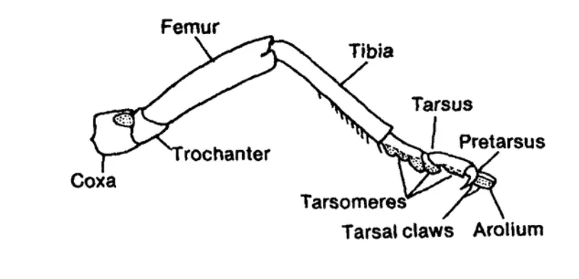
Insects, as members of the class Hexapoda, possess a unique anatomical structure characterized by three pairs of legs, with one pair situated on each of the thoracic segments. This six-legged design is a defining feature of insects and reflects their evolutionary adaptations for various ecological niches. Each leg comprises six segments that articulate through mono-condylic joints, enabling a wide range of motion. The basic segments include the coxa, trochanter, femur, tibia, tarsus, and pretarsus, all of which have undergone modifications to serve specific functions such as locomotion, foraging, and habitat adaptation.
- Basic Structure of Insect Legs:
- Coxa: The proximal segment connecting the leg to the thorax.
- Trochanter: A small segment that allows for leg articulation.
- Femur: The longest segment, often adapted for specific functions.
- Tibia: The segment that provides leverage for movement.
- Tarsus: Composed of multiple smaller segments, contributing to balance and grip.
- Pretarsus: The distal part of the leg, often equipped with claws or adhesive pads.
- Functional Adaptations of Insect Legs:
- Ambulatory Legs:
- Examples: Forelegs and middle legs of grasshoppers.
- Characteristics: Femur and tibia are elongated, adapted primarily for walking and locomotion.
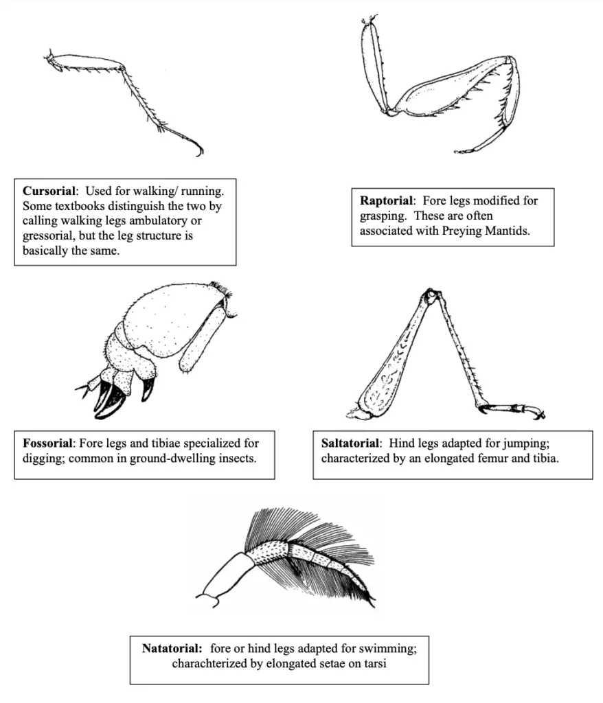
- Cursorial Legs:
- Examples: All three pairs of legs in cockroaches.
- Characteristics: Legs are suited for running, with non-swollen femurs for speed and agility.
- Saltatorial Legs:
- Examples: Hind legs of grasshoppers.
- Characteristics: Specialized for leaping, allowing for rapid escape from predators.
- Fossorial Legs:
- Examples: Forelegs of mole crickets.
- Characteristics: Adapted for digging and burrowing into the ground.
- Natatorial Legs:
- Examples: Hind legs of water bugs and water beetles.
- Characteristics: Modified for swimming, often flattened and fringed to enhance propulsion.
- Raptorial Legs:
- Examples: Forelegs of preying mantids.
- Characteristics: Equipped for grasping and capturing prey, featuring spines for better grip.
- Scansorial Legs:
- Examples: All three pairs of legs in head lice.
- Characteristics: Adapted for climbing and clinging to surfaces.
- Foragial Legs:
- Examples: Legs of honey bees.
- Characteristics: Equipped with specialized structures for collecting food and pollen.
- Ambulatory Legs:
- Structures of the Honey Bee’s Legs:
- Forelegs:
- Contain three critical components: eye brush for cleaning, antenna cleaner (strigillis), and a pollen brush for collecting pollen.
- Middle Legs:
- Feature a pollen brush formed by stiff hairs on the basitarsus and a tibial spur to assist in loosening pollen pellets from hind legs and cleaning wings.
- Hind Legs:
- Equipped with three essential structures:
- Pollen Basket (Corbicula): A shallow cavity on the hind tibia, fringed with long hairs, allowing for efficient pollen transport.
- Pollen Packer (Pollen Press): Consists of pecten (a row of stout bristles) and auricle (a small plate) to compact pollen for transport.
- Equipped with three essential structures:
- Forelegs:
- Specialized Legs in Other Insects:
- Climbing or Sticking Legs: Found in all three pairs of legs of house flies, adapted for adhesion to surfaces.
- Clasping Legs: Present in forelegs of male water beetles, facilitating grasping during mating.
- Prolegs in Caterpillars: Caterpillars possess three pairs of thoracic legs (true legs) and five pairs of abdominal prolegs.
- True Legs: Jointed and sclerotized, providing mobility.
- Prolegs: Unjointed, fleshy, and equipped with hooks (crochets) for attachment to surfaces. In some species, prolegs are absent on certain segments, leading to distinctive locomotion patterns (e.g., semi-loopers).
Modification in legs
Insects exhibit a remarkable variety of leg modifications that enhance their survival and adaptability to specific environments and lifestyles. These adaptations are primarily influenced by the ecological niches they occupy, allowing insects to perform essential functions such as locomotion, predation, and even grooming. Understanding these modifications provides valuable insight into the evolutionary biology of insects.
- Ambulatory Legs:
- Characterized by long and cylindrical structures, these legs are typically found in black field crickets. They are well-adapted for walking, allowing for efficient movement across various substrates.
- Cursorial Legs:
- Commonly seen in cockroaches, these legs are cylindrical and elongated, with well-developed coxae. This structure facilitates fast running, making it easier for these insects to escape predators and navigate their environments swiftly.
- Saltatorial Legs:
- Found in grasshoppers, these legs feature greatly enlarged hind femurs that house strong muscles, making them adept for jumping. This adaptation enables quick escapes from threats and efficient movement through their habitats.
- Scansorial (Clinging) Legs:
- Lice possess stout tibiae with a unique thumb-like process known as the tibial thumb. Their single-segmented tarsus is equipped with a curved claw called the tarsal claw, allowing them to grasp hair fibers or cloth effectively. This adaptation is crucial for their parasitic lifestyle.
- Fossorial Legs:
- Mole crickets display legs modified for digging. The tibia and tarsus are flattened and equipped with blunt spines, resembling a shovel. This adaptation facilitates burrowing into the ground, aiding in their subterranean lifestyle.
- Raptorial Legs:
- Present in mantises, these legs are specialized for catching and holding prey. The ventrally grooved femur and sickle-shaped tibia allow mantises to swiftly and effectively grasp their food, showcasing a predatory adaptation.
- Web-Spinning Legs:
- In Embia species (order Embioptera), the metatarsus is enlarged to house silk glands, enabling the secretion of silk used in constructing webs. This modification allows for the creation of protective and functional silk structures.
- Natatorial Legs:
- Found in species like Belostoma indica, these legs are broad and flattened, fringed with dense hairs, forming oar-like structures. This adaptation is essential for swimming, allowing insects to navigate through aquatic environments efficiently.
- Scooping Legs:
- Dragonflies and damselflies possess long legs with rows of stiff bristles positioned anteriorly. These adaptations are utilized to seize prey mid-flight, enhancing their hunting capabilities.
- Bladder-Footed Legs:
- Present in thrips, these legs feature distal tarsomeres with vesicles that enhance grip on feeding surfaces. This adaptation is vital for their feeding strategies.
- Antennae Cleaning Legs:
- Found in the forelegs of honey bee workers, these legs are specialized for cleaning the antennae. The metatarsus contains a semicircular bristle notch for fitting the antenna base, while a spur on the tibia helps remove pollen grains and debris effectively.
- Pollen Brushing Legs:
- Also in honey bee workers, the middle legs have bristled metatarsi that are adapted to brush pollen into heaps. The tibial spurs assist in cleaning pollen and wax from the pollen basket and the ventral surface of the abdomen.
- Pollen Collecting Legs:
- The hind legs of honey bee workers exhibit a concave cavity on the outer surface of the tibia, known as the pollen basket, fringed with hairs. This structure is critical for storing pollen grains, while the rows of hair on the inner metatarsus help in collecting and retaining pollen.
b. Wings
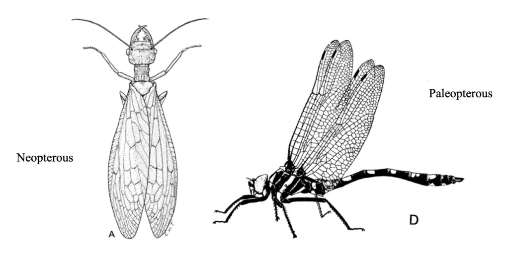
Insects are remarkable creatures, possessing a variety of wing structures that enable diverse modes of locomotion and adaptation to their environments. Fossil evidence indicates that wings have existed since the Carboniferous period, marking a significant evolutionary development in the class Insecta. Insects classified under the groups Endoterygota and Exoterygota typically have two pairs of wings—forewings and hindwings—located on the mesothorax and metathorax, collectively referred to as the pterothorax. Conversely, members of Aterygota lack wings entirely.
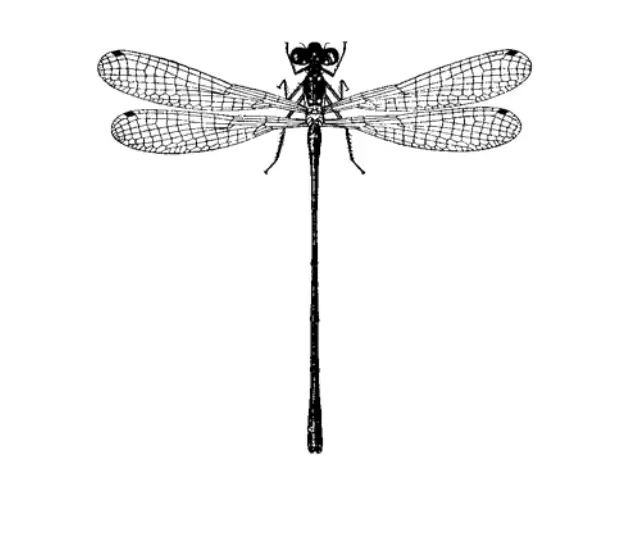
- Types of Insect Wings and Their Modifications:
- Tegmina (singular: Tegmen):
- These wings exhibit a leathery or parchment-like texture and serve primarily as protective coverings rather than for flight. They are characteristic of certain insects, such as cockroaches and grasshoppers.
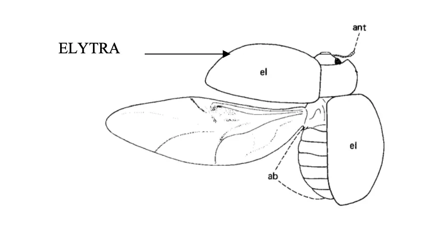
- Elytra:
- Elytra are heavily sclerotized wings, which have lost their venation and serve a protective function. They cover the hindwings and abdomen, providing durability. Beetles and weevils typically possess this wing type, which is not utilized during flight.
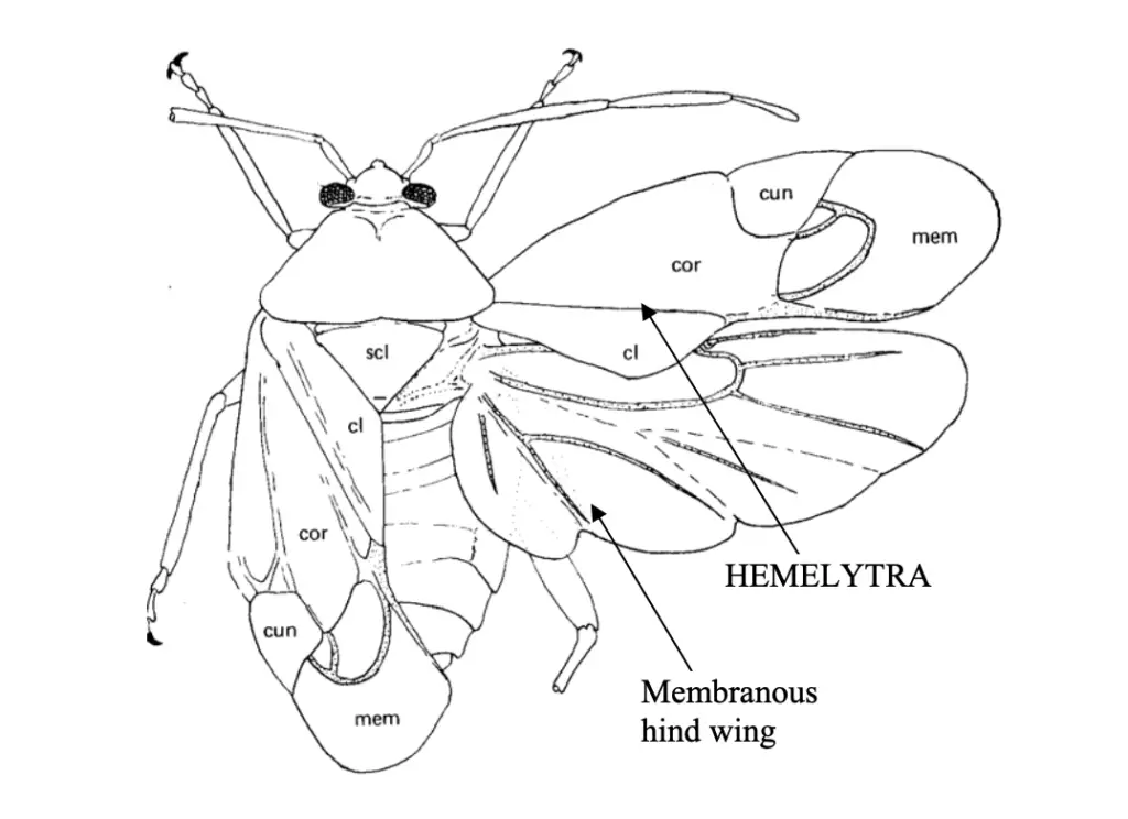
- Hemelytra:
- Hemelytra display a distinct morphology where the basal half is thick and leathery, while the distal half is membranous. This structure provides protection but is not employed in flight. It is commonly found in the forewings of heteropteran bugs.
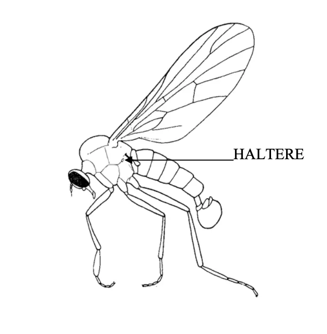
- Halteres:
- In true flies, the hindwings have been transformed into small, knobbed structures known as halteres. Each haltere consists of a slender rod with a clubbed end (capitellum) and a bulbous base (scabellum). These organs function as balancing devices, enhancing stability during flight, and are observed in true flies, mosquitoes, and male scale insects.
- Fringed Wings:
- Fringed wings are typically reduced in size, with margins adorned by long setae. This adaptation allows certain insects, like thrips, to navigate effectively through the air by swimming rather than traditional flying.
- Scaly Wings:
- The wings of butterflies and moths are covered with overlapping, small, colored scales. These unicellular outgrowths from the body wall not only provide coloration but also contribute to the smooth airflow over the wings, enhancing aerodynamics.
- Membranous Wings:
- Membranous wings are thin and transparent, characterized by a supportive network of tubular veins. These wings are crucial for flight and can be found in various insects, with either forewings, hindwings, or both being membranous.
- Tegmina (singular: Tegmen):
- Wing Coupling Mechanisms:
- In addition to the variations in wing structure, insects exhibit different coupling mechanisms that allow for coordinated movement of wings during flight:
- Hamulate: This coupling mechanism is observed in bees, enabling effective wing synchronization.
- Amplexiform: Found in butterflies, this mechanism allows for an efficient wing overlap during flight.
- Frenate: Notably seen in fruit-sucking moths, this mechanism facilitates specialized flight patterns.
- In addition to the variations in wing structure, insects exhibit different coupling mechanisms that allow for coordinated movement of wings during flight:
Modification in wings
The modification of wings in insects showcases a fascinating adaptation that has evolved to meet various ecological needs. These specialized structures not only facilitate flight but also play significant roles in defense, camouflage, and species recognition. The diversity in wing structure and function reflects the extensive evolutionary history of insects.
- Tegmina:
- In certain insects such as grasshoppers and cockroaches, the forewings are thickened and leathery, referred to as tegmina. These wings overlap each other, providing protection to the membranous hindwings while aiding in flight.
- Elytra:
- The hardened forewings of beetles are known as elytra. These structures are arched and meet at the mid-dorsal line but do not overlap. Elytra serve as protective casings for the hindwings and the body, providing both strength and rigidity.
- Hemelytra:
- In plant bugs, the wings exhibit a dual structure where the basal portion is leathery and the apical part is membranous. This type of wing is called hemelytra, facilitating both flight and protection from environmental hazards.
- Halters:
- In dipteran insects, such as mosquitoes and houseflies, the hindwings are modified into halters. These structures serve primarily as sensory organs and are crucial for maintaining body balance during flight. Halters consist of three parts: a basal lobe, a stalk, and an end knob.
- Pseudohalters:
- The forewings of strepsipteran insects are termed pseudohalters. Unlike halters, they do not significantly contribute to balance or sensory functions during flight.
- Specialized Wing Features:
- In some insects like thrips, the wings are slender with a fringe of long cilia, and the venation is almost absent. This design reduces drag during flight.
- In reproductive castes of termites and ants, wings are well-developed for nuptial flights. Post-mating, these wings are shed along sutures at their base.
- Wing Absence and Reduction:
- Ametabolous insects primitively lack wings, while in some species like fleas and lice, wings have been lost secondarily, evolving from winged ancestors. Additionally, termites may exhibit partial wing reduction (brachyptery), indicating a shift in ecological adaptation.
- Wing Margins and Angles:
- The margins of the wings are defined as costal (anterior), anal (posterior), and apical (outer edges). Angles formed at these intersections include the apical angle (between coastal and apical margins), the anal angle (between outer and anal margins), and the humeral angle at the wing base.
- Axillary Sclerites:
- The proximal region of the wing contains three axillary sclerites, which are crucial for wing articulation with the thorax. These sclerites enable a wide range of motion. The first sclerite articulates with the anterior notal process and the subcostal vein, while the second connects ventrally with the pleural wing process and the base of the radius. The third sclerite connects with the posterior notal process and the anal veins.
- Median Plates:
- At the base of the wing, two median plates give rise to the media and cubitus veins. Additionally, a humeral plate exists at the base of the costa, with a tegula located proximally. These structures collectively support wing movement and stability during flight.
7. Abdomen
The abdomen of insects is a vital component of their anatomy, typically comprising 6 to 11 segments along with a post-segmental structure known as the telson, which houses the anus. The structure and visibility of these segments can vary significantly among different insect orders and species, reflecting diverse evolutionary adaptations.
- Segment Composition:
- The abdomen is generally composed of 6 to 11 distinct segments, with the telson forming the terminal segment. For instance, in the family Acrididae (grasshoppers), all eleven segments are externally visible, while in Muscidae (house flies), only 2 to 5 segments are apparent; the remaining segments exhibit a telescopic arrangement.
- In the order Collembola, both adults and embryos consistently display six abdominal segments.
- Segmentation Variability:
- In most insects, there is a clear demarcation between the abdomen and the thorax. However, in Hymenoptera (bees, wasps, and ants), the first abdominal segment is fused with the thorax, termed the propodium.
- A notable constriction between the first and second abdominal segments is referred to as the petiole, which connects these two segments.
- Segment Structure:
- Each typical abdominal segment consists of three primary regions: the dorsal tergum, a pair of lateral pleural areas, and the ventral sternum. The posterior part of each segment typically overlaps the anterior portion of the subsequent segment, allowing for flexibility and movement.
- The segments are usually connected by a flexible membrane, although in some cases, fusion of the segments may occur.
- Appendages and Spiracles:
- In general, the first seven abdominal segments of adult insects lack appendages. However, apterygotes (wingless insects) and various immature aquatic insects may possess abdominal appendages.
- Anterior abdominal segments frequently have spiracles located within the pleural membrane, facilitating respiration.
- Anatomy Surrounding the Anus:
- The anus, located at the posterior end of the abdomen, is protected by three sclerites: one dorsal epiproct and two lateral paraprocts. Sensory organs called cerci are situated at the anterior margin of the paraprocts, enhancing the insect’s environmental awareness.
- Directly beneath the anus lies the genital opening, which is surrounded by specialized sclerites that constitute the external genitalia.
- Female Genitalia:
- In female insects, the appendages of the eighth and ninth abdominal segments collectively form an ovipositor, a structure specialized for egg-laying. This complex comprises four valvifers and six valvulae, facilitating the safe placement of eggs in the environment.
- Male Genitalia:
- In males, the genital opening is typically found within a tube-like structure known as the aedeagus or penis, which is thrust into the female’s body during copulation. Other structures associated with male external genitalia may include claspers and styli, which assist in mating.
- Additional Structures:
- The abdomen may also feature various specialized structures, such as pincers, median caudal filaments, cornicles, abdominal gills, and furcula. These adaptations serve different functions, including defense, locomotion, or respiration.
8. Male genitalia of insect
The male genitalia of insects represent a highly specialized and intricate system evolved for reproductive success. This structure plays a crucial role in mating, sperm transfer, and ensuring the continuation of species. The development and morphology of male genitalia derive from a pair of primary phallic lobes located on the ventral surface of the ninth segment during embryonic development. These lobes differentiate into various components essential for reproduction.
- Basic Structure of Male Genitalia:
- Primary Phallic Lobes: The foundational elements that give rise to the genital structures.
- Phallomeres: The division of the phallic lobes results in:
- Mesomeres: The inner pair that merges to form the aedeagus, the principal intromittent organ used during copulation.
- Parameres: The outer pair that develop into claspers, which serve various functions during mating.
- Aedeagus: The intromittent organ that facilitates sperm transfer.
- Endophallus: The inner lining of the aedeagus, continuous with the ejaculatory duct.
- Phallotreme: The opening at the tip of the aedeagus where the ejaculatory duct terminates.
- Gonopore: Located at the junction of the ejaculatory duct and the endophallus; it is internal in many insects.
- Functional Adaptations:
- Eversible Endophallus: In some species, the endophallus can be extended during copulation, allowing the gonophore to assume a terminal position.
- Claspers: Derived from parameres, these structures vary significantly among different insect groups and facilitate the grasping of the female during mating.
- Modifications Across Different Insect Orders:
- Collembola and Diplura: Lack an intromittent organ (aedeagus), relying on alternative methods for reproduction.
- Thysanura: Exhibit terminal segments that resemble those of females; sperm transfer does not occur directly to the female.
- Ephemeroptera and Dermaptera: Characterized by the presence of paired penes, enhancing reproductive strategies.
- Orthoptera and Dermaptera: In these groups, claspers can originate from parameres and cerci.
- Plecoptera: Notably absent of claspers, suggesting different mating strategies.
- Culicidae: The aedeagus is situated above the anus due to a 180-degree rotation of segment eight occurring shortly after eclosion, representing an adaptation in morphology.
- Odonata: Possess intromittent organs on segments two and three, with clasping appendages located on segment ten; however, the genital apparatus on segment nine is rudimentary.
9. Female genitalia of insect
The female genitalia of insects, particularly the ovipositor, are specialized structures designed for egg-laying and reproductive success. This complex system varies significantly across different insect orders, reflecting adaptations to their specific reproductive strategies. The position of the gonopore, where eggs exit the body, is typically located on or behind the eighth or ninth abdominal segment, although variations exist in certain groups such as mayflies and earwigs, where it may be found behind segment seven.
- Basic Anatomy of Female Genitalia:
- Gonopore: The opening through which eggs are laid, typically situated on the eighth or ninth abdominal segment.
- Ovipositor: A specialized structure formed from the elongated terminal abdominal segments, facilitating the insertion of eggs into various substrates.
- In certain species, such as house flies, the ovipositor is telescopic, formed from segments six to nine, and retracted into segment five when not in use.
- In tephritid flies, the ovipositor’s tip is hardened to enable egg deposition into fruit tissues.
- Ovipositor Structure Across Orders:
- Thysanura:
- Gonocoxae: The first and second gonocoxae (or valvifers) are located at the base of the ovipositor, associated with the eighth and ninth segments.
- Gonapophyses: The first and second gonapophyses (valvulae) articulate with the gonocoxae, forming a shaft that directs the egg during oviposition. The second gonapophyses are fused, creating a tube for egg passage.
- Gogangulum: A small sclerite attached to the first gonapophysis, articulating with the second gonocoxa and tergum of segment nine, contributes to the ovipositor’s structural integrity.
- Thysanura:
- Orthoptera:
- Gonoplac: An additional process found in the second gonocoxa, which may form a sheath around the gonapophyses. This structure is well-developed in orthopterans, serving as the dorsal valves of the ovipositor.
- The fusion of the gonangulum with the first gonocoxa is a notable adaptation in this order.
- Hymenoptera:
- The first gonocoxae are absent, and the second gonapophyses are fused, moving over the first through a tongue-and-groove mechanism.
- In groups like Symphyta, the ovipositor retains its original function for egg-laying, while in aculeate hymenopterans (like bees), it has evolved into a stinging organ. In these cases, eggs are ejected from the genital chamber’s opening rather than passing down the ovipositor shaft.
- In honeybees, the first gonapophyses are referred to as lancets, and the fused second gonapophyses form the stylet, which together create an inverted trough that culminates in a basal bulb where the poison gland’s reservoir discharges.
Practice Flashcard
[flashcard id=”59834″]
- https://egyankosh.ac.in/bitstream/123456789/85340/1/Unit-2.pdf
- https://extension.oregonstate.edu/sites/default/files/documents/9591/external-morphology.pdf
- http://cpsc270.cropsci.illinois.edu/syllabus/pdfs/lecture02.pdf
- https://core.ac.uk/download/pdf/144784808.pdf
- https://tmv.ac.in/ematerial/zoology/SEM%206%20DSE%204%20External%20Morphology%20of%20Insects.pdf
- https://genent.cals.ncsu.edu/students/lab-schedule/1455-2/
- https://www.rlbcau.ac.in/pdf/PGCourse/Entomology/Insect%20morphology%20APE%20501.pdf
- Text Highlighting: Select any text in the post content to highlight it
- Text Annotation: Select text and add comments with annotations
- Comment Management: Edit or delete your own comments
- Highlight Management: Remove your own highlights
How to use: Simply select any text in the post content above, and you'll see annotation options. Login here or create an account to get started.