What is Elastic Cartilage?
- Elastic cartilage, also known as yellow cartilage, is a specialized type of connective tissue found in various body organs that do not bear weight or mechanical stress. It plays a crucial role in providing strength, shape, and elasticity to these organs. This distinct type of cartilage is characterized by its abundance of elastic fibers, which contribute to its unique properties and appearance.
- Visually, elastic cartilage has a yellowish tint, hence its alternate name, due to the presence of elastic fibers within its extracellular matrix. This cartilage type is found in several anatomical structures, including the external ear (pinna), eustachian tube, epiglottis, and larynx. These organs rely on the elasticity and resilience of elastic cartilage to fulfill their functions effectively.
- While elastic cartilage shares similarities with hyaline cartilage in terms of its histological structure, it can be distinguished by its dense network of elastic fibers. These fibers allow the tissue to stretch and recoil, enabling it to return to its original shape after deformation. It is important to note that elastic cartilage lacks a direct blood supply and nerve innervation.
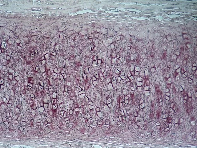
- Microscopically, elastic cartilage exhibits features similar to hyaline cartilage. The cells, known as chondrocytes, are housed within lacunae and are surrounded by a territorial capsular matrix and interterritorial matrix. The extracellular matrix of elastic cartilage contains elastic fiber networks, collagen fibers (type II), and a ground substance. These components contribute to the tissue’s resilience and strength, allowing it to withstand mechanical stress.
- The principal protein present in elastic cartilage is elastin, which plays a vital role in providing elasticity to the tissue. Elastin fibers give the cartilage its unique ability to revert to its resting form after being stretched or deformed. This property is particularly important in organs such as the external ear and epiglottis, which require flexibility and shape maintenance.
- It is worth noting that elastic cartilage is one of the three main types of cartilage found in the human body. The other two types are hyaline cartilage and fibrous cartilage. Elastic cartilage stands out due to its distinct yellowish appearance, elasticity, and the presence of elastin fibers in its extracellular matrix.
- In summary, elastic cartilage is a specialized connective tissue that imparts strength, shape, and elasticity to various body organs. It is characterized by its yellowish color, attributed to the abundance of elastic fibers. Elastic cartilage plays a crucial role in organs that require flexibility and shape maintenance, such as the external ear and epiglottis. Through its unique properties, elastic cartilage ensures the optimal functioning of these organs within the complex system of the human body.

Definition of Elastic Cartilage
Elastic cartilage is a specialized type of connective tissue characterized by its yellowish appearance and high content of elastic fibers. It is found in body organs that require flexibility and shape maintenance, such as the external ear, epiglottis, and eustachian tube. Elastic cartilage provides strength and elasticity to these organs, allowing them to maintain their shape and function effectively.
Location of Elastic Cartilage
Elastic cartilage is located in specific regions of the body that require both strength and flexibility for their normal functioning. The primary locations of elastic cartilage include:
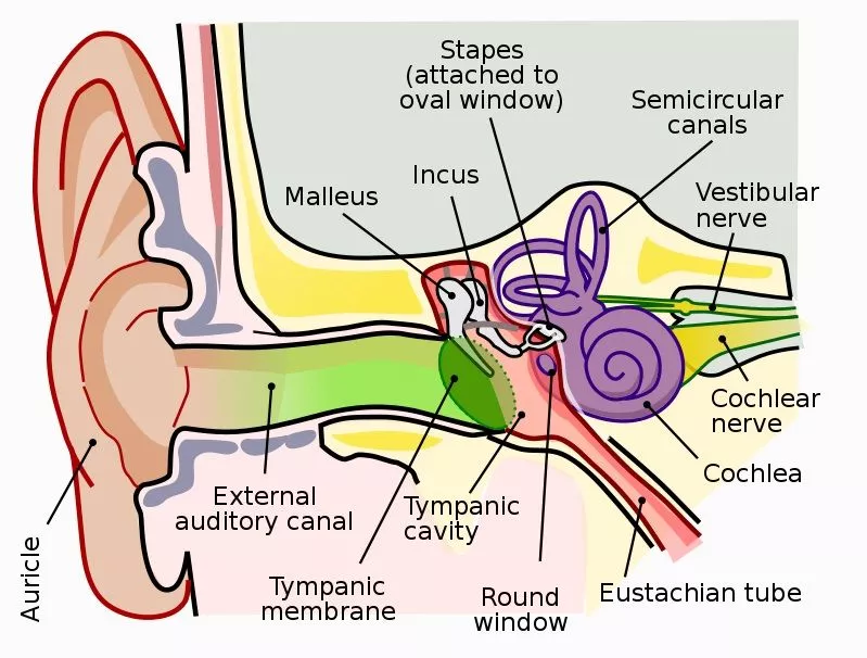
- External Ear (Ear Pinnae): Elastic cartilage shapes the outer ear, known as the pinnae or auricles. It provides structural support and flexibility, enabling the ears to maintain their shape while also allowing for movement and directional reception of sound waves.
- Eustachian Tube: The eustachian tube connects the middle ear to the back of the throat. Elastic cartilage in this tube helps maintain its shape and allows for the opening and closing of the tube to regulate pressure between the middle ear and the external environment.
- Larynx: Elastic cartilage plays a crucial role in the structure of the larynx, also known as the voice box. It forms the corniculate and cuneiform cartilages within the larynx, contributing to the stability and flexibility necessary for vocalization and protecting the airway during swallowing.
- Epiglottis: The epiglottis is a flap-like structure located at the base of the tongue that prevents food and liquid from entering the windpipe during swallowing. Elastic cartilage provides strength and flexibility to the epiglottis, enabling it to bend and cover the opening of the larynx when needed.
These anatomical sites contain elastic cartilage due to its unique properties. The extracellular matrix of elastic cartilage consists of elastin, fibrillin, glycoproteins, collagen types II, IX, X, and XI, and the proteoglycan aggrecan. Chondroblasts, located in the perichondrium, produce these components and maintain the gel-like environment of the extracellular matrix.
The elastic fibers within the extracellular matrix are responsible for the characteristic elasticity of the cartilage. Composed of elastin proteins and fibrillin, they can stretch and recoil, allowing the cartilage to return to its original shape after deformation. Collagen fibers, on the other hand, provide strength and structural support to the cartilage and are present in all types of cartilage, including elastic cartilage.

Understanding the locations and structure of elastic cartilage helps us appreciate its vital role in various organs, facilitating their proper function through a combination of strength and flexibility.
Structure of Elastic Cartilage
- The structure of elastic cartilage is characterized by its similarities to hyaline cartilage, with some notable differences. The matrix of elastic cartilage contains predominantly elastic fibers, which give the tissue its unique properties. The chondrocytes, specialized cartilaginous cells, are housed within spaces called lacunae within the elastic fiber network.
- Elastic cartilage consists of three main components: specialized cells, collagen and other fibers, and the extracellular matrix. It appears yellowish in color, distinguishing it from the white appearance of hyaline and fibrous cartilages.
- The chondrocytes and chondroblasts are the key cellular components of elastic cartilage. Compared to hyaline cartilage, elastic cartilage contains a higher number of chondrocytes. These cells secrete the extracellular matrix, and they are derived from mesenchymal progenitor cells located near the outer layer of the perichondrium.
- The extracellular matrix of elastic cartilage primarily consists of type II collagen fibers, elastic fibers, water, and proteoglycans (such as aggrecans). Aggrecans, specific glucosaminoglycans found only in cartilages, form aggregates with hyaluronic acid. The negative charge of aggrecans allows for water uptake, contributing to the cushion-like structure of the cartilage.
- Collagen fibers provide strength to the cartilage, while the elastic fibers, arranged in bundles, give the elastic cartilage its flexibility and ability to withstand repeated bending. Special staining techniques are required to visualize the elastic fibers under a microscope.
- The chondrocytes and extracellular matrix are enclosed within the perichondrium, a fibrous membrane that surrounds the cartilage. The perichondrium consists of an inner layer containing chondroblasts and an outer layer housing fibroblasts. The chondroblasts secrete the components of the extracellular matrix as they differentiate into chondrocytes.
- Overall, the structure of elastic cartilage, with its abundant elastic fibers and specialized cell population, allows for flexibility and resilience in organs such as the external ear, epiglottis, and eustachian tube.
Functions of Elastic Cartilage
Elastic cartilage serves various important functions in different parts of the body:
- Outer Ear: Elastic cartilage forms the external ear or pinnae, providing flexibility and shape to the ear. This allows the ear to fold during day-to-day activities without causing pain or discomfort. Additionally, the elasticity of the cartilage enables it to withstand external trauma and effectively funnel sound vibrations to the middle ear, contributing to normal hearing.
- Epiglottis and Larynx: The elasticity and flexibility of elastic cartilage play vital roles in the epiglottis and larynx. The epiglottis functions as a valve, covering the laryngeal inlet during swallowing to prevent food or liquid from entering the lungs. It then returns to its resting position, allowing for normal breathing. Elastic cartilage in the epiglottis maintains its structure and facilitates this valve-like function.
Within the larynx, elastic cartilage is found in the corniculate and cuneiform cartilages. These cartilages work together to control and modulate the vibrations generated in the voice box, influencing voice quality and pitch.
- Eustachian Tube: The Eustachian tube connects the middle ear to the back of the throat or nasopharynx. Elastic cartilage in the Eustachian tube provides strength and flexibility, allowing it to maintain its structure and return to its original shape after changes in air pressure. The tube functions to drain mucus from the ear and balance the air pressure between the middle ear and the external environment. It can open during activities like yawning to equalize the pressure and prevent infection.
In summary, the functions of elastic cartilage include providing flexibility and shape to the outer ear, facilitating swallowing and breathing by the epiglottis, modulating voice quality in the larynx, and maintaining the structure and pressure balance in the Eustachian tube. The unique properties of elastic cartilage enable these functions and contribute to the normal physiological processes of the body.
The Extracellular Matrix of Elastic Cartilage
The extracellular matrix (ECM) of elastic cartilage is composed of several key components that contribute to its unique properties and functions:
- Elastin: Elastin proteins are the primary protein component of elastic cartilage. These proteins co-polymerize with fibrillin, another protein type, to form fiber-like elastic chains. In their relaxed state, these chains are disorganized. However, when stretched, they become more uniform. This elastic property allows the cartilage to deform under pressure and then spring back into its original shape when the tension is released.
- Proteoglycan Aggrecan: The ECM of elastic cartilage contains the proteoglycan Aggrecan, which has a gel-like structure. Aggrecan is negatively charged, attracting water molecules and giving the cartilage its gel-like consistency. This gel-like structure enables elastic cartilage to withstand compressional forces.
- Glycoproteins: Glycoproteins are also present in the ECM of elastic cartilage. These proteins have carbohydrate chains attached to them and help organize the extracellular matrix and bind with cytokines. They contribute to the overall structure and function of the cartilage.
- Collagen Types II, IX, X, and XI: Collagen fibers are found in all types of cartilage, including elastic cartilage. They form a network that provides strength and a structural framework for other molecules within the ECM. Collagen fibers are abundant in elastic cartilage and are also present in the perichondrium, the outer layer of the cartilage that is more collagen-rich.
The interaction between these components, including elastin, proteoglycans, glycoproteins, and collagen fibers, gives elastic cartilage its unique properties. The presence of elastin fibers provides flexibility, while the proteoglycan aggrecan and its ability to bind with water molecules contribute to the cartilage’s lubricating and shock-absorbing qualities. The collagen fibers provide strength and support to the ECM, maintaining the structural integrity of the cartilage.
Overall, the extracellular matrix of elastic cartilage plays a crucial role in maintaining the elasticity, flexibility, and resilience of the tissue, allowing it to fulfill its functions in various parts of the body.
FAQ
What is elastic cartilage?
Elastic cartilage is a type of connective tissue that contains a dense network of elastic fibers, providing it with elasticity and flexibility.
Where is elastic cartilage found in the body?
Elastic cartilage is primarily found in specific locations such as the external ear (pinnae), epiglottis, Eustachian tube, and certain cartilages of the larynx.
What is the function of elastic cartilage in the ear?
In the ear, elastic cartilage provides flexibility and strength to the external ear (pinnae). It allows the ear to fold during activities without causing pain and helps in channeling sound vibrations towards the middle ear.
How does elastic cartilage contribute to voice production?
Elastic cartilage, specifically the corniculate and cuneiform cartilages in the larynx, plays a role in controlling and modulating the vibrations generated in the voice box. This affects the quality and pitch of the voice.
What is the role of elastic cartilage in the epiglottis?
The epiglottis, made of elastic cartilage, acts as a valve to cover the opening of the larynx during swallowing, preventing food or liquid from entering the airway. It helps in protecting the respiratory tract and prevents choking.
How does elastic cartilage function in the Eustachian tube?
In the Eustachian tube, elastic cartilage provides strength and flexibility. It helps in equalizing the air pressure between the middle ear and the external environment, facilitating proper hearing and preventing ear infections.
What is the extracellular matrix of elastic cartilage composed of?
The extracellular matrix of elastic cartilage consists of elastin fibers, proteoglycan aggrecan, glycoproteins, and collagen types II, IX, X, and XI. These components contribute to the tissue’s elasticity, flexibility, and strength.
How does the extracellular matrix of elastic cartilage provide its unique properties?
The elastic fibers, primarily composed of elastin proteins, allow the cartilage to stretch and return to its original shape. Aggrecan attracts water molecules, forming a gel-like structure that provides resistance to compression forces. Collagen fibers provide strength and support to the tissue.
Can elastic cartilage undergo repair or regeneration?
While cartilage has limited regenerative abilities, elastic cartilage can repair minor damage or injuries through the action of chondrocytes, the cells within the cartilage. However, extensive damage may require medical interventions such as surgeries.
What happens when elastic cartilage degenerates or becomes damaged?
Degeneration or damage to elastic cartilage can lead to functional impairments, such as hearing difficulties or voice changes. It can also contribute to conditions like ochronosis (thickening of ear cartilage) or disorders affecting the respiratory system. Proper care and treatment are necessary to manage any complications related to elastic cartilage.
- Text Highlighting: Select any text in the post content to highlight it
- Text Annotation: Select text and add comments with annotations
- Comment Management: Edit or delete your own comments
- Highlight Management: Remove your own highlights
How to use: Simply select any text in the post content above, and you'll see annotation options. Login here or create an account to get started.