What are Dendrites?
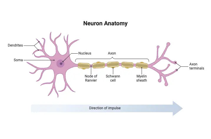
- Dendrites are complex, branched extensions of neurons, originating from the cell body, or soma. Their primary role is to receive and process incoming signals from other neurons, facilitating communication within the nervous system. This intricate structure resembles the branches of a tree, hence the name derived from the Greek word “dendron,” meaning “tree.”
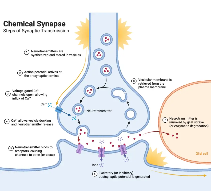
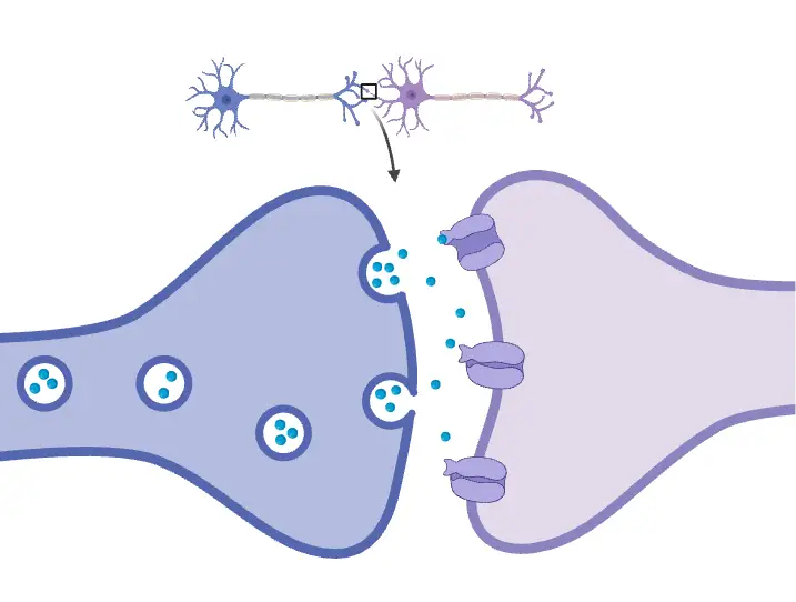
- Functionally, dendrites act as the receiving antennae for neurons. They collect electrical impulses transmitted by neighboring neurons through synapses, which are specialized junctions located at various points along the dendritic branches. The signals are typically conveyed by the axons of upstream neurons, which release neurotransmitters that bind to receptors on the dendrites. This process initiates the integration of synaptic inputs, essential for neural processing.
- The flow of information within a neuron is unidirectional. Signals travel from the dendrites to the cell body and then propagate along the axon to communicate with subsequent neurons. Dendrites play a crucial role in this sequence by processing incoming signals, summing them, and generating an electrical impulse known as an action potential. This action potential is vital for transmitting information across the nervous system.
- Furthermore, the structure of dendrites significantly influences their function. The presence of numerous branches increases the surface area available for synaptic connections, thereby enhancing a neuron’s ability to receive inputs. Dendrites also exhibit plasticity, meaning they can change in response to activity, a property important for learning and memory.

Definition of Dendrites
Dendrites are branched extensions of neurons that receive and process incoming signals from other neurons, facilitating communication within the nervous system. They transmit electrical impulses toward the cell body and play a crucial role in integrating synaptic inputs to generate action potentials.
Characteristics features of Dendrites
The following characteristics highlight the essential features of dendrites:
- Definition and Function: Dendrites function as receptors for synaptic inputs, operating much like antennas. They provide a substantial surface area, allowing a neuron to receive signals from numerous other cells. For instance, a motor neuron’s dendrites can offer the surface area of a sphere with a 340 µm diameter while only occupying the volume of a sphere with a 60 µm diameter.
- Diversity and Complexity: Dendrites exhibit a wide variety of shapes and sizes, directly linked to their specific functions and connectivity. This diversity enables neurons to integrate signals from multiple sources and perform complex computations. The configuration of a dendritic arbor is often tailored to how it connects with other neurons.
- Branching Patterns: Dendrites typically display intricate branching structures that contribute to their expansive surface area. Arborization density varies widely, ranging from selective arborizations, which target specific neurons, to space-filling arborizations that cover large areas. Many dendrites represent a compromise, characterized as sampling arborizations.
- Synaptic Specializations: Dendrites possess unique structural adaptations that enhance their synaptic input capabilities:
- Spines: These small protrusions are the primary sites of excitatory synapses and come in various forms, such as thin, mushroom, and stubby spines. The size and shape of spines influence their synaptic properties and potential for plasticity.
- Varicosities: Enlarged regions on thinner dendrites that can also form synapses.
- Filopodia: Thin, dynamic projections mainly observed during development, essential for synapse formation.
- Other Specializations: Depending on the neuron type, dendrites may feature structures like claw endings, brush endings, thorny excrescences, and coralline excrescences, which facilitate interactions with large axon terminals or specialized synaptic complexes.
- Internal Organelles: Dendrites contain various organelles critical for their functions:
- Microtubules: Provide structural support and serve as pathways for intracellular transport.
- Neurofilaments: Intermediate filaments contributing to the structural integrity of dendrites.
- Smooth Endoplasmic Reticulum (SER): Involved in calcium regulation and other cellular activities.
- Mitochondria: Supply energy necessary for dendritic function.
- Ribosomes: Sites for protein synthesis, enabling dendrites to produce the proteins needed for their operation.
- Dynamic Nature: Dendrites are not fixed structures; they display plasticity, allowing for structural changes in response to experiences and neuronal activity. These changes can include growth, retraction, branching, and alterations in spine density and morphology. This adaptability is crucial for learning and memory processes.
Structure of Dendrites
The structure of dendrites is integral to their function in neuronal communication. They exhibit complex branching patterns and specialized features that enhance their ability to receive and process synaptic inputs. The following points provide a detailed overview of dendritic structure:
- Dendritic Arbor: The dendritic arbor refers to the branching configuration of dendrites, which extends from the neuron’s cell body. This organization is not arbitrary; it reflects the neuron’s connectivity and functional requirements. The branching patterns can be categorized into three main types:
- Selective Arborization: This pattern involves dendrites that connect the cell body to a specific, distant target neuron. Such connections allow for precise communication, as seen in olfactory sensory neurons.
- Space-filling Arborization: Dendrites in this category extensively branch to occupy most of their surrounding area. This arrangement facilitates numerous synaptic connections, enhancing the neuron’s ability to integrate various signals. Cerebellar Purkinje cells are prime examples of this type.
- Sampling Arborization: This intermediate pattern covers a significant area without filling it entirely, allowing neurons to sample inputs from diverse axons. Pyramidal cells in the cerebral cortex exemplify this structure.
- Synaptic Specializations: Dendrites possess specialized structures that serve as synaptic sites where connections with axons from other neurons are formed. These specializations enhance the potential for synaptic connections:
- Spines: These small protrusions are the most common synaptic structures found on dendrites, particularly in pyramidal cells. They can vary in shape—thin, mushroom, stubby, or branched—each type suggesting different functional roles. The shape and density of spines can influence synaptic strength and plasticity.
- Other Synaptic Specializations: Various specialized structures enhance synaptic capabilities:
- Varicosities: Enlargements along thinner dendrites that often correlate with synaptic contacts, as seen in retinal amacrine cells.
- Filopodia: Long, dynamic protrusions primarily observed during development, involved in forming new synaptic contacts.
- Claw Endings: These structures form multiple synapses within glomeruli, found in cerebellar granule cells.
- Brush Endings: Similar to claw endings, these protrusions are associated with synaptic complexes in unipolar brush cells.
- Thorny Excrescences: Densely lobed protrusions that project into glomeruli, notable in CA3 pyramidal cells and dentate gyrus mossy cells.
- Racemose Appendages: Twig-like structures with synaptic varicosities, observed in neurons of the inferior olive.
- Coralline Excrescences: Complex structures with numerous synaptic protrusions found in cerebellar and vestibular nuclei.
- Intracellular Structure: The internal composition of dendrites varies depending on the neuron type and the location of the dendrites within the arbor:
- Proximal Dendrites: Located near the cell body, these larger dendrites contain organelles similar to those in the cell body, suggesting a role in protein synthesis and processing.
- Distal Dendrites: As dendrites branch further from the cell body, they become thinner and contain fewer organelles.
- Cytoskeleton: All dendrites contain a cytoskeleton made up of microtubules, neurofilaments, and actin filaments, essential for intracellular transport and structural integrity. Microtubules play a key role in the movement of mitochondria and other organelles within the dendrite.
- Synaptic Specialization Internal Structures: Smaller spines typically lack microtubules but have an actin-based cytoskeleton, facilitating rapid structural changes. Larger specializations, such as thorny excrescences, may contain microtubules and mitochondria, reflecting their functional demands. Most synaptic specializations also contain smooth endoplasmic reticulum (SER), which is continuous with the SER network in the dendrite.
Types of Dendrites
Dendrites can be categorized into various types based on their morphology and branching patterns. Each type plays a specific role in neuronal function and connectivity. The following outlines the distinct types of dendrites:
- Adendritic Dendrites: These dendrites are characterized by the absence of branches. They serve specific roles in certain neurons that do not require extensive branching for signal reception.
- Spindled Dendrites: Found in bipolar neurons, these dendrites extend in two opposite directions from the neuronal cell body, resembling a spindle shape. This structure is significant for their specialized functions in transmitting sensory information.
- Spherical Dendrites: In this type, dendritic branches encircle the neuronal cell body, creating a spherical appearance. An example includes cerebellar granule cells, where this morphology aids in receiving inputs from various sources.
- Laminar Dendrites: These dendrites extend in a planar fashion from the neuronal cell body, forming a flat layer. Retinal ganglion cells are an example, and this arrangement facilitates the processing of visual information.
- Cylindrical Dendrites: Characterized by their radial branching from the neuronal cell body in a disc-like manner, cylindrical dendrites are found in pallidal neurons. This structure supports their role in integrating signals from multiple directions.
- Conical Dendrites: These dendrites protrude in a conical shape from the cell body. Pyramidal cells exemplify this structure, which is crucial for their involvement in complex information processing and communication within the cerebral cortex.
- Fanned Dendrites: Dendrites in this category spread out in a flat, fan-like arrangement from the neuronal cell body. Purkinje cells in the cerebellum illustrate this morphology, which enhances their ability to integrate numerous synaptic inputs effectively.
Types of synaptic specializations and their respective functions
Synaptic specializations on dendrites play a crucial role in facilitating neuronal connectivity and enhancing the overall functionality of neural circuits. Each type of synaptic specialization has distinct characteristics and serves specific functions that contribute to the complexity of synaptic interactions. The following points outline the types of synaptic specializations and their respective functions:
- Simple Spines
- Description: Simple spines are the most common synaptic specializations, characterized by small protrusions typically measuring no more than 2 µm in length. They often consist of a bulbous head connected to the dendrite by a narrow neck.
- Location: Commonly found on various neurons, including cerebral pyramidal cells, striatal neurons, granule cells of the dentate gyrus, and cerebellar Purkinje cells.
- Function:
- Increased Surface Area: Simple spines enhance the dendritic surface area for synaptic connections, significantly contributing to the total dendritic surface area in certain cell types.
- Synaptic Transmission and Plasticity: They predominantly host asymmetric, excitatory synapses that utilize glutamate as a neurotransmitter, highlighting their role in excitatory neurotransmission.
- Compartmentalization: The neck constriction may isolate the spine head, facilitating localized signaling and plasticity.
- Branched Spines
- Description: Branched spines occur when two or more simple spines share a common stalk. Each branch can exhibit the same diverse shapes as simple spines.
- Location: Less frequently found, branched spines appear on CA1 pyramidal cells, dentate granule cells, and cerebellar Purkinje cells.
- Function: They may increase synaptic density, thereby contributing to the complexity of synaptic integration on dendrites.
- Varicosities
- Description: Varicosities are enlargements along thinner dendrites associated with synaptic contacts.
- Location: Present on AI amacrine cells in the retina.
- Function: They facilitate bidirectional communication by receiving synapses from rod bipolar cells and forming reciprocal synapses, containing synaptic vesicles and mitochondria to support both pre- and postsynaptic roles.
- Filopodia
- Description: Filopodia are long, thin, and transient protrusions characterized by a dense actin matrix and few internal organelles.
- Location: Observed on the dendrites of all neurons during development, but rarely in the adult brain.
- Function: They are essential for synaptogenesis, helping to establish nascent synaptic contacts and facilitating the formation of neuronal circuits during development.
- Claw Endings
- Description: Claw endings are multi-lobed protrusions at dendrite tips that create multiple synapses within synaptic complexes known as glomeruli.
- Location: Found on granule cells of the cerebellar cortex, interacting with mossy fiber axon terminals.
- Function: By forming multiple synapses within a glomerulus, claw endings strengthen the efficacy of connections between granule cells and mossy fiber inputs.
- Brush Endings
- Description: Brush endings are similar to claw endings but exhibit distinct multi-lobed protrusions.
- Location: Located on unipolar brush cells in the cerebellar cortex.
- Function: They participate in synapses with both mossy fiber rosettes and claw endings of cerebellar granule cells, highlighting their dual presynaptic and postsynaptic roles.
- Thorny Excrescences
- Description: These are densely lobed protrusions that extend into glomeruli and often contain microtubules and mitochondria.
- Location: Found on the proximal dendrites of CA3 pyramidal cells and dentate gyrus mossy cells.
- Function: Thorny excrescences integrate inputs from multiple axons within glomeruli, enhancing synaptic connections and the overall information processing capacity.
- Racemose Appendages
- Description: These are twig-like branched structures with synaptic varicosities and bulbous tips.
- Location: Observed on neurons in the inferior olive and relay cells of the lateral geniculate nucleus.
- Function: They receive both excitatory and inhibitory synapses, contributing to the balance of excitation and inhibition in their respective circuits.
- Coralline Excrescences
- Description: These are complex varicosities characterized by numerous thin protrusions and tendrils, suggesting active growth.
- Location: Found on dendrites in the cerebellar and vestibular nuclei.
- Function: Although their exact role is not fully understood, their intricate structure suggests involvement in forming multiple synaptic contacts.
Role of synaptic specializations in enhancing neuronal connectivity
The role of synaptic specializations in enhancing neuronal connectivity is fundamental to the efficient processing and transmission of information within the nervous system. These specialized structures significantly contribute to the complexity and functionality of neuronal networks. The following points outline their key roles:

- Increased Surface Area: Synaptic specializations, particularly dendritic spines, markedly increase the surface area available for synapse formation. This expansion is critical for dendrites, which serve as the primary sites for receiving synaptic inputs. By increasing surface area, a neuron can connect with a greater number of axons, thereby enhancing its connectivity potential and allowing for more extensive information processing.
- Strategic Location for Synapses: Synaptic specializations are strategically positioned on dendrites to optimize the likelihood of forming synapses with nearby axons. Although some synapses can form directly on the dendritic shaft, these shaft synapses are relatively rare. For example, in pyramidal cells, only about 5% of synapses occur on the shaft, while the remaining 95% are located on specialized structures. This positioning facilitates efficient communication between neurons.
- Specificity and Diversity of Contacts: Different types of synaptic specializations are often tailored to interact with specific types of axonal inputs. This specificity enhances the precision of neuronal circuits, allowing for preferential synapse formation with particular axons. For instance, claw endings in cerebellar granule cells are uniquely associated with mossy fiber axon terminals, while thorny excrescences on CA3 pyramidal cells engage with diverse inputs within glomeruli. Such diversity contributes to the unique functional roles of different neuronal types.
- Strengthening Synaptic Efficacy: Structures like claw endings and brush endings form large areas of synaptic contact within specialized synaptic complexes known as glomeruli. This close physical proximity between pre- and postsynaptic elements enhances the efficacy of synaptic transmission. The intimate arrangement fosters stronger connections between neurons, improving the overall efficiency of communication within the neural network.
- Enhanced Network Complexity: Synaptic specializations act as concentrated hubs for synaptic input, allowing for targeted increases in synapse formation. This targeted connectivity, along with the varied types and strategic placements of these specializations, enables neurons to establish intricate and highly interconnected networks. Such networks are essential for the computational capabilities of the brain, facilitating complex processing tasks like learning, memory, and sensory perception.

Function of Dendrites
The following points detail the primary functions of dendrites:
- Signal Reception: Dendrites are specialized for receiving chemical signals, specifically neurotransmitters, released from neighboring neurons at synapses. This capability is crucial for inter-neuronal communication.
- Signal Processing: Upon receiving inputs, dendrites process these signals by integrating both excitatory and inhibitory inputs. This summation determines whether the neuron will generate an electrical impulse known as an action potential, which is vital for transmitting information.
- Amplification of Weak Signals: Dendrites are adept at amplifying weak signals, allowing neurons to detect and respond to subtle changes in their environment. This amplification is essential for the accurate interpretation of sensory information.
- Filtering of Information: Dendrites filter incoming signals, enabling neurons to differentiate between various types of inputs. This function is crucial for discerning relevant information from background noise, enhancing the overall efficiency of neuronal processing.
- Synaptic Plasticity: Dendritic spines, small protrusions on dendrites, exhibit the ability to change shape and size in response to activity. This process, known as synaptic plasticity, is fundamental for learning and memory, as it facilitates the strengthening or weakening of synaptic connections based on experience.
- Integration of Inputs: Dendrites play a vital role in integrating multiple signals from different sources. This integration is critical for higher-order processing within the nervous system, allowing for complex decision-making and behavioral responses.
What are the primary structural and functional distinctions between dendrites and axons?
Dendrites are primarily designed to receive and process signals, while axons are specialized for transmitting those signals to other cells. Below are the key differences:
- Functionality
- Dendrites:
- Primarily function to receive and process incoming signals from other neurons.
- Facilitate the integration of synaptic inputs, contributing to the neuron’s overall response to stimuli.
- Axons:
- Responsible for transmitting electrical impulses away from the cell body to other neurons or target cells.
- Serve as conduits for long-distance communication within the nervous system.
- Dendrites:
- Structural Characteristics
- Dendrites:
- Exhibit a branched structure that increases their surface area, allowing for multiple synaptic connections.
- Often possess specialized structures known as spines, which serve as sites for synaptic input, enhancing connectivity.
- Axons:
- Typically have a more uniform, elongated structure, often extending significantly farther than dendrites.
- Can have a myelin sheath, which insulates the axon and facilitates faster signal transmission through saltatory conduction.
- Dendrites:
- Length and Reach
- Dendrites:
- Generally shorter, usually measuring 1-2 mm in length, even in larger neurons.
- Their compact size allows them to form local connections within a neural network.
- Axons:
- Can extend over considerable distances, sometimes reaching lengths of up to a meter or more.
- This capacity for long-range transmission enables communication between different regions of the nervous system.
- Dendrites:
- Synaptic Interactions
- Dendrites:
- Receive input at synapses, which are specialized junctions that facilitate communication between neurons.
- Many synapses occur on the dendritic shaft or on specialized protrusions, maximizing synaptic surface area and interaction potential.
- Axons:
- Typically do not engage in synaptic transmission themselves but terminate in synaptic boutons that release neurotransmitters to communicate with target cells.
- Dendrites:
- Signal Processing vs. Signal Transmission
- Dendrites:
- Involved in processing synaptic inputs, integrating multiple signals to generate a cumulative response.
- The presence of various synaptic specializations enhances their ability to modulate incoming signals.
- Axons:
- Focused on the efficient transmission of action potentials, which are all-or-nothing signals that propagate along their length.
- Utilize voltage-gated ion channels to facilitate rapid depolarization and repolarization during signal transmission.
- Dendrites:
Facts
- Did you know that dendrites are the “listeners” of the brain, responsible for receiving and processing information from other neurons while axons send information?
- Did you know that dendrites form intricate tree-like structures called dendritic arbors, which vary greatly in shape and size to increase the surface area for receiving signals?
- Did you know that the complexity of dendritic arbors often increases with the evolutionary advancement of species, with human cerebellar Purkinje cells exhibiting more intricate structures than those in simpler organisms like lampreys?
- Did you know that the specific shape and orientation of a dendritic arbor are related to its location in the brain and the types of inputs it receives, maximizing connections with relevant axons?
- Did you know that most dendrites are covered in tiny protrusions called spines, which are crucial for forming excitatory synapses and significantly enhance synaptic plasticity?
- Did you know that a single neuron can have thousands of spines, each potentially receiving input from different axons, resembling busy city streets in their complexity?
- Did you know that dendritic spines are dynamic structures that can change shape and size in response to neuronal activity, playing a key role in learning and memory?
- Did you know that some dendrites, such as those in AI amacrine cells in the retina, can contain synaptic vesicles and send signals back, enabling bidirectional communication?
- Did you know that dendrites undergo significant changes throughout life, with growth and retraction driven by experience and aging, highlighting the brain’s remarkable plasticity?
- Did you know that despite decades of research, many aspects of dendritic function remain a mystery, making them a fascinating area of study in neuroscience?
- Harris, Kristen M., and Josef Spacek, ‘Dendrite structure’, in Greg Stuart, Nelson Spruston, and Michael Häusser (eds), Dendrites, 3rd edn (Oxford
, 2016; online edn, Oxford Academic, 19 May 2016), https://doi.org/10.1093/acprof:oso/9780198745273.003.0001, accessed 23 Sept. 2024. - https://citeseerx.ist.psu.edu/document?repid=rep1&type=pdf&doi=2800b688a44983744d308bbacf783f0c0bcef0ec
- https://www.grc.org/dendrites-molecules-structure-and-function-conference/2023/
- https://synapseweb.clm.utexas.edu/sites/default/files/synapseweb/files/1999_dendrites_fiala_harris_dendrite_structure.pdf
- https://www.bu.edu/agingbrain/chapter-2-dendrites/
- https://www.kenhub.com/en/library/anatomy/dendrites
- https://en.wikipedia.org/wiki/Dendrite
- https://www.geeksforgeeks.org/dendrites-structure-types-function/