Muscle twitch, the brief and transient contraction of a muscle fiber, is a fundamental event in muscle physiology. Understanding the characteristics of muscle twitches provides insights into the intricate mechanisms that govern muscle function. This article delves into the key aspects of muscle twitch, focusing on motor units, summation, and tetanus.
Muscle twitches are initiated by the activation of motor units, which consist of a motor neuron and the muscle fibers it innervates. Each motor unit represents a single functional unit capable of generating a muscle contraction. The size and force of a muscle twitch can be modulated by the recruitment of different motor units. The coordinated recruitment of motor units is crucial for generating the appropriate force and controlling muscle movements.
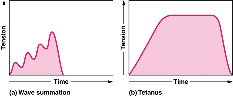
Furthermore, the phenomenon of summation plays a significant role in muscle twitches. When successive twitches occur in rapid succession, the force generated by each twitch can summate or combine, resulting in increased muscle contraction. This process, known as temporal summation or twitch summation, allows for greater force production and is essential for various activities that require strength and precision.
As muscle activity intensifies, sustained contraction can be achieved through tetanus. Tetanus occurs when a muscle fiber is stimulated at a high frequency, leading to a sustained and fused contraction without any relaxation between individual twitches. This continuous contraction allows muscles to generate maximal force and is essential for activities such as maintaining posture, lifting heavy objects, and performing powerful movements.
Understanding the characteristics of muscle twitch, including motor unit recruitment, summation, and tetanus, provides a foundation for comprehending muscle physiology and its implications in human movement, athletic performance, and rehabilitation. By exploring these aspects, researchers and practitioners can gain valuable insights into optimizing muscle function, preventing injuries, and enhancing physical performance.
In this article, we will explore the intricate details of motor unit recruitment, the phenomenon of summation, and the mechanisms underlying tetanus. By examining these characteristics of muscle twitch, we aim to deepen our understanding of muscle physiology and its implications for human movement and performance.
Whether you are an athlete, a fitness enthusiast, a healthcare professional, or simply interested in the fascinating world of muscles, this article will shed light on the key factors that shape the characteristics of muscle twitches. So, let us embark on this journey to unravel the complex mechanisms governing muscle function and delve into the remarkable world of muscle twitch physiology.
Motor Units
- Motor units play a crucial role in the functioning of skeletal muscles. They consist of an alpha motor neuron and the muscle fibers it innervates. Here is an overview of motor units and their significance in muscle control:
- Motor units are formed by the branching of alpha motor neurons as they enter a muscle. Each alpha motor neuron branches out to innervate multiple muscle fibers within that muscle. The number of muscle fibers innervated by a single motor neuron can vary, resulting in different sizes of motor units.
- The size of a motor unit is closely related to the function and control requirements of the muscle. Muscles involved in fine and precise movements, such as those controlling eye movements or movements of the hands and fingers, have small motor units. In these muscles, a single alpha motor neuron innervates only a few muscle fibers, typically ranging from 3 to 5. This arrangement allows for precise control and coordination of movements.
- In contrast, muscles involved in more powerful but less precise actions, such as the muscles of the legs and back, have larger motor units. These motor units consist of thousands of muscle fibers innervated by a single alpha motor neuron. The large number of muscle fibers per motor unit enables the generation of greater force but results in less precise control over individual muscle fibers.
- The recruitment of motor units plays a crucial role in muscle contraction. When a muscle needs to generate more force, additional motor units are recruited to join the contraction. This recruitment process follows the size principle, where smaller motor units are activated first before larger motor units are recruited as the force requirement increases. By recruiting motor units of varying sizes, the muscle can finely regulate the force and control of its contractions.
- The concept of motor units provides a framework for understanding the organization and control of skeletal muscle. The arrangement of motor units allows for both precision and strength in muscle movements, catering to the diverse functional needs of different muscle groups. The coordinated activation and recruitment of motor units contribute to the overall control, strength, and efficiency of muscle contractions.
- Understanding motor units is essential in fields such as sports performance, rehabilitation, and motor control research. By studying the characteristics and behavior of motor units, researchers and practitioners can gain insights into muscle function, movement disorders, and strategies for optimizing performance in various activities.
- In summary, motor units are comprised of alpha motor neurons and the muscle fibers they innervate. The size of motor units varies depending on the muscle’s function, with smaller units providing precise control and larger units enabling powerful contractions. The recruitment of motor units is essential for generating varying levels of force and coordinating muscle activity. The study of motor units enhances our understanding of muscle control and has implications for numerous disciplines related to human movement and performance.
Muscle Twitch
- Muscle twitch is a fundamental physiological event that occurs when a muscle fiber contracts in response to an action potential from a motor neuron. Understanding the characteristics of muscle twitch provides insights into the mechanisms underlying muscle contraction. Here are the key points about muscle twitch:
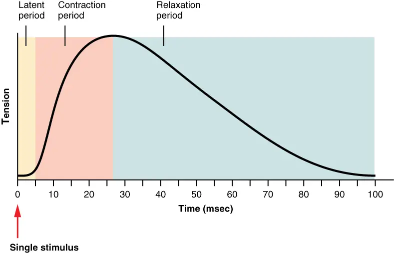
- A muscle twitch consists of three distinct phases: the latent period, the contraction phase, and the relaxation phase. The latent period is a brief delay between the arrival of the action potential and the observable tension in the muscle. During this period, several processes occur, including the release of calcium ions from the sarcoplasmic reticulum, the binding of calcium to troponin, and the formation of cross-bridges between actin and myosin filaments. The contraction phase is characterized by the generation of tension in the muscle as the cross-bridges cycle and interact. Finally, the relaxation phase is when the muscle returns to its initial length after contraction.
- The duration of a muscle twitch varies among different muscle types. It can be as short as 10 milliseconds or as long as 100 milliseconds. The specific characteristics of the muscle, its fiber type composition, and the functional requirements of the muscle influence the duration of the twitch.
- To achieve smooth and coordinated movement, motor units are activated asynchronously rather than all firing simultaneously. Asynchronous firing refers to the sequential activation of motor units, where one motor unit contracts before another has time to relax fully. This firing pattern allows for a smooth and controlled muscle contraction, preventing jerky movements.
- Even at rest, there is random firing of motor units in the muscle, contributing to muscle tone. Muscle tone refers to the continuous, partial contraction of muscles that keeps them in a state of readiness. This muscle tone helps take up the “slack” in the muscle, allowing for immediate tension generation when the muscle is called upon to contract. It also helps prevent muscle atrophy, as the continuous activation and stimulation of muscle fibers help maintain their integrity and functionality.
- Flaccid paralysis can occur when the connection between a motor neuron and muscle fibers is disrupted. In this condition, there is a loss of muscle tone, resulting in a limp and weakened muscle state.
- Understanding the characteristics of muscle twitch is essential in various fields, including exercise physiology, rehabilitation, and motor control. By studying the mechanisms underlying muscle twitches, researchers and practitioners can gain insights into muscle function, movement disorders, and strategies for optimizing performance and preventing muscle-related injuries.
- In summary, a muscle twitch is a brief contraction and relaxation cycle of a muscle fiber in response to an action potential. It consists of the latent period, contraction phase, and relaxation phase. Motor units fire asynchronously to produce smooth and controlled muscle contractions. Muscle tone, provided by random motor unit firing, helps maintain muscle readiness and prevent muscle atrophy. By exploring the intricacies of muscle twitch, we deepen our understanding of muscle physiology and its implications for human movement and function.

Summation
Muscle fibers within a motor unit can produce graded contractions by undergoing summation. Summation is the process by which multiple twitches or muscle contractions combine to produce a more forceful contraction. There are two types of summation:
- Temporal summation: This occurs when successive stimuli are applied to a muscle fiber before it has completely relaxed from the previous twitch. The second stimulus triggers a stronger contraction because the muscle fiber has not fully returned to its resting state.
- Spatial summation: This involves the recruitment of additional motor units to produce a stronger contraction. When a stronger force is needed, more motor units are activated, and the muscle fibers they innervate contract simultaneously.
Tetanus
Tetanus, in the context of muscle contraction, refers to a state of sustained muscle contraction or a prolonged series of contractions that occur when the motor nerve signals are delivered at a high frequency. Tetanus can be categorized into two types:
- Incomplete tetanus: In incomplete tetanus, the muscle fibers are stimulated at a high frequency but still have some relaxation time between contractions. This results in a sustained, yet oscillatory, contraction.
- Complete tetanus: In complete tetanus, the muscle fibers are stimulated at such a high frequency that there is no relaxation between contractions. As a result, the contractions fuse together, producing a smooth and sustained contraction without any relaxation.
Complete tetanus can be achieved by delivering stimuli at a frequency that exceeds the maximum frequency of muscle contraction. This maximal frequency is typically around 80-100 stimuli per second for most skeletal muscles. Tetanic contractions are important for activities that require sustained muscle force, such as maintaining posture or gripping objects tightly.
It’s worth noting that tetanus as a medical condition caused by the bacterial infection of Clostridium tetani is different from the tetanus described in muscle physiology.
Types Of Muscle Contraction
Muscle contractions can be categorized into different types based on force and length changes. Understanding these types of muscle contractions is crucial for comprehending muscle function and its adaptation to different forms of exercise. Here are the main types of muscle contractions:
- Isometric Contractions: In isometric contractions, muscle tension increases without a change in muscle length. The term “isometric” means “same length.” Isometric contractions are essential for maintaining posture and stabilizing joints. For example, when you hold a yoga pose or maintain a static position, your muscles are undergoing isometric contractions to generate tension without any visible movement.
- Isotonic Contractions: Isotonic contractions occur when the muscle length changes while maintaining a relatively constant tension. These contractions are further classified into two types based on how the muscle length changes:a) Concentric Contractions: In concentric contractions, the muscle generates tension and shortens in length. This type of contraction is commonly associated with muscle flexion or movements against gravity. For instance, when you perform a bicep curl and lift a weight towards your shoulder, the bicep muscle undergoes a concentric contraction.b) Eccentric Contractions: Eccentric contractions involve the generation of force by the muscle while lengthening. The muscle acts as a brake to control the movement and decelerate it. Eccentric contractions can generate more force than concentric contractions. An example of eccentric contraction is the controlled lowering of a weight from your shoulder to your waist during a bicep curl. Eccentric contractions are particularly effective for strength training, but they also cause more muscle damage and soreness compared to concentric contractions.
Muscle size and strength are influenced by the number and size of myofibrils within the muscle fibers. Resistance training, which includes both concentric and eccentric contractions, stimulates the production of more proteins within the muscle fibers. The micro-tears that occur during resistance training initiate a cascade of events leading to muscle repair and growth. This process, known as hypertrophy, involves an increase in the size of the muscle fibers as well as an increase in the amount of connective tissue within the muscle.
Endurance training, on the other hand, primarily enhances the muscle’s ability to produce ATP aerobically without significant increases in muscle size. The adaptations in endurance training focus on improving the efficiency of energy production and delivery to support prolonged physical activity.
In summary, muscle contractions can be classified as isometric or isotonic, with isotonic contractions further divided into concentric and eccentric contractions. Different types of contractions elicit distinct physiological responses and adaptations in muscle size, strength, and endurance. Understanding these types of muscle contractions provides valuable insights into the effects of exercise and training on muscle function.
Factors That Influence The Force Of Muscle Contraction
The force generated during muscle contractions can vary depending on several factors. Understanding these factors helps explain why different levels of force can be produced. Here are some key factors that influence the force of muscle contraction:
- Multiple-Motor Unit Summation or Recruitment: Motor units consist of a motor neuron and the muscle fibers it innervates. During muscle contractions, not all motor units are activated simultaneously. To increase the force generated, more motor units are recruited, meaning a greater number of motor units are firing at the same time. The heavier the load or resistance, the more motor units are recruited. However, even at maximum force, only about one-third of our total motor units are utilized. The asynchronous firing of motor units helps prevent muscle fatigue, as fatigued fibers are replaced by fresh ones to maintain force. In extreme situations, such as life-threatening emergencies, the body can recruit even more motor units to generate extraordinary force.
- Wave Summation: A single muscle twitch lasts for a relatively long period (up to 100 ms), while an action potential is much shorter (1-2 ms). If a motor unit is stimulated with increasing frequencies of action potentials, there is a gradual increase in the force generated by the muscle. This phenomenon is known as wave summation. At high frequencies, there is no time for the muscle to relax between successive stimuli, resulting in sustained contraction called tetanus. Tetanus occurs because calcium is not effectively removed from the cytosol due to the continuous stimulation. Maximum force is achieved when motor unit recruitment is at its maximum and the action potential frequency is sufficient to induce tetanus.
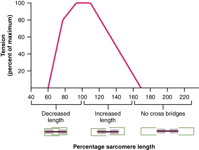
- Initial Sarcomere Length: The starting length of the sarcomeres within muscle fibers affects the force generation. Sarcomeres are the basic contractile units of muscles. If the sarcomeres start at a very short length, the thick filaments are already pushing against the Z-disc, leaving no room for further sarcomere shortening and reducing the force generated. Conversely, if the muscle is stretched to a point where the myosin heads cannot effectively contact the actin, force generation is also diminished. Maximum force occurs when the muscle is stretched to an optimal length that allows all myosin heads to fully interact with actin, providing the maximum distance for sarcomere shortening. This alignment ensures that the thick filaments are positioned at the ends of the thin filaments, optimizing force generation. It should be noted that in intact muscles in our bodies, the range of sarcomere lengths is constrained due to the arrangement of muscle attachments and joints.

In summary, the force generated during muscle contraction is influenced by factors such as motor unit recruitment, wave summation, and initial sarcomere length. By understanding these factors, we can grasp the mechanisms that contribute to different levels of force production in muscles.

Energy Source For Muscle Contraction
The energy source for muscle contraction is primarily ATP (adenosine triphosphate). ATP is essential for the functioning of myosin heads during the contraction cycle. Considering the number of myosin heads in a muscle and the number of cycles each head completes during a twitch, a significant amount of ATP is required for muscle function. In fact, it is estimated that we burn approximately our entire body weight in ATP each day, emphasizing the constant need for replenishing this energy source. There are four main ways our muscles obtain ATP for contraction:
- Cytosolic ATP: This refers to the ATP that is immediately available in the cytoplasm. It is an anaerobic source, meaning it doesn’t require oxygen, and it provides immediate energy. However, its availability is limited, making it suitable for short bursts of maximal muscle activity, such as the rapid contractions of the eye muscles.
- Creatine Phosphate: When the cytosolic ATP stores are depleted, the cell turns to creatine phosphate as a rapid energy source. Creatine phosphate is a high-energy compound that can transfer its phosphate group to ADP (adenosine diphosphate) to rapidly regenerate ATP without the need for oxygen. This process is facilitated by the enzyme creatine kinase. Creatine phosphate can replenish the ATP pool multiple times, extending muscle contraction for up to approximately 10 seconds. Creatine phosphate is a popular supplement among weightlifters, although its benefits are limited and specific to certain activities.
- Glycolysis: Glycolysis is the breakdown of glucose, and it serves as a major ATP-producing pathway during anaerobic activity. The primary source of glucose for glycolysis is glycogen stored in the muscle. Glycolysis can occur without oxygen and can rapidly provide energy for intensive muscular activity. However, its sustainability is limited to about a minute before muscle fatigue sets in.
- Aerobic or Oxidative Respiration: For activities that require a continuous supply of ATP lasting longer than a minute (e.g., walking, jogging, cycling), the muscles rely on aerobic or oxidative respiration. These metabolic processes occur within the mitochondria and utilize oxygen. Aerobic metabolism is slower than anaerobic mechanisms but is capable of providing ATP for hours. While glucose can be used in aerobic metabolism, fatty acids are the preferred fuel source. Slow-twitch and fast-twitch oxidative muscle fibers are capable of efficiently utilizing aerobic metabolism.
Fatigue
- Fatigue refers to the condition in which skeletal muscles are no longer able to contract optimally. It can be categorized into two main types: central fatigue and peripheral fatigue. Central fatigue, also known as psychological fatigue, is associated with the subjective feeling of being tired. It is believed to arise from factors released by the muscle during exercise that signal the brain to perceive fatigue. Central fatigue occurs before the muscle fiber reaches a point where it can no longer contract. Through training, athletes can learn to overcome psychological fatigue and continue performing despite feeling uncomfortable.
- Peripheral fatigue can occur at various points between the neuromuscular junction and the contractile elements of the muscle. It can be further classified into two subcategories: high frequency fatigue and low frequency fatigue. High frequency fatigue is typically experienced during activities such as circuit training and is characterized by impaired membrane excitability due to imbalances of ions. This may involve dysfunction of the Na+/K+ pump, inactivation of Na+ channels, and impairment of Ca2+ channels. Muscles affected by high frequency fatigue can recover relatively quickly, usually within 30 minutes or less.
- On the other hand, low frequency fatigue is associated with impaired Ca2+ release, possibly resulting from issues with excitation-contraction coupling. Recovery from low frequency fatigue is more challenging and can take anywhere from 24 to 72 hours.
- Several other factors can contribute to fatigue as well. These include the accumulation of inorganic phosphates, changes in pH due to hydrogen ion accumulation, glycogen depletion, and imbalances in K+ ions. It’s worth noting that ATP and lactic acid, often mistakenly associated with fatigue, do not directly contribute to its development.
- Despite ongoing research, the precise causes of fatigue are still not fully understood. Scientists continue to investigate this topic to gain deeper insights into the mechanisms underlying fatigue and potential strategies to mitigate its effects.
Skeletal Muscle Fiber Types
Skeletal muscle fibers can be categorized based on their speed of contraction and resistance to fatigue. The classic classifications include slow twitch oxidative (Type I) fibers, fast-twitch oxidative-glycolytic (Type IIA) fibers, and fast-twitch glycolytic (Type IIX) fibers. However, it’s important to note that the understanding of muscle fiber types is evolving, and these classifications may be revised in the future.
Fast-twitch fibers (Type II) contract two to three times faster than slow-twitch fibers (Type I). The speed of fiber contraction is related to the duration of the cross-bridge cycle and the ability of myosin molecules to hydrolyze ATP. Fast-twitch fibers have a more rapid ATPase activity, which contributes to their faster contraction speed. They also have efficient calcium ion (Ca2+) reuptake into the sarcoplasmic reticulum, leading to quicker twitches compared to slow-twitch fibers. This enables fast-twitch fibers to complete multiple contractions rapidly.
The characteristics of different muscle fiber types vary in terms of myosin ATPase activity, size (diameter), duration of contraction, sarcoplasmic reticulum calcium ATPase (SERCA) pump activity, fatigue resistance, energy utilization, capillary density, mitochondria numbers, and color (based on myoglobin content).
In human skeletal muscles, the ratio of fiber types varies between muscles. For example, the gastrocnemius muscle contains roughly equal proportions of slow and fast fibers, while the soleus muscle is predominantly slow twitch. The distribution of fiber types also differs between individuals, with women generally having a higher ratio of slow-twitch fibers compared to men. Certain muscles, such as eye muscles, are predominantly composed of fast-twitch fibers.
The preferred fiber type for sprinting athletes is the fast-twitch glycolytic fibers, which provide rapid contractions. However, the majority of individuals have a low percentage of these fibers. Muscle biopsies from elite sprinters have shown a high proportion of fast-twitch fibers, particularly Type IIX fibers. The ability to convert muscle fiber types from one to another is an area of ongoing research. It appears that fiber types are primarily determined embryologically by the type of neuron innervating the muscle fiber. The frequency of firing rates of the neuron also influences the muscle fiber type. Some studies suggest the presence of dual innervation in a small percentage of muscle fibers, allowing for switching between slow and fast characteristics. However, genetics is believed to play a significant role in determining muscle fiber types, with training having a more limited impact on altering the ratios.
Slow Twitch Oxidative (Type I) | Fast-twitch Oxidative (Type IIA) | Fast-Twitch Glycolytic (Type IIX) | |
| Myosin ATPase activity | slow | fast | fast |
| Size (diameter) | small | medium | large |
| Duration of contraction | long | short | short |
| SERCA pump activity | slow | fast | fast |
| Fatigue | resistant | resistant | easily fatigued |
| Energy utilization | aerobic/oxidative | both | anerobic/glycolytic |
| capillary density | high | medium | low |
| mitochondria | high numbers | medium numbers | low numbers |
| Color | red (contain myoglobin) | red (contain myoglobin) | white (no myoglobin) |
In summary, skeletal muscle fibers can be classified based on their speed of contraction and fatigue resistance. The understanding of muscle fiber types is still developing, and further research is focused on exploring the factors that determine and potentially modify these fiber types.