14 Difference Between Brightfield and Phase-contrast Microscope
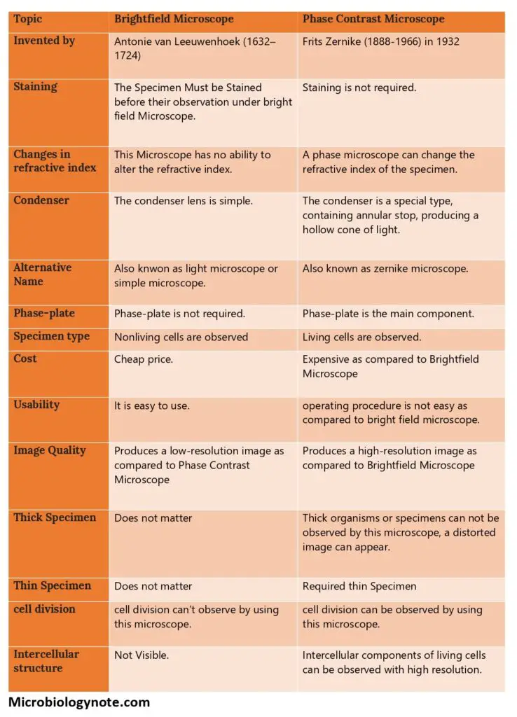
| Topic | Brightfield Microscope | Phase Contrast Microscope |
| Invented by | Antonie van Leeuwenhoek (1632–1724) | Frits Zernike (1888-1966) in 1932 |
| Staining | The Specimen Must be Stained before their observation under bright field Microscope. | Staining is not required. |
| Changes in refractive index | This Microscope has no ability to alter the refractive index. | A phase microscope can change the refractive index of the specimen. |
| Condenser | The condenser lens is simple. | The condenser is a special type, containing annular stop, producing a hollow cone of light. |
| Alternative NameAlso knwon as light microscope or simple microscope. | Also known as zernike microscope. | |
| Phase-plate | Phase-plate is not required. | Phase-plate is the main component. |
| Specimen typeNonliving cells are observed Living cells are observed. | ||
| Cost | Cheap price. | Expensive as compared to Brightfield Microscope |
| Usability | It is easy to use. | operating procedure is not easy as compared to bright field microscope. |
| Image Quality | Produces a low-resolution image as compared to Phase Contrast Microscope | Produces a high-resolution image as compared to Brightfield Microscope |
| Thick Specimen | Does not matter | Thick organisms or specimens can not be observed by this microscope, a distorted image can appear. |
| Thin Specimen | Does not matter | Required thin Specimen |
| cell division | cell division can’t observe by using this microscope. | cell division can be observed by using this microscope. |
| Intercellular structure | Not Visible. | Intercellular components of living cells can be observed with high resolution. |

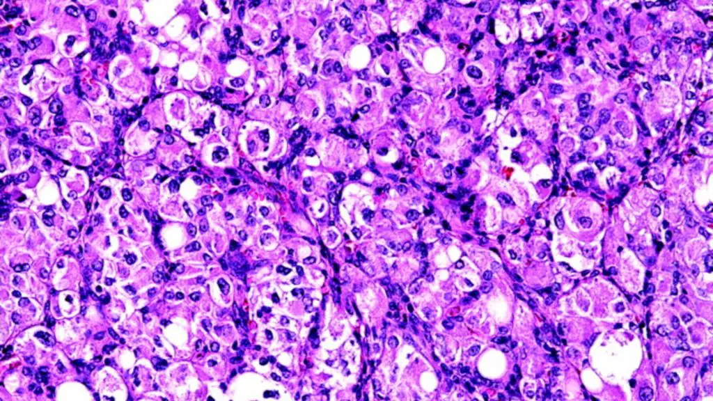
Brightfield Microscope Image

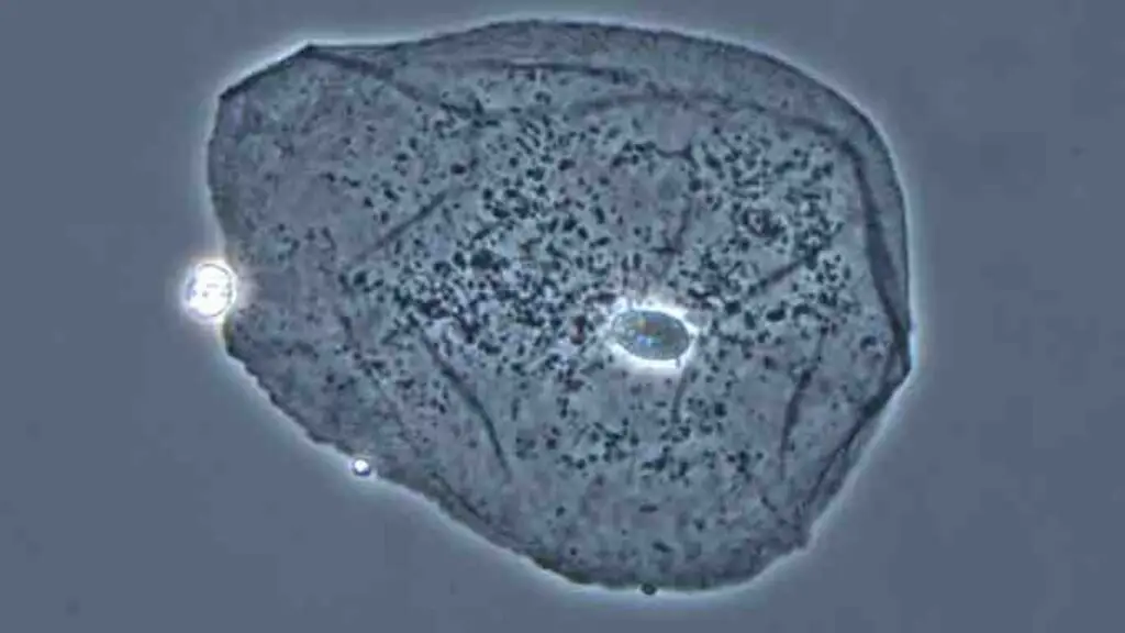
Phase Contrast Microscope Image

References
- https://microbiologynote.com/phase-contrast-microscopy/
- https://microbiologynote.com/bright-field-microscope-definition-parts-working-principle-application/
- https://en.wikipedia.org/wiki/Optical_microscope