Sourav Pan
Transcript
Hey everyone! Today, we’re diving into the fascinating world of protozoa. These amazing organisms are all around us, yet most people have never heard of them.
So what exactly are protozoa? Protozoa are single-celled eukaryotic organisms. Let me break that down for you.
Single-celled means they consist of just one cell – that’s it! Everything they need to survive happens within that single cell.
Eukaryotic means they have a true nucleus – a control center surrounded by a membrane, just like our cells.
Here’s what a typical protozoan looks like under a microscope. You can see the cell membrane that holds everything together, the nucleus that controls the cell, and various organelles that carry out different functions.
Here’s the amazing part – all the functions needed for life happen inside this single cell. Eating, breathing, moving, reproducing – everything!
Protozoa come in an incredible variety of shapes and forms. Some are blob-like and constantly changing shape, others are oval and streamlined, and some are elongated with whip-like tails.
These remarkable organisms are found everywhere on Earth – in oceans, lakes, rivers, soil, and even inside other living things. They’re truly global citizens of the microscopic world.
In our next sections, we’ll explore what makes these tiny organisms so special and how they’ve mastered the art of single-celled living.
Historically, scientists called protozoa the ‘first animals’ because of their remarkable animal-like behaviors. But what exactly makes them seem so animal-like?
The first animal-like behavior is movement. Protozoa actively move around their environment, just like animals do. They can swim, crawl, and even hunt for food.
The second animal-like behavior is hunting and predation. Protozoa actively seek out and capture food, just like predatory animals. They can engulf other microorganisms and organic particles.
Now, what makes protozoa different from plants and fungi? The key difference is that protozoa lack a rigid cell wall. This absence gives them incredible flexibility to change shape and move freely.
This flexibility allows protozoa to change shape dramatically. Watch how a protozoa can stretch, compress, and even form temporary extensions called pseudopodia for movement and feeding.
Finally, protozoa are heterotrophic, meaning they must obtain their food from organic matter. They cannot make their own food like plants do. Instead, they feed on other microorganisms, organic debris, and dissolved nutrients.
So while protozoa are not true animals, they share many animal-like characteristics: they move actively, hunt for food, lack rigid cell walls for flexibility, and obtain nutrition by consuming organic matter. These traits made early scientists classify them as the first animals.
Now we’ll meet some fascinating examples of protozoa. Think of this as meeting the main characters in our microscopic story. Each one has unique features that make them special.
First, let’s meet Amoeba, the ultimate shape-shifter. Amoebas are famous for constantly changing their form as they move and feed.
Amoebas move by extending parts of their body called pseudopodia, which means false feet. Watch how they can stretch and flow like liquid.
Next is Paramecium, often called the slipper animalcule because of its distinctive slipper-like shape. It’s covered in thousands of tiny hair-like structures called cilia.
The cilia beat in coordinated waves, allowing Paramecium to swim gracefully through water. They also help sweep food particles into its mouth-like opening.
Euglena is truly unique among protozoa. It has characteristics of both plants and animals, making it a fascinating example of nature’s versatility.
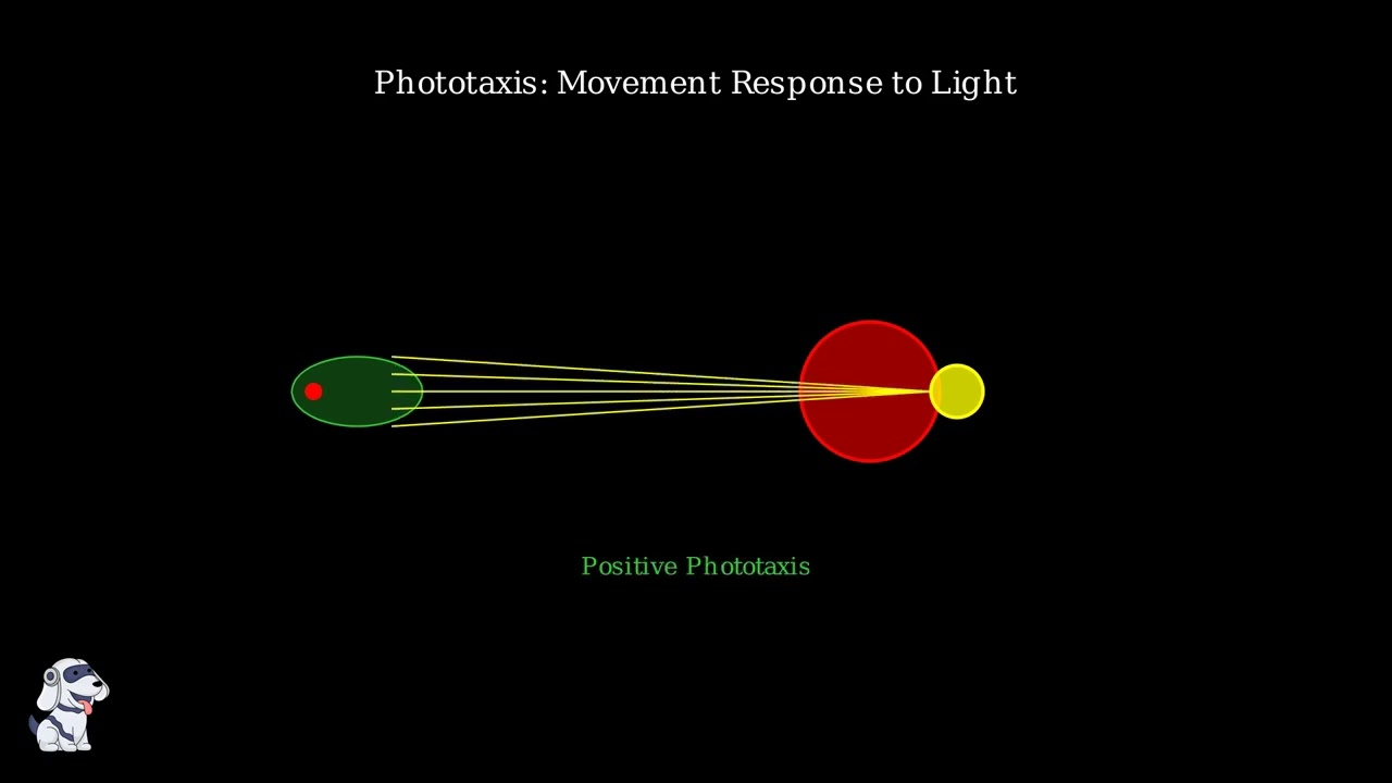
Euglena contains chloroplasts like plants, allowing it to photosynthesize when light is available. It also has an eyespot to detect light and a flagellum for movement.
Finally, we meet Trypanosoma, a parasitic protozoan with a dark side. Unlike our previous examples, this one lives inside other organisms and can cause serious diseases.
Trypanosoma has a distinctive undulating membrane that ripples as it moves through blood and other body fluids. This parasitic lifestyle makes it very different from our free-living examples.
These four examples show the incredible diversity of protozoa. From shape-shifting amoebas to light-seeking euglenoids, each has evolved unique strategies for survival in their microscopic world.
Now that we’ve met some protozoa, let’s establish the official scientific definition. What exactly makes something a protozoan?
Protozoa are unicellular, eukaryotic organisms. Let me break this down for you. Unicellular means they consist of just one cell – that’s it, just one cell doing everything.
Eukaryotic means their genetic material is contained within a membrane-bound nucleus, just like our cells. This distinguishes them from bacteria, which are prokaryotic and lack a true nucleus.
Here’s the amazing part: protozoa are structurally and functionally independent. This means each single cell can perform all the necessary life functions on its own – feeding, breathing, reproducing, moving, and responding to the environment.
Scientists describe protozoa as having a protoplasmic grade of organization. This fancy term means that different organelles within the cell have specialized jobs – there’s a division of labor happening inside that single cell.
Think of it like a tiny factory where each organelle has a specific job, but they all work together to keep that single cell alive and functioning. This is what makes protozoa so remarkable – they pack all the complexity of life into just one cell.
Scientists organize protozoa into groups based on how they move and their overall body structure. This classification system helps us understand the incredible diversity of these single-celled organisms.
There are four main groups of protozoa. Each group has its own unique way of moving around, which is the primary characteristic scientists use to classify them.
The other two groups are ciliates, which are covered in tiny hair-like structures called cilia, and sporozoans, which are typically parasitic and often lack obvious movement structures.
This classification system is based on morphology and locomotion. By studying how protozoa move and their body structures, scientists can better understand their relationships and evolutionary history.
Each of these four groups represents a different evolutionary solution to the challenge of movement and survival in microscopic environments. Understanding this classification helps us appreciate the remarkable diversity of protozoan life.
Flagellates are a fascinating group of protozoa that move using whip-like structures called flagella. These microscopic organisms literally whip their way through water and other environments.
A typical flagellate has one or more flagella extending from its cell body. These flagella act like tiny whips, beating in a wave-like motion to propel the organism through its environment.
Watch how the flagella move in a whipping motion. This undulating movement creates thrust that pushes the cell forward through the water.
Now let’s meet some important flagellates. These organisms include both harmless species and some that cause serious diseases in humans.
Trypanosoma is a dangerous flagellate that causes sleeping sickness in Africa. It has an elongated, curved shape with a single flagellum that runs along its body.
Giardia is a pear-shaped flagellate with multiple flagella. It causes giardiasis, a common intestinal infection that leads to diarrhea and stomach problems.
Trichomonas is another flagellate that can cause infections in humans. It has several flagella and is responsible for a common sexually transmitted infection.
Leishmania is a flagellate that causes leishmaniasis, a disease that can affect the skin, organs, or mucous membranes. It’s transmitted by sandfly bites.
Flagellates demonstrate the incredible diversity of protozoa. While they all share the common feature of flagellar movement, they vary greatly in shape, size, and lifestyle – from free-living organisms to serious human pathogens.
Meet the amoeboids, also known as Sarcodina – the ultimate shape-shifters of the protozoan world. These remarkable single-celled organisms have mastered the art of changing their form to move and survive.
Amoeboids move using pseudopodia, which literally means ‘false feet.’ These are temporary extensions of their cytoplasm that flow outward like liquid fingers, allowing the cell to crawl and change direction.
Watch how an amoeba extends its pseudopodia. The cytoplasm flows forward like thick honey, creating these temporary projections that act as both arms and legs for movement.
The amoeba can extend multiple pseudopodia in different directions, constantly changing its shape. This gives amoeboids their nickname as shape-shifters – they have no fixed form.
The most famous amoeboid is simply called Amoeba. Found in freshwater ponds and soil, this free-living organism demonstrates the classic amoeboid movement we just observed.
However, not all amoeboids are harmless. Entamoeba histolytica is a parasitic amoeboid that causes amoebic dysentery, a serious intestinal infection that affects millions of people worldwide.
What makes amoeboids unique among protozoa is their incredible flexibility. Unlike other protozoa with fixed shapes, amoeboids can squeeze through tiny spaces, engulf large food particles, and adapt their form to their environment.
Ciliates are a fascinating group of protozoa that earned their nickname ‘the hairy movers’ because they’re covered in thousands of tiny, hair-like structures called cilia.
These cilia are not just for show – they serve two crucial functions. First, they act like tiny oars, beating in coordinated waves to propel the organism through water.
The second function of cilia is feeding. They create water currents that sweep food particles toward the cytostome, which acts like the cell’s mouth.
Paramecium is the classic example of a ciliate. This slipper-shaped organism demonstrates all the key features of ciliates: the coordinated beating of cilia for movement and the efficient use of cilia for capturing food.
The thousands of cilia work together in perfect coordination, creating the distinctive swimming pattern that makes ciliates some of the most efficient swimmers in the microscopic world.
Sporozoans, also known as Apicomplexa, represent a unique group of protozoa that have evolved as parasitic specialists. Unlike the other protozoa we’ve seen, these organisms have adapted specifically for a parasitic lifestyle.
The first key characteristic of sporozoans is their obligate parasitic lifestyle. These organisms cannot survive outside a host and have evolved to live entirely within other organisms, depending on them for all their nutritional needs.
The second key characteristic is that mature sporozoans typically lack the locomotive organelles we’ve seen in other protozoa. They don’t have flagella, cilia, or pseudopodia for movement.
The third key characteristic, and the reason they’re called sporozoans, is their unique method of reproduction. They reproduce by forming specialized cells called sporozoites, which are the infective stage of their life cycle.
The most famous example of a sporozoan is Plasmodium, the parasite that causes malaria. This organism perfectly demonstrates all the characteristics we’ve discussed and shows why sporozoans are such successful parasites.
Sporozoans represent the ultimate parasitic adaptation among protozoa. By giving up independent movement and becoming completely dependent on hosts, they’ve become incredibly successful at what they do – unfortunately, this often means causing serious diseases in humans and other animals.
Protozoa are incredibly widespread organisms that have colonized virtually every environment on our planet. Their remarkable adaptability allows them to thrive in conditions ranging from pristine mountain lakes to the depths of the ocean.
Freshwater environments are perhaps the most common habitat for protozoa. They flourish in ponds, lakes, rivers, streams, and even temporary puddles. The abundance of organic matter and relatively stable conditions make freshwater ideal for many protozoan species.
Marine environments host countless protozoan species. From the sunlit surface waters of the ocean to the mysterious depths of the deep sea, protozoa have adapted to saltwater conditions and play crucial roles in marine food webs.
Soil provides another rich habitat for protozoa. They live in the spaces between soil particles, feeding on bacteria and organic matter. Soil protozoa are essential for nutrient cycling and maintaining healthy ecosystems.
Many protozoa have evolved to live as parasites inside other organisms. These parasitic protozoa can be found in the digestive systems of animals, within plant tissues, and even in human hosts, where they may cause diseases.
What makes protozoa so successful is their incredible adaptability. They can survive extreme temperatures, varying pH levels, different salinity conditions, and even low oxygen environments. This adaptability explains why we find them virtually everywhere on Earth.
Protozoa exist in an incredible range of sizes, creating a truly microscopic world with surprising diversity. Understanding their size and shape helps us appreciate how these single-celled organisms have adapted to different environments.
At the smallest end of the scale, most protozoa are truly microscopic, measuring just 1 to 300 micrometers. These tiny organisms are completely invisible to the naked eye and require powerful microscopes to observe.
However, some protozoa can grow surprisingly large. Foraminifera, marine protozoa with calcium carbonate shells, can reach up to 20 centimeters in diameter. That’s larger than a dinner plate!
Unlike plant cells, protozoa lack a rigid cell wall. This gives them remarkable flexibility to change shape, squeeze through tight spaces, and adapt their form for movement and feeding.
Some protozoa have developed external support structures. Shells provide protection and shape, while pellicles offer a flexible coating that maintains form without restricting movement. These adaptations show how protozoa balance flexibility with structural support.
Protozoa are eukaryotic organisms, which means their cells have a complex internal structure. Unlike bacteria, protozoan cells contain a true nucleus surrounded by a membrane, along with specialized compartments called organelles.
The cytoplasm of a protozoan is divided into two distinct regions. The outer region, called the ectoplasm, is a clear, gel-like layer that lies just beneath the cell membrane.
The inner region is called the endoplasm. This is a more granular, dense area that contains most of the cell’s organelles. The endoplasm is where the majority of cellular activities take place.
Within the endoplasm, we find various organelles that perform specific functions. These include mitochondria for energy production, endoplasmic reticulum for protein synthesis, and other specialized structures.
One of the most important organelles in many protozoa are the contractile vacuoles. These specialized structures help regulate water balance, especially in freshwater species where water constantly enters the cell.
Watch how contractile vacuoles work. They collect excess water from the cytoplasm, then contract to pump it out of the cell. This process, called osmoregulation, prevents the cell from bursting due to water pressure.
This complex cellular organization allows protozoa to carry out all the functions necessary for life within a single cell. The division between ectoplasm and endoplasm, along with specialized organelles like contractile vacuoles, makes these microscopic organisms remarkably efficient living systems.
Protozoa have fascinating ways of obtaining nutrition. Unlike plants that make their own food, protozoa are heterotrophic organisms, meaning they must consume other sources for energy and nutrients.
Most protozoa are holozoic feeders, which means they ingest solid food particles whole. They accomplish this through a process called phagocytosis, where the cell membrane surrounds and engulfs food particles.
Some protozoa have a specialized structure called a cytostome, which acts like a cell mouth. This permanent opening allows for more efficient feeding, especially in ciliates like Paramecium.
Once inside the cell, food is stored in food vacuoles where digestion occurs. These vacuoles contain digestive enzymes that break down the food into smaller molecules that the cell can absorb and use for energy.
Some protozoa, like Euglena, are mixotrophic. This means they can both photosynthesize like plants when light is available, and consume other organisms when light is scarce. This flexibility gives them a survival advantage in changing environments.
Understanding protozoan nutrition helps us appreciate how these single-celled organisms have evolved diverse feeding strategies to survive in different environments, from the flexibility of mixotrophs to the efficiency of specialized feeding structures.
Protozoa have fascinating ways of reproducing and creating new generations. Most reproduce asexually, meaning they don’t need a partner, but some can also reproduce sexually when conditions are right.
Binary fission is like cellular photocopying. One protozoan cell divides into two identical daughter cells. This is the most common way protozoa reproduce.
First, the nucleus duplicates itself, creating two identical copies of the genetic material.
Then the cell membrane pinches inward, gradually separating the cell into two parts.
Multiple fission creates many offspring at once from a single parent. Budding produces a small outgrowth that eventually separates to become a new individual.
Some protozoa can reproduce sexually. In conjugation, two cells temporarily connect and exchange genetic material, creating genetic diversity without actually reproducing.
This genetic exchange helps create variation in the population, which can be beneficial for survival in changing environments.
When conditions become harsh, many protozoa form protective cysts. This thick-walled dormant stage helps them survive drought, extreme temperatures, or lack of food.
When favorable conditions return, the protozoan emerges from its cyst and resumes normal activities. This survival strategy allows protozoa to persist through difficult times and spread to new environments.
Protozoan reproduction is remarkably diverse. While most rely on simple asexual division, the ability to reproduce sexually and form protective cysts gives these microscopic organisms incredible adaptability and survival power.
Many protozoa have evolved a remarkable survival strategy – they alternate between two distinct life stages that allow them to thrive in changing environments.
The first stage is called the cyst stage. During this phase, the protozoan becomes dormant and surrounds itself with a thick, protective wall.
The cyst stage is like a protective cocoon. The organism becomes inactive, stops feeding, and can survive extremely harsh conditions like dehydration, freezing temperatures, or lack of food.
When environmental conditions improve, the cyst transforms into the active trophozoite stage. The protective wall breaks down and the organism emerges ready to feed and multiply.
The trophozoite stage is the active, feeding form. During this phase, the protozoan actively searches for food, consumes nutrients, and reproduces rapidly through cell division.
This two-stage life cycle is incredibly advantageous. When times are tough, protozoa can hunker down as cysts and wait it out. When conditions are good, they become active trophozoites and make the most of the opportunity to grow and reproduce.
Examples of this life cycle can be seen in many disease-causing protozoa like Entamoeba, which causes dysentery, and Giardia, which causes intestinal infections. The cyst stage allows these parasites to survive outside their hosts and infect new individuals.
Unlike complex organisms with lungs or gills, protozoa have a much simpler way of breathing. These single-celled organisms exchange respiratory gases directly through their cell membrane using a process called diffusion.
Oxygen molecules from the surrounding environment move into the cell, while carbon dioxide waste products move out. This happens because molecules naturally move from areas of high concentration to areas of low concentration.
At the same time, carbon dioxide molecules move from inside the cell to the outside environment. This two-way exchange ensures the protozoan gets the oxygen it needs while removing toxic waste gases.
Diffusion works perfectly for protozoa because they are extremely small. Small cells have a high surface area to volume ratio, meaning there’s plenty of membrane surface for gas exchange relative to the cell’s internal volume.
Some protozoa, especially parasitic forms that live inside other organisms, can survive without oxygen. They use anaerobic respiration, breaking down nutrients without needing oxygen from the environment.
This flexibility in respiration methods allows protozoa to thrive in many different environments, from oxygen-rich pond water to the oxygen-poor interior of host organisms.
The structure of a protozoan cell is remarkably complex for a single-celled organism. The cell membrane forms the outer boundary, providing both protection and flexibility that allows the cell to change shape as needed.
Some protozoa have an additional structure called a pellicle. This is a cytoskeletal framework that lies just beneath the cell membrane, providing extra support and helping maintain the cell’s shape.
Inside the cell, we find mitochondria – the powerhouses that generate energy for all cellular activities. These oval-shaped organelles have internal folds called cristae that increase their surface area for energy production.
The Golgi bodies, also called Golgi apparatus, appear as stacked membranes. They modify and package proteins received from other parts of the cell, acting like a cellular post office.
Lysosomes are small, membrane-bound organelles filled with digestive enzymes. They break down waste materials, worn-out organelles, and harmful substances, acting as the cell’s cleanup crew.
Contractile vacuoles are specialized organelles that regulate water balance. They collect excess water from the cell and pump it out, preventing the cell from bursting due to osmotic pressure.
In ciliates, we find a special structure called the gullet or cytopharynx. This funnel-shaped opening allows the cell to take in food particles from the environment, directing them into food vacuoles for digestion.
Together, these structures work in harmony to maintain the protozoan cell. The flexible membrane and supportive pellicle provide structure, while the various organelles carry out specialized functions essential for survival.
Protozoa have evolved fascinating ways to move around their microscopic world. These single-celled organisms use four main types of locomotion, each perfectly adapted to their lifestyle and environment.
Each type of movement serves different purposes and allows protozoa to navigate their environments effectively. Let’s explore each method in detail.
Flagellated protozoa use long, whip-like structures called flagella. These rotate like tiny propellers, creating a spiral motion that pushes the cell through water. This is similar to how a boat propeller works.
Ciliated protozoa are covered with thousands of tiny hair-like structures called cilia. These beat in coordinated waves, like a field of wheat swaying in the wind, propelling the cell smoothly through water.
Amoeboid protozoa move by extending temporary projections called pseudopodia. The cell’s cytoplasm flows like thick liquid, creating these extensions that pull the rest of the cell forward. It’s like the cell is constantly changing shape to crawl.
Sporozoans use a different approach. They have internal structures called subpellicular microtubules that create slow, gliding movements. This method is less obvious but allows these parasitic protozoa to move through host tissues efficiently.
Each type of locomotion represents millions of years of evolution, perfectly adapted to help protozoa survive and thrive in their microscopic world. From the powerful propulsion of flagella to the subtle gliding of microtubules, these movement strategies showcase the incredible diversity of single-celled life.
Now that we understand the basics of protozoan classification, let’s take a closer look at our five key examples to see how their unique characteristics make them perfectly adapted to their environments.
First, let’s examine Amoeba, our shape-shifting champion. This remarkable organism lives in freshwater ponds, lakes, and moist soil, constantly changing its form as it moves and feeds.
Next is Paramecium, the slipper-shaped ciliate that glides gracefully through freshwater environments. Its entire surface is covered with thousands of tiny hair-like cilia that beat in coordinated waves.
Euglena is our unique mixotrophic example – it can both photosynthesize like a plant and consume food like an animal. This green flagellate is often found swimming alone in freshwater environments.
Trypanosoma represents the darker side of protozoa – this parasitic flagellate causes sleeping sickness in humans and animals. Its distinctive undulating membrane helps it swim through blood and tissue fluids.
Finally, Plasmodium – the sporozoan responsible for malaria, one of humanity’s deadliest diseases. This complex parasite has a sophisticated life cycle involving both mosquitoes and humans.
These five examples showcase the incredible diversity of protozoa – from the shape-shifting Amoeba to the disease-causing Plasmodium. Each has evolved unique adaptations that make them perfectly suited to their specific environments and lifestyles.
While many protozoa are harmless, some species pose serious threats to human health. These pathogenic protozoa cause diseases that affect millions of people worldwide, making them a critical concern for public health officials.
Giardia lamblia is a flagellated protozoan that causes giardiasis, one of the most common waterborne diseases worldwide. This parasite has a distinctive pear shape with two nuclei and eight flagella that help it move through the intestinal tract.
Giardiasis symptoms include persistent diarrhea, abdominal cramps, nausea, and fatigue. The disease is typically contracted by drinking contaminated water or through poor sanitation practices.
Entamoeba histolytica is an amoeboid protozoan that causes amoebic dysentery. Unlike Giardia, this parasite moves using pseudopodia and can actually invade and destroy intestinal tissue.
Amoebic dysentery is more severe than giardiasis, causing bloody diarrhea, intense abdominal pain, and fever. In severe cases, the parasite can spread to the liver, forming dangerous abscesses.
The public health impact of these protozoan diseases is enormous. Giardia infects approximately 200 million people annually worldwide, while amoebic dysentery affects around 50 million people each year.
Understanding these protozoan diseases is crucial for public health because it enables us to develop effective prevention strategies, improve water treatment systems, promote proper sanitation, and ensure early diagnosis and treatment.
These diseases disproportionately affect developing countries with poor sanitation infrastructure, making global health education and intervention programs essential for reducing their impact on vulnerable populations.
Scientists around the world are fighting back against protozoan diseases with innovative research and new treatment strategies.
The biggest challenge we face today is drug resistance. Many protozoan parasites have evolved to survive treatments that once worked effectively.
Researchers are developing four main types of new treatments to overcome this resistance.
First, protease inhibitors target specific enzymes that parasites need to break down proteins for survival.
Second, topoisomerase inhibitors target enzymes that help parasites copy their DNA during reproduction.
Third, antimicrobial peptides are small proteins that can punch holes in parasite cell membranes.
Finally, RNA interference uses small RNA molecules to silence specific genes that parasites need to survive.
These research efforts represent hope in our ongoing battle against drug-resistant protozoan diseases. Scientists continue working to develop these treatments into effective medicines.
The future of protozoa research holds exciting possibilities as scientists work to combat these microscopic challenges through innovative approaches and collaborative strategies.
Researchers are developing novel drug targets to combat drug-resistant protozoan parasites. These include protease and topoisomerase inhibitors, antimicrobial peptides that can penetrate cell membranes, and RNA interference techniques that can silence specific genes in parasites.
Systems biology approaches help researchers understand the complex interactions between hosts and parasites by integrating data from multiple sources. This comprehensive view allows scientists to identify new therapeutic targets and develop more effective treatment strategies.
Vaccine development efforts are targeting major protozoan parasites including Trypanosoma cruzi which causes Chagas disease, various Leishmania species, and Plasmodium vivax which causes malaria. These vaccines could prevent millions of infections worldwide.
The One Health perspective recognizes that human, animal, and environmental health are interconnected. This approach is crucial for controlling protozoan diseases, especially those transmitted between animals and humans, and for preventing future outbreaks through environmental monitoring.
The future of protozoa research focuses on four key priorities: combating drug resistance through novel therapeutic approaches, using systems biology for comprehensive understanding of host-parasite interactions, developing effective vaccines for major parasitic diseases, and applying the One Health approach for integrated disease prevention strategies.
So, what have we learned about protozoa? Let’s summarize the key points from our journey through the microscopic world of these fascinating single-celled organisms.
First, protozoa are single-celled eukaryotic organisms. Unlike bacteria, they have a true nucleus and various organelles, making them structurally complex despite being just one cell.
Second, protozoa use various methods to move around. Some use whip-like flagella, others use hair-like cilia, and some extend temporary projections called pseudopodia to crawl along surfaces.
Third, protozoa play crucial roles in ecosystems. They decompose organic matter, help control bacterial populations, and serve as food for larger organisms, forming an essential part of aquatic food webs.
Fourth, while many protozoa are harmless, some cause serious diseases in humans and animals. Malaria, sleeping sickness, and giardiasis are just a few examples of protozoan infections that affect millions worldwide.
Finally, ongoing research is essential for combating protozoan diseases. Scientists are developing new treatments, vaccines, and diagnostic tools while working to overcome the growing problem of drug resistance.
Thank you for joining us on this journey through the microscopic world of protozoa! You now have a solid understanding of these remarkable single-celled organisms and their vital roles in our world.
Study Materials
Protozoa - Definition, Classification, Characteristics, Structure, Locomotion, Examples
Helpful: 100%
Related Videos


- Text Highlighting: Select any text in the post content to highlight it
- Text Annotation: Select text and add comments with annotations
- Comment Management: Edit or delete your own comments
- Highlight Management: Remove your own highlights
How to use: Simply select any text in the post content above, and you'll see annotation options. Login here or create an account to get started.