Sourav Pan
Transcript
What exactly is a Paramecium? These fascinating creatures are single-celled microorganisms that live all around us in freshwater environments.
Paramecium, also spelled Paramoecium, belongs to a group called protists. These are neither plants nor animals, but form their own unique category of life.
These oval-shaped organisms are incredibly small, yet they contain all the machinery needed for life within just one cell.
Where can we find these remarkable organisms? Paramecia thrive in freshwater environments like ponds, lakes, and stagnant water basins.
Scientists often call Paramecia the white rats of the microbial world because they are so easy to grow and study in laboratories.
This makes Paramecia incredibly valuable for understanding fundamental biological processes, from cell division to feeding behaviors.
So in summary, Paramecium are single-celled protists that serve as perfect windows into the microscopic world of life.
So, what exactly defines a Paramecium? Let’s break down the key characteristics that make these organisms unique and fascinating.
Paramecia are microscopic, single-celled organisms. This means each individual Paramecium is made up of just one cell that contains everything needed for life.
One of their most distinctive features is their slipper-like shape. This elongated, curved form is instantly recognizable under a microscope.
What really sets Paramecia apart are the hair-like structures covering their bodies called cilia. These tiny projections are found all around the cell surface.
These cilia aren’t just decorative – they serve two crucial functions. First, they help the Paramecium move through water by beating in coordinated waves.
Second, the cilia help capture food particles from the surrounding water, directing them toward the cell’s feeding structures.
Paramecia are commonly found in three types of aquatic environments: freshwater like ponds and lakes, marine environments in the ocean, and brackish water where fresh and salt water mix.
In summary, Paramecia are defined by their single-celled structure, distinctive slipper shape, and most importantly, their cilia-covered surface that enables both movement and feeding in aquatic environments.
Every living organism has a scientific address that tells us exactly where it belongs in the tree of life. For Paramecium, this classification system helps us understand its relationships to other organisms.
We start at the broadest level with Domain Eukaryota. This means Paramecium belongs to organisms whose cells contain a true nucleus, unlike bacteria which are prokaryotes.
Next comes Kingdom Protista, a diverse group of mostly single-celled eukaryotic organisms. This kingdom includes many microorganisms that don’t fit neatly into plant, animal, or fungal categories.
The Phylum Ciliophora groups all organisms that have cilia – those tiny hair-like structures used for movement and feeding. This is a key defining characteristic of Paramecium.
Class Oligohymenophorea represents a specific group within the ciliates, distinguished by particular cellular and genetic features that scientists use to organize these organisms.
Moving down to Order Peniculida and Family Parameciidae, we get increasingly specific groupings that share more detailed structural and behavioral similarities.
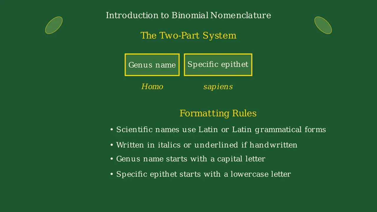
Finally, we reach Genus Paramecium – this is our specific group of slipper-shaped, ciliated protists. The genus name is what we commonly use when referring to these fascinating microorganisms.
This classification system is like a detailed address that tells us exactly where Paramecium fits in the vast diversity of life on Earth, showing its evolutionary relationships and shared characteristics with other organisms.
Understanding this classification helps scientists communicate precisely about Paramecium and reveals the evolutionary story of how these remarkable single-celled organisms are related to all other life forms.
Where exactly do paramecia live? These microscopic organisms have specific habitat preferences that determine where we can find them thriving in nature.
Paramecia are most commonly found in freshwater environments. They thrive in ponds, lakes, streams, and any body of fresh water where conditions are right for their survival.
They are especially abundant in stagnant water and scums – those greenish films you might see on the surface of still water bodies. These environments provide rich feeding opportunities.
While freshwater is their primary home, paramecia can also be found in marine and brackish water environments, showing their adaptability to different salinity levels.
An important behavior of paramecia is that they often attach themselves to surfaces. You’ll find them clinging to rocks, plant material, and other submerged objects in their aquatic homes.
Paramecia prefer warm, stagnant water conditions. These environments provide optimal temperatures for their metabolism and abundant food sources like bacteria and organic matter.
So next time you’re near a pond, lake, or any body of water, remember that there might be an entire microscopic world of paramecia thriving just beneath the surface, playing their important role in aquatic ecosystems.
Did you know there are different types of Paramecium? The microscopic world of these single-celled organisms is far more diverse than you might expect.
Currently, scientists have identified and classified 19 recognized morphospecies of Paramecium. Each morphospecies represents a distinct form with its own unique structural characteristics.
Among these 19 species, there are five particularly common examples that scientists frequently study. Let me show you these fascinating varieties.
First, we have Paramecium aurelia, shown here in light blue. This species is part of a complex group and is widely used in laboratory research.
Next is Paramecium caudatum, the most well-known species. Notice its elongated, slipper-like shape that tapers to a point at the posterior end.
Paramecium bursaria is particularly interesting because it forms symbiotic relationships with green algae, giving it a distinctive greenish appearance.
Paramecium woodruffi represents another distinct morphospecies, with its own unique size and structural characteristics that distinguish it from its relatives.
Finally, Paramecium trichium completes our common examples. Each of these species demonstrates the remarkable diversity within this single genus.
What makes this diversity so remarkable is that each species has evolved its own unique characteristics. These differences include variations in size, shape, feeding preferences, and even reproductive behaviors.
This incredible diversity within the Paramecium world demonstrates how evolution has shaped these microscopic organisms to thrive in different environments and ecological niches.
How big are these fascinating microscopic creatures? Paramecium species vary significantly in size, ranging from 50 micrometers to 330 micrometers in length.
To visualize this range, imagine a scale where the smallest Paramecium measures just 50 micrometers, while the largest can reach 330 micrometers.
Here we can see the dramatic size difference. The smallest Paramecium is quite tiny, while a typical Paramecium caudatum falls in the middle range, and the largest species can be over six times bigger than the smallest.
Now let’s focus specifically on Paramecium caudatum, one of the most commonly studied species.
Paramecium caudatum typically measures between 170 and 330 micrometers in length, with most specimens falling in the 200 to 300 micrometer range.
To put this in perspective, a typical Paramecium caudatum is about one-fourth the width of a human hair. While definitely microscopic, they’re large enough to be easily visible under a light microscope. At 0.2 to 0.3 millimeters, they’re roughly the size of a very small grain of sand.
Despite their microscopic size, these single-celled organisms are incredibly complex and fascinating creatures, packed with specialized structures that allow them to thrive in their aquatic environments.
Among the many species of Paramecium, two stand out as the most well-known and studied: Paramecium aurelia and Paramecium caudatum.
Paramecium caudatum is typically more elongated and cigar-shaped, while Paramecium aurelia has a more oval, slipper-like appearance.
But here’s where it gets really interesting. What we call Paramecium aurelia is actually not just one species, but a complex of fifteen different sibling species.
These fifteen sibling species look almost identical under a microscope. To the naked eye, they appear to be the same organism.
However, when scientists examine their DNA and biochemistry, they discover these species are genetically quite different from each other.
Most importantly, these sibling species cannot reproduce with each other through conjugation, even though they look nearly identical.
It’s like a secret society of Paramecia! They all wear the same disguise and look identical, but each has its own unique genetic identity and can only reproduce within its own secret group.
This discovery revolutionized our understanding of species identification and showed that microscopic appearance alone isn’t enough to determine if organisms are truly the same species.
Paramecium have several defining characteristics that make them unique among single-celled organisms. Let’s explore each of these key features that help us identify and understand these fascinating microorganisms.
First, paramecium are unicellular organisms. This means their entire body consists of just one cell that performs all the functions necessary for life.
Second, paramecium have a distinctive slipper-shaped or elongated oval form. This asymmetrical shape helps them move efficiently through water.
Third, paramecium are covered with thousands of tiny hair-like structures called cilia. These cilia beat in coordinated waves to propel the organism through water and help capture food.
Fourth, paramecium have a pellicle – a flexible but firm membrane that covers their entire body. This pellicle maintains their shape while allowing for some flexibility during movement.
Fifth, paramecium have a unique dual nucleus system. They contain one large macronucleus that controls daily functions, and one or more small micronuclei involved in reproduction.
Sixth, paramecium have contractile vacuoles that act like tiny pumps. These structures collect excess water and pump it out of the cell, maintaining proper water balance.
Seventh, paramecium have an oral groove – a funnel-shaped depression that channels food particles into the cell. This structure is essential for their feeding process.
Finally, paramecium are heterotrophic organisms, meaning they cannot make their own food and must consume other organisms like bacteria, algae, and small organic particles to survive.
These eight characteristics work together to make paramecium highly successful single-celled organisms. Their unique combination of structure and function allows them to thrive in aquatic environments worldwide.
Now we’ll examine the key structural components of a paramecium, starting with two essential features that define how this microscopic organism interacts with its environment.
The pellicle is a flexible, firm, and thin membrane that covers and protects the entire body of the paramecium. Think of it as the cell’s skin – it’s elastic and gives the paramecium its definite slipper-like shape.
Covering the entire surface of the paramecium are thousands of tiny hair-like structures called cilia. These microscopic fibers act like tiny oars, essential for both movement through water and gathering food particles.
Watch how the cilia work together in coordinated waves. They beat in a synchronized pattern, creating currents that propel the paramecium forward and sweep food particles toward its mouth.
Together, the pellicle and cilia form a perfect partnership. The pellicle maintains the paramecium’s shape and protects its internal structures, while the cilia provide the power for movement and feeding – making the paramecium a highly efficient microscopic swimmer.
These two structures work together to make the paramecium one of nature’s most successful single-celled organisms.
Now we’ll examine two crucial feeding structures of the Paramecium: the oral groove and the cytostome. These work together like a sophisticated food collection system.
The oral groove is a large, shallow depression located on the ventro-lateral side of the Paramecium. Think of it as a funnel-shaped collection area that captures food particles from the surrounding water.
At the deepest part of the oral groove, we find the cytostome, also called the cell mouth. This is the actual opening where food particles enter the Paramecium’s interior.
The feeding process works like a tiny conveyor belt system. Cilia around the oral groove beat in coordinated waves, creating water currents that sweep food particles toward the cytostome.
Watch as food particles are swept along the oral groove toward the cytostome. The coordinated beating of cilia creates a current that efficiently guides bacteria and other small organisms into the cell.
Once food reaches the cytostome, it passes through the cytopharynx, a short tube that leads deeper into the cell where digestion begins. This elegant system ensures the Paramecium can efficiently capture and process nutrients from its environment.
This feeding system demonstrates the remarkable efficiency of single-celled organisms. The oral groove and cytostome work together to ensure the Paramecium can thrive in its aquatic environment.
Now we’ll examine three important structural components of the Paramecium: the cytopyge, ectoplasm, and endoplasm. Each plays a vital role in the cell’s daily functions.
First, let’s look at the cytopyge, also called the anal pore. This small opening is located on the ventral surface of the Paramecium, positioned behind the oral groove.
The cytopyge serves as the cell’s waste disposal system. When food particles have been digested and nutrients absorbed, any undigested material is expelled through this opening, keeping the cell clean and healthy.
Next is the ectoplasm, the thin, clear outer layer of the cytoplasm. This transparent region lies just beneath the pellicle and contains important cellular structures.
The ectoplasm houses the cilia that you see extending from the cell surface, as well as fibrillar structures and defensive organelles called trichocysts. This layer is essential for movement and protection.
The endoplasm is the inner region of the cytoplasm, containing the majority of the cell’s organelles and metabolic machinery. This is where most of the cell’s life processes occur.
Within the endoplasm, you’ll find mitochondria for energy production, food vacuoles for digestion, various granules for storage, and most importantly, the nuclei that control all cellular activities.
Together, these three components work in harmony. The cytopyge eliminates waste, the ectoplasm provides structure and movement, and the endoplasm houses the essential organelles that keep the Paramecium alive and functioning.
Now we’ll examine two crucial internal structures of the paramecium: the trichocysts and the nucleus system. These components work together to protect the cell and control its vital functions.
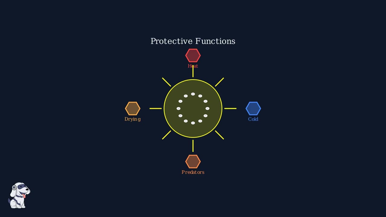
First, let’s look at the trichocysts. These are small, bottle-shaped structures scattered throughout the paramecium’s cytoplasm. Each trichocyst is filled with a special fluid and acts like a tiny defensive weapon.
When the paramecium is threatened by predators or harmful chemicals, these trichocysts can rapidly discharge their contents, creating a protective barrier or deterrent around the cell.
Now let’s focus on the nucleus system. Unlike most cells that have just one nucleus, paramecium has two distinct types: the large macronucleus and the smaller micronucleus.
The macronucleus is the larger, kidney-shaped structure that controls the day-to-day activities of the cell. It manages metabolism, feeding, and movement – essentially running the cell’s daily operations.
The micronucleus is much smaller and rounder. This structure is essential for reproduction, containing the genetic material that gets exchanged during sexual reproduction processes like conjugation.
Together, these two nuclei create a division of labor – the macronucleus handles immediate cellular needs while the micronucleus preserves and manages genetic information for future generations.
These structures – the defensive trichocysts and the dual nucleus system – demonstrate the remarkable complexity and organization found within this single-celled organism.
Now we focus on one of the most important structures for the Paramecium’s survival: the contractile vacuoles. These specialized organelles are essential for maintaining the cell’s water balance.
Paramecium typically has two contractile vacuoles located near opposite ends of the cell. These structures are like tiny water pumps that work continuously to keep the cell healthy.
The primary function of contractile vacuoles is osmoregulation. Since Paramecium lives in freshwater environments, water constantly enters the cell through osmosis. Without a way to remove this excess water, the cell would swell and burst.
Watch as water molecules continuously enter the Paramecium from its freshwater environment. This creates a constant challenge that the contractile vacuoles must address.
The contractile vacuoles work in a rhythmic cycle. First, they collect excess water from the cytoplasm, gradually filling up like tiny balloons.
Then, the vacuoles contract forcefully, expelling the collected water through temporary pores in the cell membrane. This prevents the cell from taking in too much water and bursting.
Think of contractile vacuoles as tiny water pumps working around the clock. Just like a pump removes excess water from a basement, these organelles remove excess water from the cell to maintain the perfect internal environment.
This pumping cycle repeats continuously throughout the Paramecium’s life, ensuring proper water balance and cell survival in its freshwater habitat.
Without contractile vacuoles, Paramecium could not survive in freshwater environments. These remarkable structures demonstrate how even single-celled organisms have sophisticated mechanisms to maintain their internal balance.
Paramecia are remarkable swimmers, propelling themselves through water using thousands of tiny hair-like structures called cilia. These microscopic oars work together in perfect coordination to create one of nature’s most efficient swimming systems.
Each individual cilium performs a two-phase beating cycle. First comes the effective stroke – a fast, powerful movement where the cilium stays relatively stiff and pushes against the water.
Then comes the recovery stroke – a slower, more flexible movement where the cilium curls and sweeps forward in a counter-clockwise motion, preparing for the next effective stroke.
When thousands of cilia beat together in coordination, they create waves of activity that sweep across the cell surface like wind blowing across a field of grain. This coordinated movement is called the ciliary carpet.
This coordinated ciliary action allows paramecia to move in multiple ways. They can swim forward at impressive speeds, rotate around their axis, and even reverse direction when they encounter obstacles.
Paramecia are surprisingly fast swimmers, capable of reaching speeds up to 2 millimeters per second. That might not sound like much, but for a microscopic organism, it’s equivalent to a human swimming at about 6 miles per hour!
When a paramecium encounters an obstacle or unfavorable conditions, it performs an avoidance reaction. The cilia reverse their beating direction, causing the organism to stop, spin, or turn away from the obstacle before resuming forward movement.
This sophisticated movement system makes paramecia highly effective at navigating their aquatic environment, finding food, and avoiding dangers. The coordinated beating of thousands of cilia represents one of nature’s most elegant solutions to microscopic locomotion.
Paramecia are like tiny living vacuum cleaners, constantly searching for food in their aquatic environment. But what exactly do these microscopic organisms eat?
Paramecia primarily feed on three main types of food: bacteria, algae, and yeasts. These tiny organisms make up the perfect meal for a hungry paramecium.
Most paramecia are heterotrophic, meaning they cannot make their own food and must consume other organisms to survive. They are like the carnivores of the microscopic world.
However, some paramecium species are mixotrophic. These clever organisms have formed partnerships with tiny algae that live inside their cells, providing them with additional nutrients through photosynthesis.
Paramecia use a feeding method called holozoic feeding. They use their hair-like cilia to create water currents that sweep food particles directly into their oral groove, like a tiny conveyor belt system.
Think of a paramecium as having its own tiny buffet! The cilia act like little hands, constantly sweeping and gathering food from the surrounding water, ensuring the paramecium never goes hungry in its microscopic world.
The feeding mechanism of paramecium is a fascinating process that demonstrates how a single-celled organism can efficiently capture, process, and digest food. Let’s explore each step of this remarkable biological system.
First, the paramecium uses its cilia to create water currents that sweep food particles toward the oral groove. These hair-like structures beat in coordinated waves, acting like tiny oars to direct bacteria and other small organisms into the feeding apparatus.
The oral groove is a deep depression lined with specialized cilia that funnel food particles inward. This structure acts like a collection channel, concentrating the captured food before it enters the cell’s digestive system.
From the oral groove, food particles pass into the buccal cavity, also called the gullet. This funnel-shaped structure narrows as it leads deeper into the cell, concentrating the food stream toward the cytostome.
The cytostome, or cell mouth, is a small opening where food particles actually enter the cell’s interior. This is the gateway between the external feeding structures and the internal digestive system.
Once inside the cell, food particles are packaged into food vacuoles – membrane-bound compartments that act like temporary stomachs. These vacuoles form at the cytostome and then begin circulating through the cell’s cytoplasm.
As the food vacuoles circulate through the cytoplasm in a process called cyclosis, digestive enzymes enter the vacuoles to break down the food particles. The vacuole contents become increasingly acidic as digestion proceeds.
The food vacuoles continue to circulate through the cell, allowing nutrients to be absorbed into the cytoplasm. Meanwhile, undigested waste materials accumulate in the vacuoles and must be eliminated from the cell.
Finally, undigested waste is eliminated through the cytopyge, a temporary opening that forms on the cell surface. This completes the feeding cycle, allowing the paramecium to efficiently extract nutrients while removing waste materials.
This sophisticated feeding mechanism allows paramecium to thrive in aquatic environments rich with bacteria and organic matter. The entire process – from capture to elimination – demonstrates the remarkable efficiency of single-celled organisms.
Paramecia have fascinating reproductive abilities that allow them to multiply rapidly in favorable conditions. Understanding how these single-celled organisms reproduce helps explain their success in aquatic environments.
The primary method of reproduction in Paramecium is asexual reproduction through a process called binary fission. This means one parent cell divides to create two identical daughter cells.
During binary fission, the paramecium first elongates and then develops a constriction in the middle. The cell gradually pinches inward until it completely separates into two independent cells.
The speed of reproduction is remarkable. A well-fed Paramecium can complete one cycle of binary fission every six hours. This means the population can double every six hours under optimal conditions.
This exponential growth pattern means that from a single Paramecium, you could theoretically have over 16 million cells in just one day! This rapid reproduction helps Paramecia quickly colonize new environments and recover from population declines.
While binary fission is the primary reproductive method, Paramecia also have sexual reproduction processes called conjugation. However, this is less common and serves mainly to increase genetic diversity rather than population growth.
In summary, Paramecium reproduction is dominated by rapid asexual binary fission, allowing populations to double every six hours under favorable conditions. This efficient reproductive strategy makes Paramecia highly successful microorganisms in aquatic ecosystems.
Binary fission is how Paramecium reproduces asexually, creating two identical offspring from a single parent cell. This process is called transverse binary fission because the cell divides across its width.
The process begins when the Paramecium cell starts to elongate and prepare for division. The cell becomes longer as it prepares to split into two parts.
During division, the two types of nuclei behave very differently. The small micronucleus undergoes mitosis, which is the normal process of cell division where chromosomes are carefully separated.
Meanwhile, the larger macronucleus divides through a different process called amitotic division. This means it splits without the organized chromosome separation seen in mitosis.
The gullet, which is the feeding structure of the Paramecium, also divides into two parts so that each new cell will have its own feeding apparatus.
As the division continues, a constriction forms in the middle of the cell, gradually pinching the cell into two separate parts. This creates the characteristic transverse division.
Finally, the division is complete! The single Paramecium has become two genetically identical daughter cells, each with its own micronucleus, macronucleus, and gullet. This process ensures the continuation of the Paramecium lineage.
Conjugation is a fascinating sexual reproduction process in Paramecium that creates genetic diversity. Unlike simple cell division, conjugation involves two separate Paramecium cells working together to exchange genetic material.
The process begins when two compatible Paramecium cells of different mating types encounter each other. These cells are attracted to one another and begin to move closer together.
When the two cells make contact, they form a cytoplasmic bridge between them. This bridge allows the cells to exchange genetic material while remaining as separate organisms.
Now we need to focus on what happens inside the nuclei. Each Paramecium has two types of nuclei: a large macronucleus and a smaller micronucleus. The micronucleus is crucial for conjugation.
During conjugation, the micronucleus in each cell undergoes meiosis, a special type of cell division that reduces the chromosome number by half. This creates haploid micronuclei ready for exchange.
Next comes the crucial exchange step. Each Paramecium sends one of its haploid micronuclei across the cytoplasmic bridge to the other cell. This is where the actual genetic recombination occurs.
After the exchange is complete, the two Paramecium cells separate. Each cell now contains genetic material from both original parents, creating genetic diversity that wouldn’t exist through simple cell division.
Finally, each cell destroys its old macronucleus and creates a new one from the exchanged genetic material. This completes the conjugation process, resulting in two genetically recombined Paramecium cells.
When we place a paramecium under a microscope, we enter a fascinating miniature world. Even though paramecia are microscopic, they’re actually large enough to be seen with the naked eye under ideal conditions.
At first glance, we might see just a tiny moving speck. But as we adjust our focus and increase magnification, the incredible details of this single-celled organism become visible.
Under high magnification, the paramecium’s distinctive slipper shape becomes clearly visible. This elongated, oval form is perfectly adapted for swimming through water.
All around the cell’s surface, we can observe thousands of tiny hair-like structures called cilia. These cilia beat in coordinated waves, propelling the paramecium through the water like microscopic oars.
The characteristic slipper shape and the beating cilia are two of the most obvious features we can identify when observing a paramecium under the microscope.
Looking inside the cell, we can identify several important internal structures. The contractile vacuoles appear as clear, circular areas that rhythmically expand and contract to regulate water balance.
The nucleus appears as a large, dense oval structure, often the most prominent feature inside the cell. This contains the paramecium’s genetic material.
We can also observe the oral groove, a distinctive depression that leads to the cell’s mouth, where the paramecium captures food particles.
Under the microscope, we can clearly identify these key structures that make the paramecium such a remarkable single-celled organism.
What makes observing paramecia so fascinating is that we’re watching a living, active organism. The cilia beat continuously, the contractile vacuoles pulse rhythmically, and the entire cell glides gracefully through the water – it’s truly like peering into a miniature world teeming with life and activity.
Paramecia, like all living organisms, experience aging. Unlike humans who age over decades, paramecia have a much shorter but fascinating aging process tied directly to their reproduction cycles.
A paramecium can only undergo between one hundred to two hundred cycles of binary fission before it reaches the end of its lifespan and dies. This aging process is called clonal aging.
The key to understanding paramecium aging lies in the macronucleus, the large nucleus that controls daily cellular functions. Each time the paramecium divides, the macronucleus accumulates DNA damage.
Let’s watch what happens as a paramecium goes through multiple fission cycles. With each division, more DNA damage accumulates in the macronucleus, gradually weakening the cell.
However, paramecia have a remarkable solution to this aging problem: sexual reproduction through conjugation. When two paramecia conjugate, they exchange genetic material and rebuild their macronuclei, essentially resetting their biological clocks.
This aging mechanism highlights the crucial importance of sexual reproduction in maintaining healthy paramecium populations. Without conjugation, entire lineages would eventually die out from accumulated DNA damage.
Two species perfectly demonstrate the diversity within the Paramecium genus: Paramecium caudatum and Paramecium aurelia. Each species has unique characteristics that showcase different aspects of paramecium biology.
Paramecium caudatum is the most common and well-known species of the genus. It has a distinctive spindle-shaped body that is rounded at the front and tapers to a blunt point at the posterior end.
Notice the characteristic spindle shape – wider in the middle and narrowing toward both ends, but especially toward the rear. This streamlined form helps it move efficiently through water.
Now let’s clear the screen and examine Paramecium aurelia, which demonstrates remarkable reproductive diversity within the genus.
Paramecium aurelia reproduces asexually through binary fission, like other paramecia. However, it also undergoes sexual processes including conjugation between two cells and autogamy within a single cell.
Conjugation involves two paramecia of compatible mating types coming together. They form a bridge between their cells and exchange genetic material through their micronuclei.
Autogamy is even more remarkable – it’s a sexual process that occurs within a single cell. The paramecium undergoes meiosis and genetic reorganization without needing a partner.
These two species perfectly demonstrate the incredible diversity within the Paramecium genus. From the common spindle-shaped caudatum to the reproductively complex aurelia, each species has evolved unique strategies for survival and reproduction.
The Paramecium genome represents a remarkable achievement in molecular biology. Scientists have successfully sequenced the complete genetic code of these fascinating microorganisms.
The sequencing was completed using Paramecium tetraurelia as the model species. This groundbreaking work revealed something extraordinary about the Paramecium genome.
The genome analysis provided evidence for three whole genome duplications that occurred during Paramecium evolution. This means the entire genetic code was copied three separate times throughout evolutionary history.
The micronucleus of Paramecium aurelia species contains between thirty and sixty-three chromosomes. This wide range reflects the genetic diversity within the species complex.
This genetic information is crucial for understanding the evolution and adaptation of Paramecia. The genome data helps scientists trace how these organisms developed their unique characteristics and adapted to different environments over millions of years.
Study Materials
Paramecium Under Microscope
Paramecium Cell Definition, Characteristics, Classification, Movement, Diagram.
Helpful: 0%
Related Videos



- Text Highlighting: Select any text in the post content to highlight it
- Text Annotation: Select text and add comments with annotations
- Comment Management: Edit or delete your own comments
- Highlight Management: Remove your own highlights
How to use: Simply select any text in the post content above, and you'll see annotation options. Login here or create an account to get started.