- The skeletal system in vertebrates is a vital structural component that provides support, protection, and facilitates locomotion. Unlike soft-bodied animals, which can easily twist and contort their bodies, vertebrates require a rigid internal framework to support their larger and more complex structures. In soft-bodied animals, the ability to bend and move through tight spaces is a significant evolutionary advantage. However, this flexibility also limits their size, as larger, soft bodies are prone to collapsing under their own weight. Therefore, a supportive structure is essential for larger organisms to maintain their form and integrity.
- In vertebrates, the need for internal support is met by the endoskeleton, a framework made of bone, cartilage, or both. The endoskeleton, unlike the exoskeletons found in arthropods or mollusks, is located inside the body. These structures are composed of connective tissue, which is mesodermal in origin and grows with the organism.
- This internal skeleton not only provides rigidity and a distinct shape but also serves as a protective casing for the body’s soft, vital organs. Additionally, it provides attachment points for muscles, which is crucial for efficient locomotion. In vertebrates, the skeletal system supports movement by allowing muscles to contract against a solid base, ensuring coordinated motion.
- The vertebrate skeleton is primarily categorized into two types of tissue: bone and cartilage. Both are connective tissues, but they differ in their structural properties and functions. Bone is rigid and dense, providing strong support and protection, whereas cartilage is more flexible and provides cushioning in areas that require flexibility, such as joints. In vertebrates, these tissues work in tandem to ensure that the skeletal system functions optimally. The bones, especially, are critical for long-term support, while cartilage maintains flexibility where necessary.
- To understand the evolutionary significance of vertebrate skeletons, we can compare the skeletal structures of two vertebrate groups: amphibians and mammals. Amphibians, such as frogs, are anamniotes, meaning they lay eggs in water, and their skeletons reflect this primitive lifestyle. In contrast, mammals are amniotes, with adaptations for terrestrial life, including the presence of more advanced skeletal features, such as stronger limb bones and modifications that support efficient movement on land. The changes observed in the skeletal system from amphibians to mammals are a testament to the evolutionary pressures that have shaped vertebrate anatomy over millions of years.
Cartilage and Bone: Key Components of the Vertebrate Skeletal System
The vertebrate skeletal system is primarily composed of two types of connective tissue: cartilage and bone. Both play crucial roles in providing structural support, protection, and aiding in locomotion. Although both tissues are essential for the overall functioning of the skeleton, they differ significantly in their structure, composition, and properties.
- Cartilage:
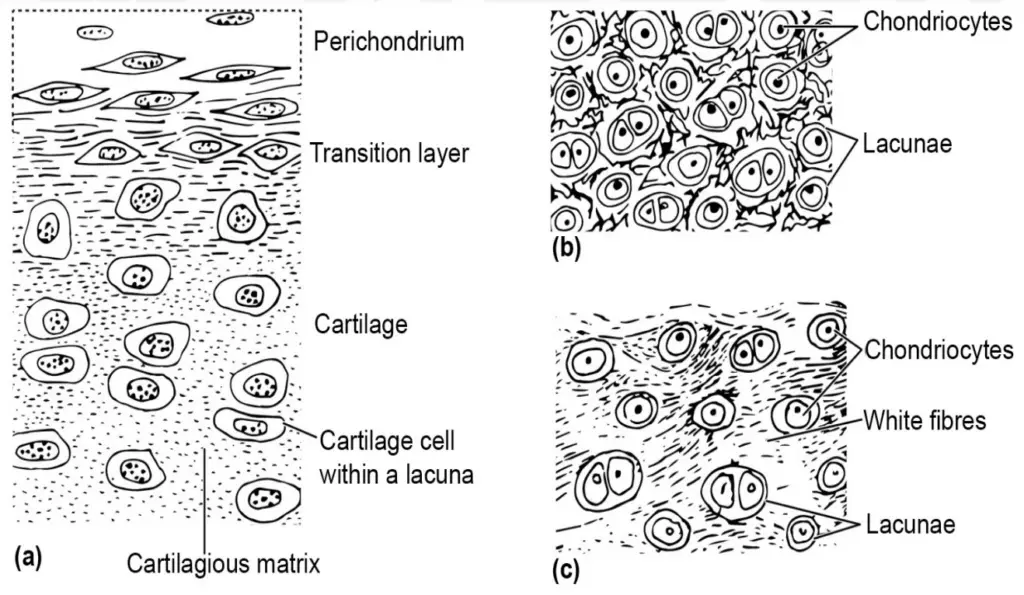
- Cartilage is a soft, elastic, and flexible connective tissue. It is less rigid than bone and allows for greater flexibility in certain areas of the body, such as joints.
- This tissue is made up of a non-living ground substance called the matrix, within which living cells are embedded.
- The matrix of cartilage contains elastic or tough white fibers, which contribute to its resilience and elasticity.
- The protein component in cartilage is called chondrin, which gives it a rubbery texture and allows it to maintain its shape under pressure.
- The living cells in cartilage are referred to as chondroblasts, which secrete the matrix and later become trapped in it, transforming into chondrocytes (mature cartilage cells).
- One key characteristic of cartilage is the absence of blood vessels and nerves. Nutrients are supplied through diffusion within the matrix, which limits the thickness of cartilage since diffusion becomes less efficient over large distances. This is why cartilage is typically thin in nature.
- Cartilage is believed to have appeared first during the embryonic development of vertebrates and is later replaced by bone in most species, except in certain groups like elasmobranch fishes (sharks, rays) and cyclostomes (lampreys and hagfishes), where cartilage remains the primary skeletal material.
- Bone:
- Bone is a more ossified, rigid connective tissue that provides strength and rigidity to the body.
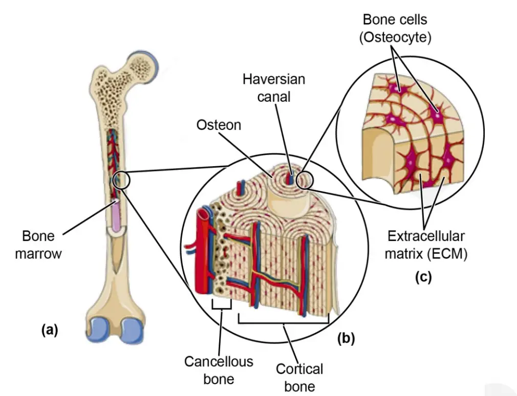
- Similar to cartilage, bone has a matrix that contains collagen, a protein that provides flexibility, preventing the bone from being too brittle. The matrix is also rich in inorganic salts, particularly calcium phosphate and calcium carbonate, which combine to form calcium hydroxyapatite. This compound gives bone its hardness and strength.
- The bone matrix is organized into concentric rings that form a structure called an osteon, or Haversian system, which is critical for maintaining bone integrity and function.
- Within the osteon, the bone cells, called osteocytes, are located in small cavities known as lacunae. These cells are interconnected by tiny passages called canaliculi, which allow for the distribution of nutrients and communication between osteocytes.
- Bone-secreting cells, called osteoblasts, are responsible for producing the collagen and calcium salts that make up the bone matrix. Once the osteoblasts become trapped in the matrix they produce, they mature into osteocytes.
- Types of Bone Formation:
- Endochondral Ossification: Some bones, such as the long bones (e.g., vertebrae, ribs, limbs) and the bones at the base of the skull, develop through this process. In this type of ossification, cartilage serves as a template or precursor that is gradually replaced by bone tissue. The cartilage does not become bone but is entirely replaced by it during development.
- Intramembranous Ossification: In other cases, bones are formed directly from membrane coverings. These bones, called membrane bones or investing bones, are formed from sheets of mesenchymal tissue. This process occurs in the flat bones of the face, most cranial bones, and the collarbone (clavicle). In this method, no cartilage template is involved; instead, bone forms directly from the mesenchymal tissue.
Classification of Skeleton in Vertebrates
The vertebrate endoskeleton is divided into two major categories based on its position and function within the body. These are the axial skeleton and the appendicular skeleton. Each plays a critical role in providing support, structure, and facilitating movement in vertebrates.
- Axial Skeleton:
- The axial skeleton is situated along the body’s anteroposterior axis, meaning it runs from the head to the tail, serving as the central framework for the organism.
- It includes the skull, which forms the head skeleton and protects the brain and sensory organs.
- The vertebral column (spine) is another key component of the axial skeleton, providing structural support and protecting the spinal cord.
- Additionally, the sternum (commonly referred to as the breastbone) is part of the axial skeleton. It is located in the chest region and provides attachment points for the ribs and muscles of the chest.
- Appendicular Skeleton:
- The appendicular skeleton is located on the sides of the body and is composed of the limbs and their associated girdles.
- The limb girdles serve as attachments between the limbs and the axial skeleton. These include the pectoral girdle (shoulder girdle) and the pelvic girdle (hip girdle).
- The limbs themselves, which consist of the bones of the arms and legs, are also part of the appendicular skeleton, allowing for mobility and interaction with the environment.
A. Axial Skeleton
The axial skeleton is an essential component of the vertebrate endoskeleton, primarily responsible for providing structural support and protecting vital structures such as the brain, spinal cord, and blood vessels. It consists of two main parts: the skull and the vertebral column.
1. The Skull
The skull, also referred to as the cephalic skeleton or head skeleton, forms the foundational skeletal structure of the head in craniate vertebrates. Vertebrates can be categorized based on the presence or absence of a head skeleton into two subdivisions: Acraniata, which lack a head skeleton, and Craniata, which possess one. Urochordates and cephalochordates are examples of Acraniata, while the majority of vertebrates are classified as Craniata.
- Structure and Composition:
- The skull of craniate vertebrates is composed of various parts that collectively enclose the brain, cranial nerves, and other sensory organs within the head.
- The portion of the skull that encases the brain is known as the cranium, which consists of multiple bones that provide structural integrity and protection.
- Sensory capsules, which are parts of the skull that enclose the sense organs, are critical for the functioning of various sensory modalities. For instance, the paired auditory capsules protect the hearing apparatus, while the olfactory capsules safeguard the structures involved in the sense of smell. Additionally, the orbits, which are cup-like bony structures, house the eyeballs.
- Orientation and Functionality:
- The cranium, often referred to as the brain box, is oriented along the anteroposterior axis. The olfactory capsules are located at the anterior end, while the auditory capsules are positioned on the posterolateral sides of the cranium.
- This configuration facilitates the passage of blood vessels that supply the brain and sensory organs, highlighting the skull’s role in providing essential support and protection to these critical systems.
- Visceral Skeleton and Evolution:
- In contemporary fish, gill slits are present on the pharyngeal wall, supported by a skeletal framework known as the visceral skeleton. This structure, while vital for aquatic respiration, lost significance in terrestrial vertebrates as lungs replaced gills.
- However, elements of the visceral skeleton have been retained and repurposed in the formation of jaws. Additionally, these skeletal elements contribute to the middle ear’s structure, facilitating the transmission of sound waves to the internal ear and forming support for the buccal cavity.
- Classification of Vertebrates:
- Cyclostomes, which are jawless vertebrates, lack jaws and are classified under the group Agnatha, meaning “without jaws.” In contrast, the majority of vertebrates possess jaws and are categorized as Gnathostomata, which translates to “mouth bound by jaws.”
- Developmental Stages:
- In vertebrate embryonic development, the brain and sensory capsules are initially surrounded by a tough membranous skull known as the membranocranium.
- This membranous structure is later reinforced by the development of cartilage, leading to the formation of the chondrocranium, which serves as the brain case.
- The chondrocranium, composed of cartilage or its replacement bone, forms the anterior portion of the axial skeletal system. Throughout development, the chondrocranium is modified in accordance with the shape and requirements of the brain and specialized sense organs.
- Evolutionary Progression:
- In most vertebrates, the brain case ultimately fuses with dermal and visceral skeletal materials, culminating in a complex and definitive skull structure. This skull is then subject to ossification, whereby cartilage is replaced by bone, or new bones are formed from membrane-covered tissues.
2. Visceral Arches
The visceral arches, integral components of the vertebrate skull, are structures derived from the visceral skeleton. These arches, initially cartilaginous, contribute to the formation of the cranium and other associated features. The visceral skeleton, consisting of a series of cartilaginous rods, provides dermal support along the pharyngeal wall, situated between the gill slits in fishes. Over time, these arches have undergone modifications in various vertebrate groups, largely based on the presence or absence of gills and the type of jaw suspension.
- Formation and Structure:
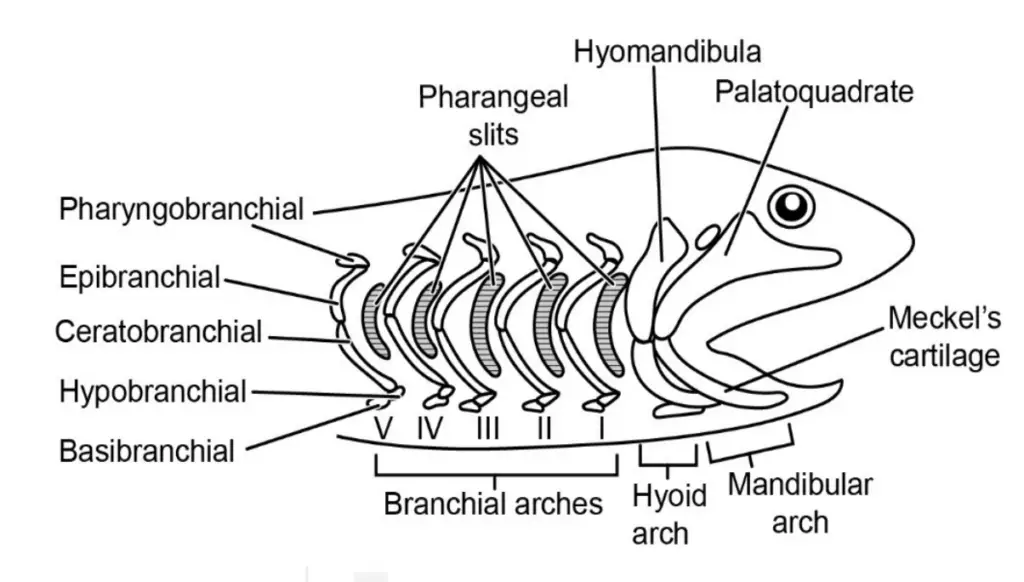
- The visceral skeleton forms a series of cartilaginous rods that encircle the pharynx, creating paired loops on the lateral walls.
- These rods form structural supports, particularly between gill slits in aquatic vertebrates, contributing to respiratory processes.
- In most vertebrates, the left and right sides of the corresponding visceral arches are connected by an unpaired midventral cartilage that spans the floor of the pharynx.
- Number of Visceral Arches:
- In the hypothetical vertebrate ancestor, it is assumed there were seven visceral arches corresponding to the seven gill slits.
- The number and function of these arches, however, vary among different vertebrate species, depending on their evolutionary adaptations, such as the presence or absence of gills and their jaw structures.
- Classification of Visceral Arches:
- The anterior-most arch, referred to as the mandibular arch, is pivotal in the formation of the jaw.
- The palatoquadrate forms the upper jaw, while Meckel’s cartilage forms the lower jaw.
- This arch is located just behind the mouth opening.
- The second arch, known as the hyoid arch, is made up of the hyomandibular and ceratohyal cartilages.
- Following the hyoid arch, the remaining arches are termed branchial arches, which primarily serve to support the gill slits in aquatic vertebrates.
- The anterior-most arch, referred to as the mandibular arch, is pivotal in the formation of the jaw.
- Function of Branchial Arches:
- The branchial arches provide structural support for gill slits in fishes and other aquatic vertebrates, playing a vital role in respiration.
- Evolution of the Visceral Skeleton:
- In cyclostomes (jawless vertebrates like lampreys), there are numerous gill slits, and the visceral skeleton forms a structure called the branchial basket. This structure holds the gill slits and supports respiratory functions.
- During vertebrate evolution, a significant modification occurred when the mouth shifted backward to establish contact with the first gill slit. In the first jawed fishes, the skeletal support of the first gill slit underwent further adaptation, transforming into the foundational support for the mouth, thereby leading to the development of jaws.
3. Jaw Suspensorium
The jaw suspensorium is a critical anatomical structure that connects the jaws to the skull in vertebrates. It evolved as a specialized support mechanism in jawed vertebrates, or gnathostomes, allowing them to utilize their jaws for feeding and other functions. The jaw suspensorium, while varying in different vertebrate groups, is derived from modifications of the visceral arches, particularly the mandibular and hyoid arches.
- Formation of Jaws and Their Connection to the Skull:
- The two palatoquadrates (dorsal part of the first visceral arch, also called the mandibular arch) grow along the upper margin of the mouth, eventually uniting in the midline to form the upper jaw.
- The Meckel’s cartilages (ventral part of the first visceral arch) extend along the lower margin of the mouth and unite anteriorly in the midline to form the lower jaw.
- The Role of the Quadrate and Meckel’s Cartilage:
- The quadrate portion of the palatoquadrate plays a pivotal role in suspending the jaws to the skull. This supportive function of the quadrate is why it is referred to as the suspensorium.
- Evolutionary Development of Jaw Suspension:
- In the earlier stages of gnathostome evolution, jaws were connected to the skull primarily by ligaments. This form of jaw attachment is known as the autodiastylic type of suspensorium.
- Types of Jaw Suspensorium:
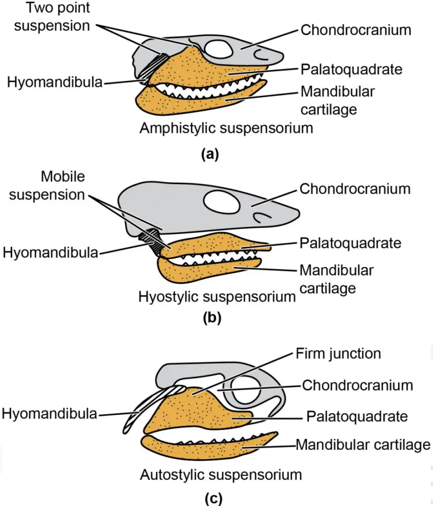
- Amphistylic Suspensorium:
- In some sharks, the upper jaw is braced against the skull and attached to the hyomandibula at its posterior end, which in turn is also braced against the cranium. This configuration is known as amphistylic suspensorium.
- Hyostylic Suspensorium:
- In most living fishes, the upper jaw is not directly connected to the anterior part of the cranium. Instead, it is attached by ligaments, while the posterior part of the upper jaw is attached to the hyomandibula of the hyoid arch. The lower jaw also attaches to the hyomandibula, leading to the hyostylic suspensorium configuration.
- Autostylic Suspensorium:
- In tetrapods, chimeras, and lungfishes, the upper jaw is fused directly with the skull. The hyomandibula is no longer involved in suspending the jaws. This is known as autostylic suspensorium. In this condition, the jaw is ‘self-braced,’ meaning the jaw’s support comes directly from its fusion to the skull.
- Amphistylic Suspensorium:
- Evolutionary Shift in the Role of the Hyoid Arch:
- As jaw suspension evolved, the elements of the hyoid arch—which were previously involved in jaw suspension—no longer played a part in this function. Instead, these elements became repurposed into other functions, particularly in tetrapods, where they are incorporated into the middle ear as ear ossicles. This transformation underscores the adaptability of skeletal structures in vertebrate evolution.
- Branchial Arches and Respiration:
- The branchial arches, while not directly involved in jaw suspension, play a role in respiration. In fishes, these arches allow the pharynx to dilate and contract, facilitating the intake and expulsion of water during respiration.
- Over time, in tetrapods, these branchial arches became obsolete for respiration, as they were replaced by pulmonary respiration (lungs). This led to the loss of branchial respiration and the functional adaptations that came with it.
4. The Vertebral Column
The vertebral column, also referred to as the backbone or spine, is a fundamental structural component of vertebrates, providing both support and flexibility. It is a segmented chain of individual units known as vertebrae (singular: vertebra), which extends from the posterior of the skull to the tip of the tail in many vertebrates. The vertebral column forms the longitudinal axis of the body, playing a critical role in maintaining stability and allowing movement.
- Basic Structure of Vertebrae:
- Each vertebra is composed of several distinct parts that enable it to connect securely with adjacent vertebrae while permitting a controlled degree of movement.
- Articulation of Vertebrae:
- Articulation between vertebrae is achieved through convexities (bulges) and concavities (depressions).
- The bulge of one vertebra fits into the depression of an adjacent vertebra, allowing the vertebrae to be securely connected while still permitting some movement.
- Additional structures further strengthen these connections, ensuring that vertebrae stay in place and minimizing the risk of dislocation or separation during movement.
- Tube-Like Structure:
- The vertebral column forms a tube-like structure that is located dorsally (along the back) in the body.
- It serves the crucial function of enclosing and protecting the spinal cord, a key component of the central nervous system.
- Tail Region:
- In many vertebrates, including some animals with tails, the vertebrae in the tail region form a ventral (lower) tube that encloses and protects blood vessels.
- Key Components of a Typical Vertebra:
- A vertebra is a complex structure composed of several distinct components, each serving specific functions:
- Centrum:
- The centrum is the central body of the vertebra and is in line with the notochord present during early development.
- The centrum has bulges and depressions at its anterior and posterior ends, which articulate with the adjacent vertebrae.
- Neural Arch:
- The neural arch is a dorsal, arch-like structure that encloses the spinal cord.
- The neural arches of all vertebrae together form the neural canal, a protective tube surrounding the spinal cord.
- The neural arches are formed by the vertical neural plates, which arise from the dorsolateral sides of the centrum. These plates meet at the top to form the neural arch, with a backwardly directed neural spine often present dorsally above the neural arch.
- Transverse Processes:
- A pair of transverse processes extends laterally from the sides of the centrum.
- These processes serve as attachment points for muscles, helping with the movement and stabilization of the spine.
- Haemal Arch (in Certain Vertebrates):
- In fishes, salamanders, most reptiles, and long-tailed mammals, the caudal vertebrae (tail vertebrae) typically feature a haemal arch on their ventral side.
- The haemal arch is made up of a pair of plates called haemapophyses. This arch encloses a haemal canal, which protects blood vessels. In some species, the haemal arch also features a haemal spine projecting ventrally.
- Prezygapophyses and Postzygapophyses:
- The centrum bears plate-like projections at its anterior and posterior ends, known as prezygapophyses and postzygapophyses, respectively.
- These structures help firmly articulate adjacent vertebrae and limit movement between them to prevent excessive flexibility or instability.
- Diapophyses and Parapophyses:
- In higher vertebrates, such as mammals, a pair of lateral processes called diapophyses arise from the base of the neural arch.
- Additionally, parapophyses are found on each side of the centrum. These processes are critical for articulating with the ribs, as the bifurcated heads of the ribs attach to the diapophyses and parapophyses.
Types of Vertebrae
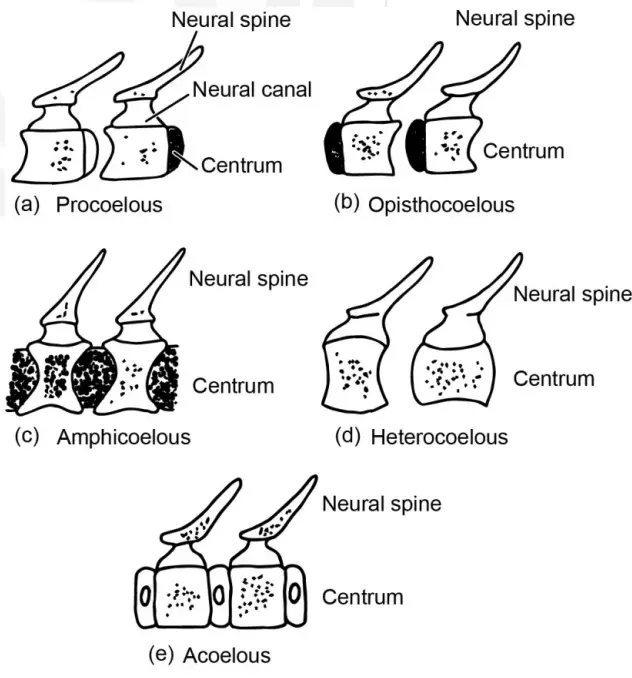
The vertebral column comprises various types of vertebrae, which are classified based on the structure of their centromere. Each type exhibits unique characteristics, facilitating specific functions and movements within the vertebral column. Understanding these different types enhances comprehension of vertebrate anatomy and locomotion.
- Procoelous Vertebrae:
- The procoelous vertebra features a concavity at the anterior end and a convexity at the posterior end.
- The convex anterior surface fits into the concave posterior surface of the adjacent vertebra, facilitating articulation.
- Examples of procoelous vertebrae can be observed in frogs, lizards, and other living reptiles.
- Opisthocoelous Vertebrae:
- Opposite to procoelous vertebrae, the opisthocoelous vertebra has a convex anterior end and a concave posterior end.
- This configuration allows the anterior convexity to articulate with the concave surface of the following vertebra.
- Most salamanders exhibit opisthocoelous vertebrae.
- Amphicoelous Vertebrae:
- The amphicoelous vertebra displays concavities at both the anterior and posterior ends.
- When viewed laterally, these vertebrae appear dumbbell-shaped, contributing to flexibility in the vertebral column.
- Amphicoelous vertebrae are commonly found in most fishes, some salamanders, and caecilians.
- Heterocoelous Vertebrae:
- The heterocoelous vertebra exhibits a unique shape, appearing concave from side to side while being convex when viewed from above and below.
- This saddle-shaped configuration allows for extensive lateral and dorsoventral rotation.
- Heterocoelous vertebrae are characteristic of birds, facilitating their aerial movements.
- Acoelous or Amphiplatyan Vertebrae:
- The acoelous vertebra, also known as amphiplatyan, is characterized by flat surfaces on both ends, lacking any significant bulging or depressions.
- This type of vertebra provides stability and support without compromising flexibility.
- Acoelous vertebrae are typically found in mammals, reflecting their need for a robust yet flexible spine.
Vertebral Column of Vertebrates
The vertebral column, also known as the spine, is an essential component of the skeleton in vertebrates, providing structural support, protection for the spinal cord, and facilitating movement. While the basic structure of the vertebral column is present across all vertebrate groups, the region-specific specialization and types of vertebrae vary among different species. These adaptations allow vertebrates to move efficiently in their respective environments. Below is a detailed explanation of the vertebral column and its structural variations in different vertebrate groups.
- Presence of Notochord:
- All vertebrates initially possess a notochord during their embryonic development. In most vertebrates, the notochord is eventually replaced by the vertebral column, although remnants of it may remain.
- In hagfish and lampreys (cyclostomes), the notochord is retained throughout their life, supported by lateral neural cartilages. This represents a primitive feature that is only transient in other vertebrates.
- In other vertebrate species, the notochord is present only in embryonic stages, eventually being replaced by more complex structures such as the vertebral column.
- Vertebral Structure in Teleost Fishes:
- In teleost fishes, the vertebrae are characterized by ossified amphicoelous vertebrae, where the vertebrae have concave surfaces at both ends, facilitating their articulation.
- Teleost vertebrae can be classified based on their location into two main categories: trunk vertebrae and tail vertebrae, depending on their position in the body.
- Vertebral Regions in Tetrapods:
- In tetrapods (amphibians, reptiles, birds, and mammals), the vertebral column is specialized into distinct regions, each serving specific functions:
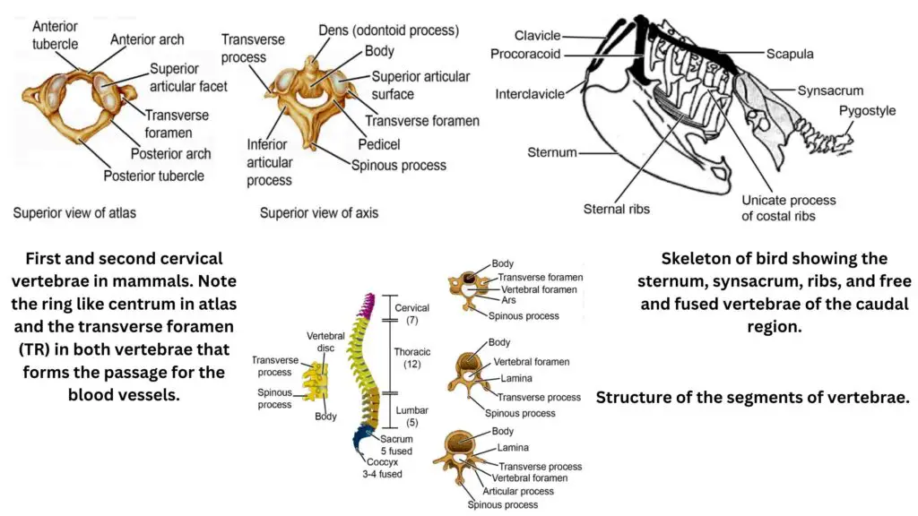
- Cervical Region: The cervical vertebrae form the neck region. In amphibians, only a single cervical vertebra is present, which limits neck movement. However, in most reptiles and mammals, there are seven cervical vertebrae, offering greater flexibility for head and neck movement. The first cervical vertebra, called the atlas, articulates with the back of the skull, allowing multidirectional movement of the head. The second cervical vertebra, the axis, features an odontoid process that fits into the atlas, serving as a pivot to facilitate head rotation.
- Thoracic Region: The thoracic vertebrae are located in the middle part of the vertebral column and are connected to the ribs, providing structural support and aiding in respiratory movements.
- Lumbar Region: The lumbar vertebrae are more robust than the vertebrae of other regions. These vertebrae support the body’s weight and are found in the lower back.
- Sacral Region: In tetrapods, the sacral vertebrae are often fused to form the sacrum, which is connected to the pelvic girdle, contributing to the stability required for locomotion. In birds, a specialized structure called the synsacrum is present. The synsacrum results from the fusion of the last thoracic vertebrae with the lumbar, sacral, and a few caudal vertebrae, providing enhanced support for bipedal movement.
- Caudal (Tail) Region: The vertebrae in the tail region are known as caudal vertebrae. In certain species, these vertebrae are fused, forming structures such as the urostyle in frogs and toads, or the pygostyle in birds. In humans and apes, the last few caudal vertebrae fuse to form the coccyx or tailbone, a vestigial structure with limited function.
- In tetrapods (amphibians, reptiles, birds, and mammals), the vertebral column is specialized into distinct regions, each serving specific functions:
- Specialized Vertebrae and Adaptations:
- The specialized vertebrae in tetrapods reflect the diverse modes of locomotion and support required by each species. In birds, the synsacrum provides stability for bipedal locomotion, while the fusion of vertebrae in mammals and reptiles supports their specific postural and movement requirements.
- In frogs and toads, the terminal vertebrae fuse to form the urostyle, aiding in the rigidity of the tail region for swimming or jumping. In birds, the fusion of the last four to five caudal vertebrae forms the pygostyle, which supports tail feathers and helps in steering during flight.
- In humans and apes, the last three to five caudal vertebrae fuse to form the coccyx, which provides attachment for muscles and ligaments and serves as a rudimentary tail structure.
- Intervertebral Discs:
- Between the vertebrae, intervertebral discs made of fibrous cartilage and remnants of the notochord are present in mammals. These discs act as shock absorbers, allowing for movement between vertebrae while providing cushioning for the spinal cord.
B. Appendicular Skeleton
The appendicular skeleton consists of the bones that form the limbs and girdles of vertebrates. This section of the skeleton plays a vital role in locomotion and supports the vertebrate body during movement. The appendicular skeleton is primarily composed of the pectoral and pelvic girdles, along with the limbs they support. These structures, while varying among different vertebrate species, share many similarities in their skeletal elements, which are essential for movement.
- Paired Appendages:
- Most vertebrates possess two pairs of appendages: an anterior pectoral pair and a posterior pelvic pair. In fishes, the paired appendages take the form of fins, while in other vertebrates, these appendages evolve into jointed limbs, known as podia.
- During embryonic development, the paired appendages in fishes appear as horizontal folds, and in other vertebrates, they develop as limb buds.
- Both forelimbs and hind limbs articulate and are supported by their respective girdles, which are the pectoral girdle (anterior) and pelvic girdle (posterior). Together, the girdles and the skeleton of the limbs form the appendicular skeleton.
- Pectoral Girdle:
- The pectoral girdle is comprised of both dermal and endochondral (replacement) bones, and each half of the girdle contains three components: the clavicle, scapula, and coracoid.
- In humans, the pectoral girdle includes only the clavicle and scapula, while some mammalian species, such as dogs and horses, possess only the scapula.
- Each part of the pectoral girdle features an acetabular cavity, which facilitates the articulation of the head of the humerus bone of the forelimb. This cavity allows for a secure connection between the girdle and the forelimb, enabling efficient movement.
- Pelvic Girdle:
- The pelvic girdle is made up of two dermal bones: a dorsal iliac and a ventral ischio-pubic part. It consists of two identical appendicular hip bones, which are fused together to form a ring.
- Each half of the pelvic girdle is formed by the ilium, ischium, and pubis, and it contains a glenoid cavity, where the head of the femur bone of the hind limb articulates.
- The pelvic girdle also includes the pelvic spine, which consists of the sacrum and coccyx—these structures support the vertebral column and contribute to the overall stability of the pelvic region.
- Forelimbs and Hind Limbs (Pentadactyl Limbs):
- The extremities of both the forelimbs and hind limbs are typically pentadactyl, meaning they are equipped with five digits each, a trait that is conserved across many vertebrate species.
- Despite differences in function and appearance, the forelimbs and hind limbs have a similar skeletal structure, which follows a basic pattern. This similarity is evident in the corresponding bones and joints of the fore and hind limbs, allowing for parallel functions in support and movement.
- Skeletal Elements and Joints:
- The general skeletal pattern of the appendicular skeleton includes the following bones and joints (Table 2.1 highlights the corresponding structures in the anterior and posterior limbs):
- Shoulder Joint (pectoral girdle) ↔ Hip Joint (pelvic girdle)
- Pectoral Girdle ↔ Pelvic Girdle
- Upper Arm Bone (humerus) ↔ Upper Thigh Bone (femur)
- Lower Arm Bones (radius and ulna) ↔ Lower Hind Limb Bones (tibia and fibula)
- Elbow Joint ↔ Knee Joint
- Wrist (Carpal Bones) ↔ Ankle (Tarsals)
- Palm (Metacarpals) ↔ Sole (Metatarsals)
- Fingers (Phalanges) ↔ Toes (Phalanges)
- The general skeletal pattern of the appendicular skeleton includes the following bones and joints (Table 2.1 highlights the corresponding structures in the anterior and posterior limbs):
- Functional Adaptations:
- Despite their structural similarities, the forelimbs and hind limbs serve different functions depending on the species. For example, in terrestrial vertebrates, the forelimbs often play a more dominant role in supporting the body’s weight and movement, while the hind limbs provide power and propulsion.
- In aquatic animals like fishes, the pectoral and pelvic fins are used for stabilization, steering, and locomotion, while in terrestrial vertebrates, the limbs are adapted for walking, running, or flying, depending on the species.
1. Ribs and Sternum
The ribs and sternum are integral components of the vertebral thoracic skeleton, serving both protective and structural roles in vertebrate species. They are part of the larger system that encases and protects the vital organs in the thorax, including the heart and lungs. The ribs are paired, slender, curved bones that extend from the vertebral column and contribute to the formation of a rib cage, while the sternum, often referred to as the breastbone, forms a central support for the attachment of the ribs and other components of the thoracic skeleton.
- Ribs:
- Ribs are primarily found in the thoracic region of most vertebrates. They are paired structures that attach to the vertebrae on one end and the sternum on the other. The ribs are composed of two parts: a bony vertebral portion that connects to the vertebrae, and a ventral portion that can be either cartilaginous or bony, depending on the species.
- The ribs, together with the vertebral column and sternum, form a skeletal cage that surrounds and protects the thoracic organs.
- Evolution of Ribs: The presence of ribs first appeared in Gnathostomes (jawed vertebrates). Ribs evolved in two distinct types:
- Dorsal or Intramuscular Ribs: These ribs grow as extensions of the vertebrae and extend into the horizontal septa. In elasmobranchs (sharks and rays) and amphibians, these ribs are short and incompletely developed. However, they are well-developed in amniotes (reptiles, birds, and mammals), forming a more complete rib cage.
- A typical dorsal rib has two heads: the capitulum, which attaches to the vertebrae’s centrum, and the tuberculum, which connects to the transverse processes of the vertebrae. Together, these two heads enclose a space called the vertebro-arterial canal or vertebrarterial foramen, through which the vertebral artery passes.
- Ventral or Haemal Ribs: These ribs are believed to have originated from the haemal arches. They encircle the body but are located inside the peritoneal cavity. Ventral ribs are seen in many teleost fishes, ganoid fishes, and Dipnoi (lungfishes).
- Both dorsal and ventral ribs can be present in teleost fishes.
- Dorsal or Intramuscular Ribs: These ribs grow as extensions of the vertebrae and extend into the horizontal septa. In elasmobranchs (sharks and rays) and amphibians, these ribs are short and incompletely developed. However, they are well-developed in amniotes (reptiles, birds, and mammals), forming a more complete rib cage.
- Attachment and Structure: In most vertebrates, including reptiles, birds, and mammals, ribs are attached by movable joints only to the thoracic vertebrae. However, snakes possess ribs attached to all of their vertebrae.
- In birds, the ribs are fully ossified and firmly attached to the sternum. In contrast, in mammals, the sternal part of the rib is typically cartilaginous.
- Some ribs are false ribs, which are shorter and not directly attached to the sternum but to other ribs. In humans, ribs 8 to 10 are false ribs, and this characteristic is also found in crocodiles.
- Floating ribs are ribs that are not attached to the sternum at all and are free. In humans, these are ribs 11 and 12.
- Sternum:
- The sternum is a shield-shaped or rod-shaped bone located on the midventral side of the thorax in most vertebrates. It is commonly known as the breastbone.
- In amniotes, the ventral ends of the ribs attach to the sternum. This attachment provides structural support for the rib cage and contributes to the protection of internal organs in the thorax.
- The sternum of birds is distinct because it is broad and develops a ventral keel-like projection, which provides a surface for the attachment of wing muscles. This keel is crucial for flight, offering the necessary surface area for muscle attachment during wing movement.
- In snakes, which have lost their limbs, the sternum is also lost, as it is no longer necessary for their body structure and movement.
Axial skeleton of frog and rabbit
1. Skull of Frog and Rabbit
The skull structure of both frogs and rabbits offers an insightful comparison of vertebrate anatomy, reflecting the evolutionary and functional differences between amphibians and mammals. While the frog skull is more simplistic, reflecting its lifestyle and evolutionary stage, the rabbit skull is more complex, aligned with mammalian adaptations for terrestrial living and specialized feeding.
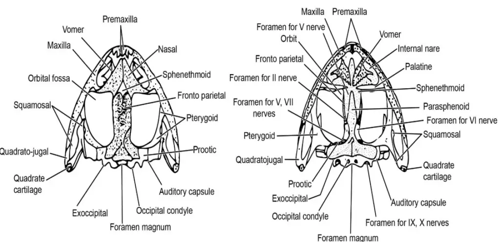
Skull of Frog
The frog skull is relatively simple in structure compared to other vertebrates. Its fewer bones and areas of ossification make it a subject of study for understanding more primitive vertebrates.
- Cranium: The cranium of the frog is elongated anteroposteriorly, composed of six bones. At the posterior end, there is a large foramen magnum surrounded by a pair of exoccipitals, which bear the occipital condyles. These condyles articulate with the atlas, the cervical vertebra. The roof and sides of the cranium are formed by the frontoparietals, which are composite bones.
- Sphenethmoid and Parasphenoid: At the front of the cranium lies a single sphenethmoid bone, almost entirely covered by nasal bones, leaving only a triangular portion visible on the dorsal side. Below it, the parasphenoid forms the floor of the cranium, taking the shape of a dagger.
- Auditory and Olfactory Capsules: Each auditory capsule, situated at the posterior-lateral side of the cranium, is composed of the pro-otic bone. The capsule transmits sound via a cartilaginous extra stapes and a bony stapes. The olfactory capsules, which house the sense of smell, are located anteriorly, covered by the nasal and vomer bones.
- Eye Orbits: The eye orbits are spaces on either side of the cranium containing the eyeballs, with the upper and lower jaw bones forming the boundaries.
- Jaws:
- Upper Jaw: The upper jaw of the frog is horse-shoe shaped and made up of the premaxilla, maxilla, and quadratojugal. These are fixed to the skull and help hold prey in place, but not for mastication.
- Lower Jaw: The lower jaw is also horse-shoe shaped and consists of the mentomeckelian, dentary, and angulosplenial bones. The angulosplenial articulates with the quadratojugal of the upper jaw, allowing some movement.
- Teeth: Frogs have teeth on the premaxilla, maxilla, and vomer (vomerine teeth). These teeth are curved and fused to the bones, aiding in holding prey rather than in chewing.
- Hyoid Apparatus: The hyoid apparatus is a cartilaginous structure supporting the floor of the buccal cavity, with two pairs of hyoid cornua (anterior and posterior) providing attachment points for the tongue and connecting to the auditory region.
Skull of Rabbit
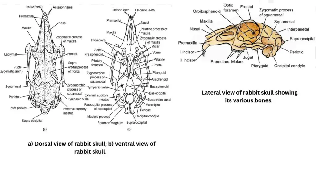
The rabbit skull is more complex than that of the frog, reflecting the adaptations of mammals to a terrestrial and more advanced evolutionary life form. It has an elongated shape due to the prolongation of the jaws for forming a snout, which allows for greater jaw muscle attachment.
- Cranium:
- Occipital Segment: The cranium is divided into an occipital segment, which surrounds the foramen magnum. This segment is more complex than that of the frog, consisting of four bones: the supraoccipital, exoccipital (pair), and basioccipital. The foramen magnum in rabbits is large and directed downward rather than backward as in frogs, and it accommodates two occipital condyles for articulation with the first vertebra, similar to the frog’s bicondylar condition.
- Parietal and Frontal Segments:
- The parietal segment consists of six bones, including the basisphenoid, parietals, and squamosal bones, with the squamosal forming part of the zygomatic arch.
- The frontal segment includes the frontal bones, presphenoid, and orbitosphenoid, which surround the brain and contribute to the structure of the eye orbits.
- Sensory Capsules:
- Auditory Capsules: The auditory capsules in rabbits consist of a fused complex called the periotic bone, which is formed from the prootic, epiotic, and opisthotic bones. These capsules house the tympanic bone and auditory ossicles, which are involved in hearing. The tympanic bone encloses the tympanic cavity and auditory ossicles (malleus, incus, and stapes), which convey sound from the external environment to the inner ear.
- Olfactory Capsules: The olfactory capsules, like in frogs, are located in front of the cranium and enclosed by nasal bones. A mesethmoid bone divides the nasal cavity, and turbinal bones inside help to condition the air.
- Jaws:
- Upper Jaw: The upper jaw of the rabbit is more specialized, consisting of premaxilla and maxilla bones. The premaxilla forms the anterior part of the snout and bears the incisor teeth, while the maxilla carries premolar and molar teeth. The palate, which separates the nasal and buccal cavities, is formed by the horizontal palatine processes.
- Lower Jaw: Unlike the frog’s lower jaw, which is made up of three bones, the rabbit’s lower jaw consists of a single dentary bone. The dentary bears incisors, premolars, and molars, with a gap (diastema) between the incisors and other teeth. The dentary bone articulates with the glenoid fossa of the squamosal and zygomatic bones.
- Teeth: Rabbits have heterodont dentition, meaning they possess different types of teeth (incisors, premolars, molars) adapted for various functions, such as cutting and grinding. Additionally, rabbits have diphyodont dentition, meaning they form two sets of teeth: deciduous (milk) teeth and permanent teeth.
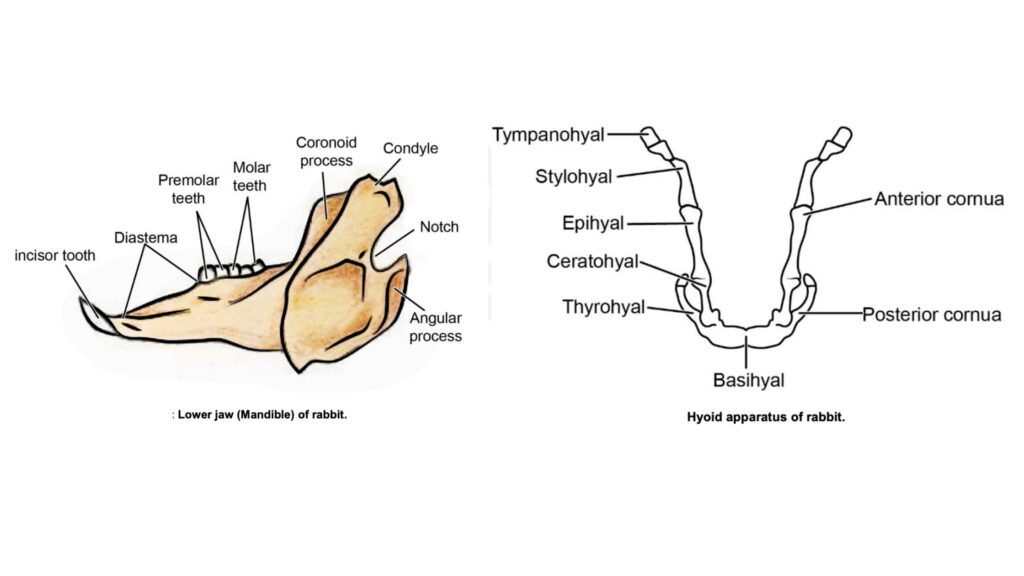
Key Differences between the Skull of Frog and Rabbit:
- Cranial Structure: The rabbit’s skull is more elongated and complex, reflecting its mammalian nature. In contrast, the frog’s skull is simple and compact.
- Teeth and Dentition: Frogs have homodont teeth, all of which are of the same type. Rabbits, however, have heterodont dentition with different tooth types for specialized functions.
- Jaws and Feeding Adaptations: The frog’s jaws are immovable and primarily serve to hold prey, while the rabbit’s jaws are adapted for chewing and processing food, with specialized dentition.
- Sensory Capsules: The auditory and olfactory capsules of the rabbit are more complex, reflecting the increased functionality and refinement in sensory systems of mammals.
2. Vertebral Column of Frog and Rabbit
The vertebral column is an important skeletal structure that serves as the central axis of the body and provides support and protection for the spinal cord. The vertebral columns of frogs and rabbits differ significantly due to their distinct lifestyles and body plans. Below is a detailed comparison of the vertebral columns of frogs and rabbits, highlighting their structure, functions, and unique features.
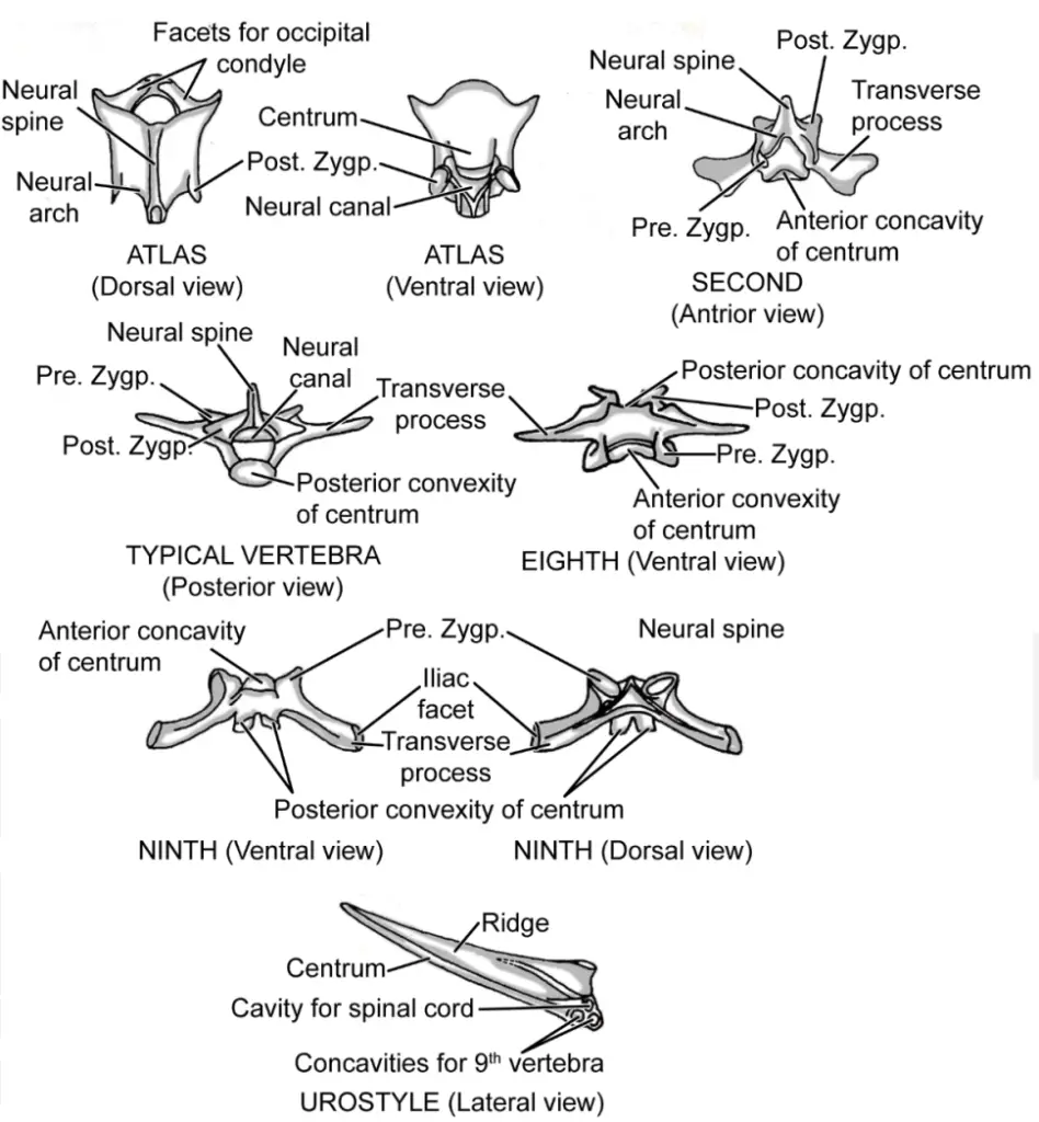
- Frog Vertebral Column:
- The vertebral column of the frog is short and simple, comprising only nine vertebrae.
- At the posterior end of the column is the urostyle, a rod-like structure that represents the fused caudal vertebrae of the tadpole. The urostyle serves to provide support to the frog’s body after it undergoes metamorphosis, during which the tail is lost.
- The first vertebra, known as the atlas vertebra, is ring-shaped and lacks both transverse processes and pre-zygapophyses. It has a small centrum and two concavities on its anterior surface to articulate with the skull’s condyles.
- The second vertebra, called the axis vertebra, follows the atlas. This vertebra is similar to those of other vertebrates, with typical vertebral features.
- Vertebrae 3 to 7 are procoelous, meaning they are concave in front and convex behind. These vertebrae have transverse processes, as well as both pre- and post-zygapophyses. Additionally, they feature a neural arch that encloses the neural canal and a backwardly directed neural spine.
- The eighth vertebra is biconcave, with concavities on both the anterior and posterior surfaces. This structure allows for greater movement between the vertebrae.
- The ninth vertebra has a convexity in front, and its posterior end is marked by two knob-like bulges that articulate with the anterior end of the urostyle. This vertebra also has transverse processes pointing backward, but lacks post-zygapophyses.
- The urostyle is a long, slender rod-like structure that is as long as the entire vertebral column. It is characterized by a dorsal ridge and two concavities at its anterior end for articulation with the ninth vertebra.
- Between the vertebrae are paired apertures known as intervertebral foramina, which allow the passage of spinal nerves. Ligaments bind the vertebrae together, limiting their movement but providing some flexibility.
- Rabbit Vertebral Column:
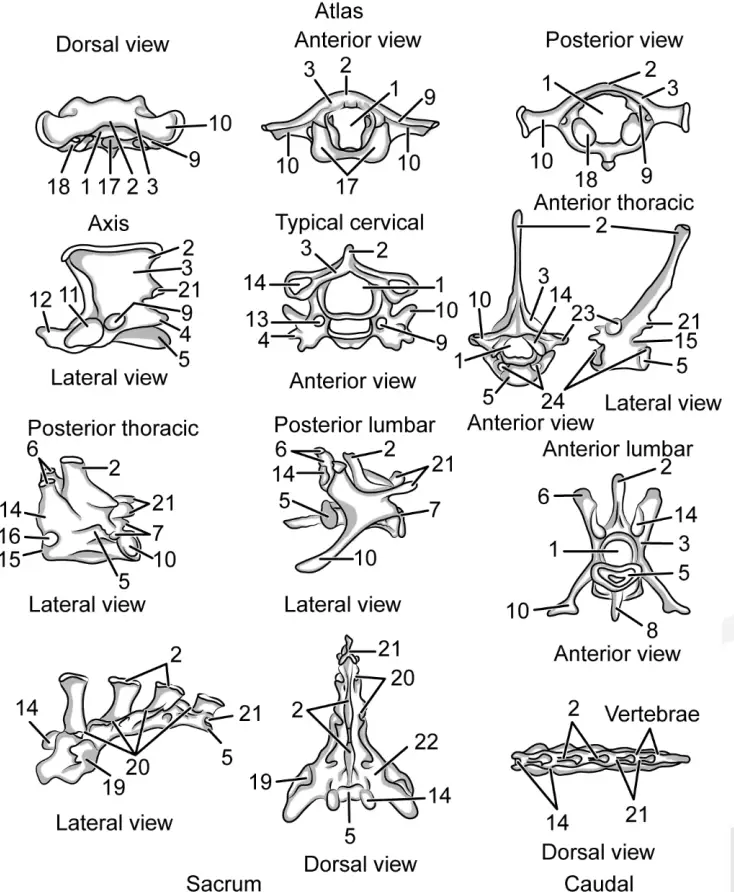
- The vertebral column of the rabbit is considerably longer and more complex than that of the frog. It is divided into five regions: cervical, thoracic, lumbar, sacral, and caudal.
- The rabbit’s vertebral column consists of 45 vertebrae, divided as follows: seven cervical vertebrae, twelve thoracic vertebrae, seven lumbar vertebrae, four sacral vertebrae, and fifteen caudal vertebrae.
- The vertebrae are separated by fibrous cartilage plates known as intervertebral discs, with the central part of each disc, called the nucleus pulposus, being a remnant of the notochord.
- The cervical region of the rabbit consists of seven vertebrae. Despite the varying neck lengths in mammals, such as giraffes and elephants, the number of cervical vertebrae remains constant across all mammals. The cervical vertebrae have short centra, small neural spines, and vertebral arterial foramina that allow the passage of vertebral arteries, except in the seventh vertebra.
- The atlas vertebra, the first cervical vertebra, is ring-shaped and lacks a distinct centrum. It has large, flattened transverse processes and concavities on its anterior surface for articulation with the condyles of the skull. Small facets on the posterior surface of the atlas allow articulation with the axis vertebra, the second cervical vertebra.
- The axis vertebra is notable for its large neural arch, which encloses a large neural canal. It also has an odontoid process, or plough-like structure, that fits into the lower part of the neural canal of the atlas. This process allows for the rotation of the head.
- Cervical vertebrae 3 to 7 exhibit a typical structure with broad centra and large neural arches. Transverse processes of all cervical vertebrae, except the seventh, are bifurcated, and the seventh vertebra features a long neural spine.
- The thoracic vertebrae are characterized by a centrum, a neural arch, a backwardly directed neural spine, and stout transverse processes. The ribs articulate with the thoracic vertebrae via these processes.
- The lumbar vertebrae are large, with shorter neural spines and longer transverse processes. In the first two lumbar vertebrae, a ventral process called hypapophyses is present, and metapophyses and anapophyses are well developed.
- The sacral vertebrae are fused to form a composite structure called the sacrum, which is wedged between the two halves of the pelvic girdle. The sacral vertebrae have large neural spines but lack hypapophyses and anapophyses. Small metapophyses are present.
- The caudal vertebrae gradually decrease in size toward the posterior end. More posterior caudal vertebrae consist of centra only.
3. Sternum and Ribs of Frog and Rabbit
The sternum, also known as the breastbone, plays a crucial role in protecting internal organs and providing support for the attachment of muscles and ribs. While both frogs and rabbits possess a sternum, their ribs exhibit notable differences. Frogs, being amphibians, lack ribs altogether, while rabbits, as mammals, have a well-developed rib cage. This distinction is reflected in the structure and function of the sternum and rib attachments in each species. Below is a detailed comparison of the sternum and ribs in both animals.
- Sternum of Frog:
- The frog’s sternum is composed of both cartilage and bone, reflecting its amphibian nature.
- It is divided into four segments that are situated in front and behind the epicoracoid of the pectoral girdle.
- Anteriorly, the sternum has a homosternum that is connected to a cartilaginous episternum at the front.
- The posterior end of the homosternum is attached to the epicoracoid of the pectoral girdle.
- A mesosternum lies behind the epicoracoid, followed by a cartilaginous plate-like structure called the xiphisternum.
- The frog’s sternum is connected to the pectoral girdle along the midventral line, where the two halves of the pectoral girdle meet. This connection provides stability to the pectoral region.
- Ribs are completely absent in frogs, which is typical of amphibians. The absence of ribs allows for a more flexible and less encumbered body, aiding in locomotion and breathing.
- The sternum in frogs, especially in connection with the pectoral girdle, serves primarily to stabilize the forelimbs during movement.
- Sternum and Ribs of Rabbit:
- The rabbit’s sternum is attached to the pectoral girdle only on the ventral side, and it is not connected to the ribs.
- The rabbit has twelve pairs of ribs located in the thoracic region. These ribs are associated with the thoracic vertebrae, forming part of the rib cage.
- The first seven pairs of ribs are known as true ribs, as they are directly connected to the sternum on the ventromedian side.
- The remaining five pairs of ribs are called false ribs or floating ribs. These do not connect to the sternum, and they offer more flexibility to the rib cage.
- The rabbit’s sternum is made up of several segmental bones, a feature unique to mammals.
- The manubrium sterna, the anterior-most and longest part of the sternum, forms the presternum and features a keel. The keel serves as an anchor for muscles, aiding in the rabbit’s breathing and movement.
- The xiphisternum, at the posterior end of the sternum, is cartilaginous and rounded, ending in a small piece of xiphoid cartilage.
- Between the manubrium and the xiphisternum lie five elongated bony pieces, collectively called the sternebrae, which make up the mesosternum or body of the sternum.
- The ribs attach to the sternebrae via their costal cartilage, forming a functional rib cage that is connected to the thoracic vertebrae for added protection and structural support.
Appendicular skeleton of frog and rabbit
1. Pectoral Girdle of Frog and Rabbit
The pectoral girdle serves as a vital structure in terrestrial vertebrates, supporting the forelimbs and facilitating movement. This anatomical feature varies significantly between species, reflecting their unique evolutionary adaptations. In this discussion, the pectoral girdle of frogs and rabbits will be compared, highlighting their structural differences and functional implications.
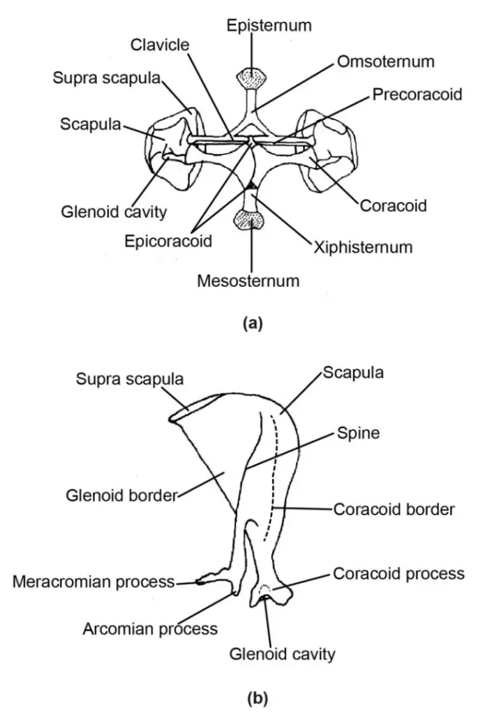
- Pectoral Girdle of Frog:
- The frog’s pectoral girdle comprises two halves that are united at the midventral line but remain separate dorsally.
- The outer ends of each half are bent upwards, forming an arch-like structure. This design helps to enclose and protect the thoracic organs.
- Each half features a cartilaginous, triangular suprascapula, which is partially ossified. This component plays a role in the support and movement of the forelimbs.
- Attached to the inner end of the suprascapula on the ventral side is a stout, flat scapula bone. The scapula is essential for the attachment of muscles and provides a surface for articulation with the humerus.
- From each scapula, two bones extend toward the midventral side:
- Clavicle: A slender rod-like bone located anterior to the coracoid, separated by a space called the coracoid fenestra.
- Coracoid: This bone also extends toward the midventral side and meets the epicoracoid cartilage at the midline.
- At the junction of the clavicle, coracoid, and scapula is a depression known as the glenoid cavity. This cavity is crucial for the articulation of the head of the humerus, allowing for the movement of the forelimb.
- The pectoral girdle is further joined to the sternum at the mid-ventral line, emphasizing the structural integration of these elements for stability and support during locomotion.
- Pectoral Girdle of Rabbit:
- In contrast to frogs, the rabbit’s pectoral girdle consists of fewer bones, reflecting its more advanced evolutionary stage.
- The primary components are a flat, thin, triangular scapula and a rod-like clavicle.
- The glenoid cavity is located at the narrow end of the scapula, articulating with the head of the humerus. This articulation is crucial for the forelimb’s range of motion.
- Instead of the prominent coracoid found in frogs, rabbits possess a small curved structure known as the coracoid process, which is fused to the scapula at the narrow end, positioned in front of the glenoid cavity.
- A thin strip-like suprascapula is present at the dorsal edge of the scapula, aiding in the stability and support of the girdle.
- The outer surface of the scapula features a ridge called the spine, which provides additional surface area for muscle attachment.
- The scapula’s free ventral end terminates in a process called the acromian process, which serves as an attachment point for muscles involved in limb movement.
- A branch-like process known as the metacromian extends posteriorly from the scapula, further enhancing the muscle attachment sites.
- The clavicle lies obliquely between the presternum and the scapula, contributing to the overall structure of the pectoral girdle.
2. Pelvic Girdle of Frog and Rabbit
The pelvic girdle, a critical structure in terrestrial vertebrates, provides support for the hind limbs and is integral to locomotion. It connects the hind limbs to the vertebral column and assists in bearing the weight of the body during movement. Both frogs and rabbits exhibit distinct pelvic girdle structures, reflecting their unique evolutionary adaptations. This section compares the pelvic girdle of frogs and rabbits, highlighting the similarities and differences between these two species.
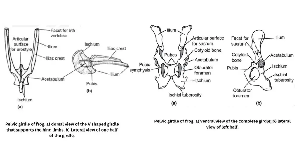
- Pelvic Girdle of Frog:
- The frog’s pelvic girdle is simpler and consists of fewer bones compared to its pectoral girdle.
- It is composed of two halves, which are joined at one end and free at the other, forming a V-shaped structure.
- Each half of the pelvic girdle is made up of three bones: the ilium, ischium, and pubis.
- Ilium: This bone is elongated and its bar-like, free end articulates with the transverse process of the 9th vertebra, providing stability.
- The other end of the ilium, along with the pubis and ischium, forms a disc-like structure known as the acetabulum, which houses a cavity.
- The acetabulum is the point where the proximal head of the thigh bone, the femur, articulates with the pelvic girdle, allowing for movement of the hind limbs.
- The disc-like structure of each half of the pelvic girdle is fused together, creating a solid foundation for the frog’s hind limb movements.
- During early development, the three bones (ilium, ischium, and pubis) are distinct and separate, but they later become fused in the adult frog. This fusion is assisted by cartilage or bone and is called symphysis.
- The frog exhibits pubic symphysis and ischiatic symphysis, where the pubis and ischium are closely joined, forming immovable joints that contribute to the stability of the pelvic girdle.
- Pelvic Girdle of Rabbit:
- Like frogs, the rabbit’s pelvic girdle is also composed of two halves, which are united by symphysis at the midline.
- The three main bones present in the rabbit’s pelvic girdle are the ilium, ischium, and pubis (similar to the frog).
- The acetabular cavity is located at the union of the ischium and ilium on the outside of the pelvic girdle.
- In young rabbits, the three bones are distinct, but as they mature, these bones fuse to form a single structure known as the innominatum in the adult.
- Ilium: In the rabbit, the ilium is positioned dorsally, in front of the acetabulum, and has a rough, expanded, wing-like inner surface. This structure articulates with the transverse processes of the 1st sacral vertebra, further supporting the pelvic girdle.
- Ischium: Located posteriorly and dorsally, the ischium extends downwards and forms the ischial tuberosity at the ischiatic symphysis, providing a stable anchor for muscles.
- Pubis: The smallest of the three bones, the pubis lies anteriorly and points downward. It is separated from the ischium by a wide opening called the obturator foramen.
- The two pubes unite at the midventral line through a pubic symphysis, similar to the frog’s pelvic girdle.
- The acetabulum in the rabbit is surrounded by a small bone called the cotyloid bone, which provides additional structural support.
3. Limbs of Frog and Rabbit
The limb skeleton of vertebrates is largely conserved across species, although adaptations to specific environments and lifestyles result in subtle variations between animals. Both frogs and rabbits exhibit a similar skeletal pattern for their limbs, but there are notable differences in structure that reflect their diverse modes of locomotion and evolutionary adaptations. This section explores the structure of the forelimbs and hindlimbs in both frogs and rabbits, offering an in-depth comparison based on the given content.
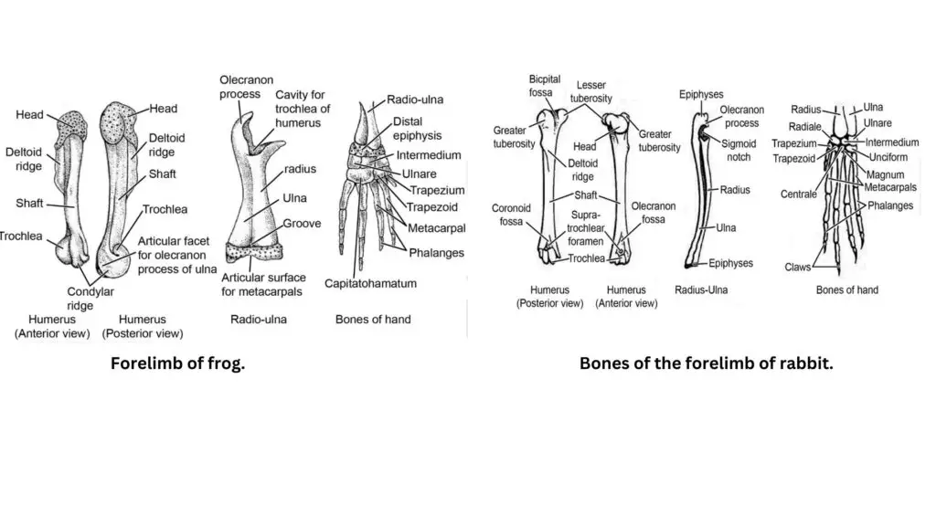
- Forelimb of Frog:
- The frog’s forelimb is composed of three main bones: the humerus, radio-ulna, and the carpals.
- Humerus: The upper arm of the forelimb is formed by a stout, long, and curved bone called the humerus. At its proximal end, the humerus fits into the glenoid cavity of the pectoral girdle. The distal end is round and has two projections, with a characteristic ridge known as the deltoid ridge.
- The humerus articulates with the radio-ulna, which is a fused bone formed by the radius and ulna. This fusion is present in frogs and results in the radio-ulna.
- The radio-ulna at its proximal end has a concavity that allows it to form the elbow joint with the distal end of the humerus. A backwardly directed process, known as the olecranon process, is present at the elbow joint.
- The wrist (or carpals) consists of several bones, with three proximal carpal bones—radiale, central, and ulnare—articulating with the radius and ulna. The distal carpal bones are fused and articulate with the metacarpals of the hand.
- The metacarpals in the frog are long, with the innermost metacarpal being reduced. The fingers are numbered 2 to 5, with two phalanges in fingers 2 and 3, and three phalanges in fingers 4 and 5. Notably, the thumb, corresponding to the first finger in other vertebrates, is absent in frogs.
- The frog’s forelimb is composed of three main bones: the humerus, radio-ulna, and the carpals.
- Forelimb of Rabbit:
- The rabbit’s forelimb follows a similar pattern to the frog, with slight differences in bone structure and morphology.
- Humerus: In rabbits, the humerus also forms the upper arm of the forelimb, but it has two tuberosities on its outer border that serve as attachment points for the bicep muscles. The tendons of these muscles are inserted between these tuberosities in a groove called the bicipital groove.
- The deltoid ridge is also present on the anterior surface of the humerus, similar to the frog’s forelimb.
- The distal end of the humerus has two distinct articular surfaces: the trochlea, which articulates with the ulna, and the capitulum, which articulates with the radius.
- Two fossae (depressions) are present near the trochlea: the anterior coracoid fossa and the posterior olecranon fossa. These depressions allow for articulation with the radius and ulna.
- The radius and ulna are connected, with the ulna being the longer of the two. The epiphysis of the radius and ulna articulates with the wrist, which consists of nine carpal bones. These bones are arranged in two rows, with the proximal row consisting of radiale, ulnare, and an intermediate intermedium, and the distal row containing the trapezium, trapezoid, magnum, and centrale.
- The rabbit’s metacarpals are long and narrow, with five fingers in total, as opposed to the frog’s four. All fingers have three phalanges, except for the first, which has two phalanges. The distal phalanges in the rabbit have grooves for claw insertion.
- The rabbit’s forelimb follows a similar pattern to the frog, with slight differences in bone structure and morphology.
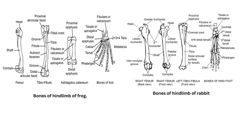
- Hindlimb of Frog:
- The hindlimb of the frog is specially adapted for jumping and locomotion in water. It consists of the femur, tibio-fibula, tarsal bones, and metatarsals.
- Femur: The femur is the uppermost bone of the hindlimb and is long, slightly curved, and swollen at both ends. Its proximal end fits into the acetabulum of the pelvic girdle.
- The distal end of the femur articulates with the tibio-fibula, a fused bone formed by the tibia and fibula. This structure acts as the lower bone of the hindlimb.
- A longitudinal groove is present on the tibio-fibula, and near its proximal end is a tibial crest.
- The tibio-fibula articulates with the tarsal bones, forming the ankle joint. The tarsal bones are arranged in two rows: the proximal row consists of the tibiale (or astralagus) and the fibulare (or calcaneum), which are united at their ends with a gap in between. These bones enhance the frog’s jumping ability.
- The metatarsals in the frog are elongated, with five toes in total. The first and second toes each have two phalanges, while the third, fourth, and fifth toes each have three phalanges. A supplementary sixth toe, in the form of a calcar made of two short bones, is present for additional support during jumping.
- The hindlimb of the frog is specially adapted for jumping and locomotion in water. It consists of the femur, tibio-fibula, tarsal bones, and metatarsals.
- Hindlimb of Rabbit:
- The rabbit’s hindlimb structure reflects its need for terrestrial locomotion and movement on solid ground.
- Femur: The femur is long and stout, with a prominent head at its proximal end that fits into the acetabulum of the pelvic girdle. The head of the femur has three trochanters: the greater trochanter, lesser trochanter, and third trochanter, which aid in muscle attachment.
- The shaft of the femur ends in a pair of expanded condyles that articulate with the tibia and fibula, forming the knee joint.
- The tibia and fibula are the lower bones of the hindlimb. The tibia and fibula are free at their proximal ends but fused at their distal ends. The knee joint also contains a patella, or knee cap, for additional support.
- The distal end of the fused tibia and fibula articulates with six tarsal bones, arranged in two rows, which form the ankle joint. The astralagus and calcaneum are the largest bones in the proximal row.
- The rabbit’s metatarsals are long, and the foot has four toes, with each toe possessing three phalanges, unlike the frog, which has five toes.
- The rabbit’s hindlimb structure reflects its need for terrestrial locomotion and movement on solid ground.
- Text Highlighting: Select any text in the post content to highlight it
- Text Annotation: Select text and add comments with annotations
- Comment Management: Edit or delete your own comments
- Highlight Management: Remove your own highlights
How to use: Simply select any text in the post content above, and you'll see annotation options. Login here or create an account to get started.