- The secretory system in plants plays a crucial role in the release of various substances from plant cells to the external environment. This process involves specialized structures known as secretory cells or secretory tissues, which are responsible for producing and transporting secretions.
- Secretions can be classified into beneficial and non-beneficial categories. Beneficial secretions include those that enhance plant growth, aid in reproduction, or protect against herbivores and pathogens. In contrast, non-beneficial secretions may include waste products or substances that do not contribute positively to the plant’s health.
- Various types of secretions are produced by plants, including water, nectar, salts, tannins, resins, latex, gums, digestive enzymes, and hormones. Each of these substances serves specific functions, contributing to the overall well-being and survival of the plant. For instance, nectar attracts pollinators, while resins can act as a defense mechanism against pests.
- The secretory cells can be found in different tissues throughout the plant. These cells may be dispersed among other cell types or grouped together in specialized structures, allowing for efficient secretion and transport of substances. The mechanisms behind secretion can involve various physiological processes, including diffusion, active transport, and exocytosis, depending on the nature of the substance being secreted.
- Overall, the secretory system in plants is a complex and vital component that facilitates interactions between the plant and its environment. Understanding these processes not only highlights the adaptive strategies of plants but also underscores the importance of these secretions in ecological relationships, such as those between plants and pollinators or plants and herbivores.
The classification of secretory tissues in plants
The classification of secretory tissues in plants is essential for understanding how they function and interact with their environment. These tissues are broadly divided into two main categories based on their position within the plant body: external secretory tissues and internal secretory tissues. Each category serves specific purposes and involves different mechanisms for secretion.
- External Secretory Tissue
- This category includes tissues located on the outer surfaces of the plant, such as epidermis and glandular structures.
- Functions: External secretory tissues are primarily involved in interactions with the environment, such as attracting pollinators, repelling herbivores, or protecting the plant from pathogens.
- Examples:
- Nectar-producing glands: These structures secrete nectar to attract pollinators, playing a vital role in reproduction.
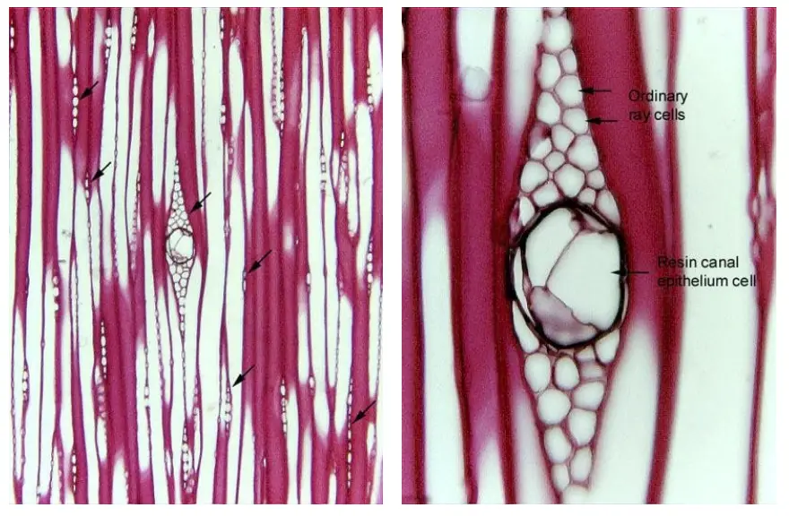
- Resin canals: Found in various plants, these canals secrete resin that acts as a defense mechanism against herbivores and pathogens, sealing wounds and deterring potential threats.
- Glandular trichomes: Hair-like structures that may secrete a variety of substances, including oils and mucilage, contributing to plant protection and moisture retention.
- Internal Secretory Tissue
- Internal secretory tissues are located within the plant, often associated with vascular or ground tissues.
- Functions: These tissues typically play roles in the synthesis and storage of substances that are crucial for the plant’s metabolic processes and growth.
- Examples:
- Laticifers: Specialized internal tubes that transport latex, a milky fluid containing rubber and other compounds. This secretion has protective functions and can deter herbivory.
- Glands in the cortex and pith: These may produce and store various secretions, including oils, resins, or hormones, contributing to growth regulation and defense mechanisms.
- Secretory cells in parenchyma: Parenchyma tissues can contain secretory cells that release substances like digestive enzymes, playing a role in nutrient breakdown and assimilation.
External secretory tissues in plants
1. Glandular trichomes
Glandular trichomes are specialized structures found on the epidermis of various plant species. These unicellular or multicellular appendages, which can take the form of hairs, scales, or other configurations, play a crucial role in the secretion of a wide range of substances that are essential for plant defense and interaction with the environment.
- Structure and Composition:
- Glandular trichomes consist of a head made up of secretory cells, which can be either unicellular or multicellular.
- These secretory cells are supported by a stalk composed of non-glandular cells known as endodermal cells.
- The endodermal cells serve a vital function by preventing the secreted solutions from re-entering the plant tissue through the apoplast, thereby ensuring that the secretions perform their intended functions effectively.
- Functions of Glandular Trichomes:
- Glandular trichomes are involved in the secretion of various substances, including:
- Water: Contributing to hydration and cooling of the plant surface.
- Salts: Playing a role in nutrient regulation and osmotic balance.
- Nectar: Attracting pollinators and other organisms that aid in reproduction.
- Mucilage: Providing a protective coating and aiding in water retention.
- Terpenes: Serving as chemical defenses against herbivores and pathogens, and contributing to the aromatic qualities of certain plants.
- Adhesives: Assisting in the trapping of insects or other small organisms, enhancing the plant’s ability to deter herbivory.
- Digestive Enzymes: Facilitating the breakdown of organic matter, particularly in carnivorous plants.
- Irritants: These substances can cause stinging or irritation to herbivores, serving as a deterrent to feeding.
- Glandular trichomes are involved in the secretion of various substances, including:
- Cellular Characteristics:
- The secretory cells within glandular trichomes are often rich in organelles, particularly mitochondria, which provide the energy necessary for the secretion process.
- The presence of these organelles indicates the high metabolic activity required for the production and release of secretory products.
2. Hydathodes
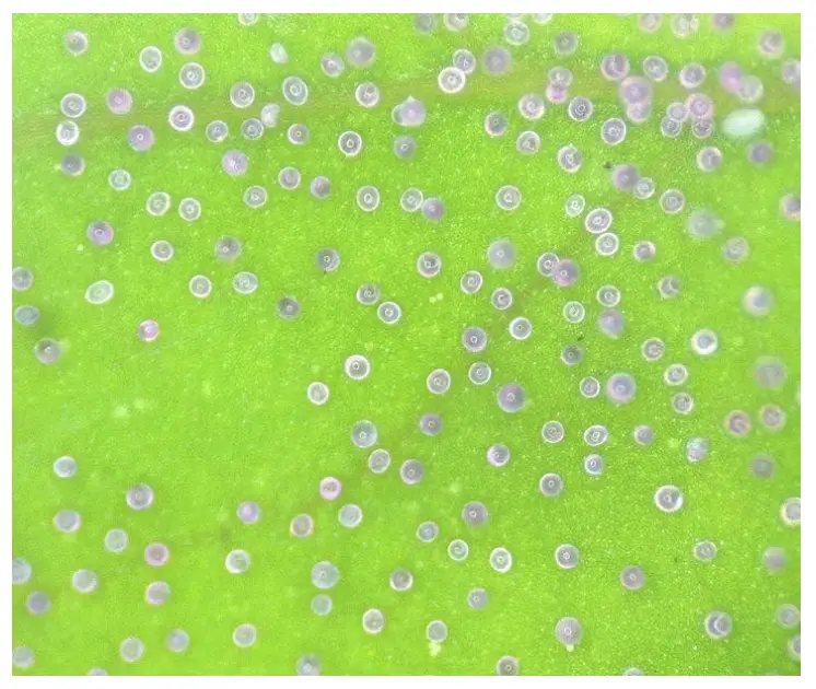
Hydathodes are specialized structures located on the leaf epidermis that play a critical role in the secretion of aqueous solutions. Through a process known as guttation, hydathodes release liquid water containing dissolved organic and inorganic substances, which is particularly important during periods of low transpiration.
- Mechanism of Guttation:
- Guttation occurs when root pressure elevates the pressure within the xylem sap, leading to water exuding from hydathodes, especially when atmospheric transpiration is minimal.
- This process is most notable in certain plant species, such as tropical plants like Colocasia, which can release up to 100 milliliters of water in a single night.
- Active Hydathodes:
- In some plants, hydathodes are classified as active, meaning that the secretion of water is energetically supported by the glandular cells themselves.
- Experimental evidence suggests that when living cells of the hydathode are killed—such as by treating the leaf surface with an alcoholic solution of mercuric chloride—no liquid emerges, even when water is forced into the leaf under pressure. This illustrates the dependence of hydathodes on living cells for their function.
- Structural Variations:
- There is significant structural variation in hydathodes across different plant species. During their development, the procambium near the lobes or serrations of the leaf differentiates, leading to slight swelling.
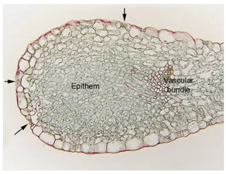
- Cells near the procambium may proliferate to form loose parenchyma tissue known as the epithem, which often contains an extensive system of intercellular spaces.
- Unlike adjacent mesophyll cells, the cells within the epithem typically lack chloroplasts, making them distinct in function and structure.
- Cellular Composition:
- The epithem is usually surrounded by a sheath of tightly fitting cells that contain tannin-like substances, providing structural integrity and possibly playing a role in regulating secretion.
- In certain plants, the walls of sheath cells may be suberized and feature Casparian bands, which add an additional layer of complexity to water movement regulation.
- The epidermis overlying the epithem houses mother cells that develop into guard cells for water pores. These guard cells are typically larger than those found in stomata, and they lose the ability to regulate stomatal movement, remaining permanently open to facilitate water discharge.
- Functional Roles:
- The hydathodes in species like Populus are considered unspecialized because they release water containing varying concentrations of sugars, which can be perceived as nectar, thus attracting pollinators.
- In some cases, trichome hydathodes actively secrete salts and other solutions, which may accumulate and lead to plant injury through their interactions with pesticides.
3. Salt Glands and Chalk Glands: The Salt secreting trichomes
Salt glands and chalk glands represent specialized structures in certain plant species that play a crucial role in the secretion of inorganic salts and other compounds. These adaptations are particularly prevalent in plants that thrive in saline environments, often referred to as halophytes.
- Function and Composition:
- Salt glands are responsible for the secretion of inorganic salts, primarily sodium (Na⁺) and chloride (Cl⁻) ions. Other ions commonly secreted include potassium (K⁺), magnesium (Mg²⁺), nitrate (NO₃⁻), phosphate (PO₄³⁻), bromide (Br⁻), and bicarbonate (HCO₃⁻).
- The secretions are primarily derived from the transpiration stream, allowing the plants to manage excess salt accumulation.
- Comparative Structure:
- While both salt glands and hydathodes facilitate secretion, they differ significantly in origin. Salt glands are formed from multicellular trichomes that perform glandular functions, whereas hydathodes are of stomatal origin.
- Salt glands exhibit several sophisticated characteristics that enhance their secretory efficiency:
- They possess a large surface area for absorbing materials.
- There are specialized pathways for transporting materials within the glands, such as transfer walls or fields of plasmodesmata.
- A distinct group of cells is dedicated to the actual secretion process.
- Mechanisms are in place to isolate the secretions and prevent leakage back into the symplasm.
- Types of Salt Glands:
- Bladder-like Hairs (Salt Bladders):
- A characteristic feature of the family Chenopodiaceae, particularly in Atriplex (saltbush) species, these glands consist of a narrow stalk cell topped by a large secretory bladder cell.
- The bladder cell has a large central vacuole at maturity, and both bladder and stalk cells are covered by a cuticle layer.
- The connection between bladder cells and mesophyll cells is established through symplasm, allowing the transpiration stream’s ions to be delivered effectively. The bladder cell secretes ions into the central vacuole, eventually lysing and depositing salts as a white powder on the leaf surface. This process is classified as holocrine secretion.
- Two-Celled Glands:
- Found in species such as Cynodon, Spartina, Sporobolus, and Distichlis, these simple glands comprise a basal cell and a cap cell, with both containing a continuous cuticle cover.
- The cap cell features a large nucleus and is involved in direct salt secretion onto its surface. This glandular structure does not have a large vacuole, and the basal cell contains partitioning membranes that are crucial for secretion.
- The secretion type for these glands is eccrine, and the uptake of ions from the basal cell, followed by their secretion from the cap cell, involves active processes facilitated by ATPase activity.
- Bladder-like Hairs (Salt Bladders):
- Multicellular Glands:
- In Tamarix aphylla (Tamaricaceae), the salt glands consist of eight cells: two basal collecting cells and six secretory cells. The transfusion cells connect the uncutinized wall portions of the lowest secretory cells and the underlying collecting cells, ensuring symplastic continuity with neighboring mesophyll cells.
- The secretory cells’ plasma membrane surface area is enhanced by wall ingrowths, and salty water is exuded through pores located on the cuticle.
- Chalk Glands:
- The salt glands of mangrove species such as Avicennia contain two to four collecting cells, a stalk cell, and eight to twelve secretory cells. In the chalk glands of Plumbago capensis and Armeria maritima (family Plumbaginaceae), there are four secretory cells surrounded by four subsidiary cells, with surface depressions corresponding to the glandular pores.
- X-ray analysis of secretions from these glands reveals the presence of calcite (CaCO₃) and nesquehonite (MgCO₃·3H₂O). Distinguishing between chalk glands and salt glands can be challenging due to their similar structures.
4. Glandular trichomes secreting Lipophilic Substances
Salt glands and chalk glands are specialized trichomes that play an essential role in the secretion of salts and other compounds, particularly in plants that thrive in saline environments. These adaptations enable halophytes to manage excess salts, facilitating their survival and growth in challenging conditions.
- Glandular Trichomes and Their Functions:
- Many families of eudicots exhibit glandular trichomes that secrete lipophilic substances, primarily terpenoids, including essential oils and resins.
- These lipophilic compounds serve various functions, such as:
- Deterring herbivores, acting as a defense mechanism.
- Attracting pollinators, which aids in reproduction.
- Facilitating the dispersal of certain fruits due to their sticky nature.
- Types of Glandular Trichomes in the Mint Family (Lamiaceae):
- Within the mint family, two primary types of glandular trichomes are identified for their role in secreting essential oils:
- Peltate Trichomes:
- Composed of a basal cell, a short stalk cell, and a broad head consisting of 4 to 18 secretory cells arranged in one or two concentric rings.
- Both the lateral wall of the basal cell and the short stalk cell are completely cutinized.
- A large sub-cuticular space forms due to the detachment of the cuticle and the outer layer of the cell wall, which serves as a storage area for secretory products.
- The secretory products remain within this space until external forces rupture the cuticle, allowing for the release of the substances.
- Capitate Trichomes:
- These trichomes consist of a basal cell, a uniseriate stalk (which can be one or several cells long), and a head formed from one or several secretory cells.
- In the Lamiaceae family, there are typically two types of capitate trichomes: short-stalked and long-stalked variants.
- During the active secretion phase, there is a notable increase in the number of dictyosomes and smooth endoplasmic reticulum, leading to the dilation of their cisternae.
- The cytoplasm retracts irregularly from the cell wall, and a split occurs between the pectic and cuticular layers, resulting in the formation of a characteristic subcuticular space.
- As secretion occurs, vacuoles within the trichomes lose their contents, facilitating the release of essential oils.
- Peltate Trichomes:
- Within the mint family, two primary types of glandular trichomes are identified for their role in secreting essential oils:
- Salt Glands:
- Salt glands primarily occur in halophytes, enabling them to secrete inorganic salts to cope with saline environments.
- The secretions consist mainly of Na⁺ and Cl⁻ ions, with other ions such as K⁺, Mg²⁺, NO₃⁻, PO₄³⁻, Br⁻, and HCO₃⁻ also being present.
- The process of secretion is derived from the transpiration stream, although the origin and structure of salt glands differ from that of hydathodes.
- Chalk Glands:
- Chalk glands, found in some plant species like Plumbago capensis and Armeria maritima, secrete compounds such as calcium carbonate (CaCO₃) and magnesium carbonate (MgCO₃·3H₂O).
- The mechanisms involved in chalk gland secretion are closely related to those in salt glands, leading to similarities in structure and function.
5. Nectaries: Nectar – secreting trichomes
Nectaries are specialized secretory structures responsible for the production and secretion of nectar, an aqueous fluid characterized by a high concentration of sugars. They play a crucial role in attracting pollinators and facilitating various ecological interactions. Nectaries can be classified into two main types based on their location and function: floral and extrafloral nectaries.
- Types of Nectaries:
- Floral Nectaries:
- Found within flowers, these nectaries are directly associated with the pollination process.
- Various floral organs, including sepals, petals, stamens, ovaries, and receptacles, may bear these nectaries.
- They reward pollinators, such as insects and other animals, by secreting nectar.
- Extrafloral Nectaries:
- Located on vegetative structures or peripheral floral parts, extrafloral nectaries are typically not involved in pollination or reproduction.
- They serve to attract insects, especially ants, which help protect the host plants against herbivorous mammals and foliage-eating insects.
- Some extrafloral nectaries act as safety valves for releasing excess sugars during rapid photosynthetic activity. For instance, nectar dripping from the tips of Ailanthus leaflets has been observed during periods of high photosynthesis.
- Trees such as Tilia may also produce sugary droplets that accumulate on nearby surfaces, like parked automobiles, particularly on sunny and humid days. This “safety-valve” release can occur due to local ruptures in the cuticle.
- Floral Nectaries:
- Nectary Structure and Function:
- Depending on the taxon, a nectary may consist of either secretory epidermal cells or glandular trichomes.
- The epidermis typically features specialized parenchymatous cells, which are common in most nectaries.
- Nectar secreted by the underlying nectariferous tissue is released onto the surface, aided by modified stomata.
- Modes of Nectar Translocation:
- The mechanisms for sugar translocation in nectaries are diverse, with granulocrine and eccrine secretion being the two primary modes.
- In some cases, the sugar-rich solution exuded from the phloem may travel along the cell wall (apoplasm) without re-entering the protoplast.
- For example, in Acer plantanoides, as the solution moves along the cell wall, enzymes may act on the sugars, and certain non-sugar components may be resorbed by the cell.
- In nectaries with specialized tissues, sucrose is unloaded from nearby phloem and undergoes enzyme-catalyzed changes within the symplasm before being secreted into the cell wall (apoplasm).
- If the secretion is of the eccrine type, the cell may adopt the structure and function of a transfer cell. Granulocrine secretion, however, is the most common mode observed.
- In some short-lived flowers, nectar may be released through holocrine secretion, where cellular breakdown of the secretory tissue occurs, as seen in Turnera and certain Helleborus species.
- The mechanisms for sugar translocation in nectaries are diverse, with granulocrine and eccrine secretion being the two primary modes.
- Nectar Composition:
- The nectar produced may be predominantly sucrose, glucose, or exhibit an equal ratio of sucrose to glucose/fructose.
- Nectar secreted from nectarines that originate from phloem sap typically shows a higher concentration of sugars compared to those secreted by both xylem and phloem sap or primarily by xylem sap.
- Research on Arabidopsis thaliana has identified the gene CRABS CLAW (CRC) as necessary for the development of nectaries, with crc mutant flowers lacking these structures.
- Variations in nectar composition can occur even between male and female flowers of the same plant species; for instance, in Cucurbita pepo, the female flower produces nectar that is sweeter and has lower protein content than that of the male flower.
- In some plants, the prenectar derived from phloem accumulates as starch in plastids of nectariferous cells, which upon hydrolysis serves as the primary source of sugars during anthesis.
- Specific Nectary Structures:
- Nectaries of Lonicera japonica:
- Nectar is secreted by short unicellular trichomes located on the inner epidermis of corolla tubes.
- The mode of secretion is granulocrine, characterized by the stacking of endoplasmic reticulum-lined cisternae that produce vesicles that fuse with the plasma membrane to release nectar.
- Nectaries of Abutilon striatum:
- Nectar is secreted by multicellular trichomes or hairs located on the lower inner side of the fused sepals.
- This secretion does not follow either the eccrine or granulocrine modes; rather, nectar is released through short-lived pores in the cuticle at the tips of the trichomes.
- Nectaries of Lonicera japonica:
6. Colleters: Multicellular appendages that produce sticky secretions
Colleters are specialized multicellular appendages predominantly found on young leaves and buds, renowned for their role in secreting sticky, glue-like substances. These secretions are primarily mucilaginous or resinous and are notable for being insoluble in water. The presence of colleters serves important protective functions within plant structures, particularly concerning dormant buds and developing meristems.
- Function of Colleters:
- Protection: Colleters provide a protective coating to dormant buds and developing meristems, shielding them from environmental stressors and potential damage. This is crucial during the early growth phases of plants when they are most vulnerable.
- Secretion: The secretions produced by colleters may deter herbivory and inhibit microbial infections, contributing to the overall health and survival of the plant.
- Characteristics of Colleters:
- Colleters typically appear on young leaves and buds, reflecting their primary role in the early development stages of plants.
- The secretory material produced is sticky and serves multiple protective and possibly ecological functions.
- Types of Secretions:
- The nature of the secretion from colleters can vary. In many instances, the secretion is classified as granulocrine, wherein secretory materials accumulate beneath the cuticle of the colleters. Eventually, the cuticle ruptures, allowing the sticky substances to be released.
- Although granulocrine is the most common mode of secretion observed, other modes may also be present, indicating variability in function and secretion processes among different plant species.
- Colleters in the Rubiaceae Family:
- The Rubiaceae family exhibits a standard type of colleters, which is widely recognized as the most common form. This family includes a diverse range of plant species that demonstrate the significance of colleters in plant development.
7. Osmophores: The fragrance producing glands
Osmophores are specialized glands responsible for producing fragrances in certain plants, playing a critical role in attracting pollinators and enhancing reproductive success. The term “osmophore” derives from the Greek words “osmo,” meaning odor, and “pherein,” meaning to bear, reflecting the primary function of these glands in fragrance production. They produce volatile substances, including terpenoids and aromatic compounds, which contribute to the characteristic scents of various flowers.
- Occurrence of Osmophores:
- Osmophores are present in a diverse range of plant families, including:
- Araceae
- Asclepiadaceae
- Orchidaceae
- Solanaceae
- Aristolochiaceae
- Saxifragaceae
- Burmanniaceae
- This wide distribution highlights the evolutionary significance of fragrance production across different plant lineages.
- Osmophores are present in a diverse range of plant families, including:
- Cellular Structure of Osmophores:
- The glandular tissues that constitute osmophores typically consist of several cell layers, allowing for efficient secretion and storage of fragrant compounds.
- Cells within osmophores are characterized by:
- Numerous Amyloplasts: These organelles store starch, which may serve as a precursor for fragrance compounds.
- Mitochondria: These are vital for energy production, supporting the metabolic processes involved in synthesizing aromatic compounds.
- Abundant Smooth-Surfaced Endoplasmic Reticulum (ER): The smooth ER is involved in the biosynthesis of lipids, including terpenoids.
- Scarce Golgi Bodies: While Golgi bodies typically play a role in processing and packaging proteins, their limited presence in osmophores suggests that the synthesis and secretion processes may primarily rely on other cellular mechanisms.
- Secretion Mechanisms:
- Osmophores exhibit both granulocrine and eccrine secretion modes, reflecting the complexity of fragrance production.
- Granulocrine Secretion: In this mode, secretory products accumulate within vesicles and are released when these vesicles fuse with the plasma membrane.
- Eccrine Secretion: This involves the direct release of secretions through the cell membrane into the external environment, allowing for rapid dispersal of volatile compounds.
- Osmophores exhibit both granulocrine and eccrine secretion modes, reflecting the complexity of fragrance production.
8. The Glandular structures of Carnivorous plants
The glandular structures of carnivorous plants are fascinating adaptations that enhance their ability to attract, capture, and digest insects. These specialized glands not only facilitate the secretion of digestive enzymes but also enable the absorption of nutrients, underscoring the complex interactions between plant physiology and nutrient acquisition. The various types of glands found in these plants serve specific roles in trapping and digesting prey, contributing to the overall functionality of these unique organisms.
- General Overview of Glandular Structures:
- Carnivorous plants possess glandular trichomes that serve as critical sites for both secretion and absorption.
- These glands allow for two-way transport, where secretions exit the gland to attract and digest prey, while digestive products enter the plant for nutrient absorption.
- A key requirement for this dual function is the loss of control over fluid movement within the cytoplasm of the basal endodermal cell, allowing for efficient transport mechanisms.
- Types of Glandular Structures:
- Alluring Glands: These glands secrete substances that attract insects to the plant.
- Mucilage Glands: These produce a sticky secretion that aids in trapping prey.
- Digestive Glands: These secrete enzymes that break down prey into absorbable nutrients.
- Examples of Carnivorous Plants and Their Glandular Mechanisms:
- Sundew (Drosera species):
- Trap Mechanism: Adhesive trap.
- The tentacles of Drosera act as stalked glands composed of multicellular structures that facilitate secretion and absorption.
- Each tentacle features a multicellular stalk containing tracheids, surrounded by an endodermal layer with Casparian strips.
- The head of the tentacle secretes mucilage and digestive enzymes while also absorbing the resulting nutrients.
- Research has demonstrated rapid calcium transport within these glands, suggesting a complex mechanism for nutrient reabsorption that operates against mucilage flow.
- Pitcher Plants (Nepenthes):
- Trap Mechanism: Pitcher trap.
- The pitcher structure contains diverse glands with distinct roles.
- Multicellular alluring glands located on the underside of the pitcher lid secrete nectar, attracting prey.
- Numerous digestive glands line the inner surface of the pitcher, while others at the base may assist in digestion and absorption.
- These glands secrete enzymes such as phosphatase and esterases, facilitating the breakdown of trapped insects.
- Nepenthes pitchers can hold substantial amounts of fluid, enhancing their capacity for trapping and digesting prey.
- Bladderwort (Utricularia):
- Trap Mechanism: Suction trap.
- Utricularia features specialized trichomes, including bifid and quadrifid hairs, which assist in prey capture.
- Dome-shaped external glands secrete mucilage and sugars to attract insects, while short-stalked internal glands create tension by extracting water.
- The trap functions by releasing tension when an insect touches the bristles, causing water influx and trapping the prey.
- Venus Flytrap (Dionaea):
- Trap Mechanism: Snap trap.
- The closure of the trap is triggered by electrical signals from multicellular sensory hairs.
- Each hair contains associated nectaries that respond to touch, facilitating rapid trap closure.
- Digestion begins shortly after the trap closes, with enzymes like protease breaking down the insect within 36 hours.
- Sundew (Drosera species):
Internal secretory tissues in plants
The internal secretory cells play a critical role in plant physiology, providing essential functions that contribute to the plant’s survival, defense, and adaptation. These specialized cells are responsible for the secretion of various substances, including crystals, oils, mucilage, and tannins. Each type of secretory cell exhibits distinct structural features and functionalities, highlighting the diversity of plant adaptations.
- Crystals and Silica-Secreting Cells:
- Many plants deposit inorganic materials, primarily calcium salts and silicon oxides, within their cells.
- Calcium salts often form crystals, while silicon oxide contributes to silica bodies.
- Crystal-containing cells are regarded as secretory idioblasts, and various forms of calcium oxalate crystals are present in plant cells:
- Solitary Crystals: These are rhomboidal or prismatic crystals found in leaves of plants such as Citrus, Begonia, Hyoscyamus, and Vicia sativa.
- Druses: These spheroidal aggregates of prismatic crystals are located in the leaves of Datura stramonium and Ruta graveolens, as well as in the fleshy cortex of Anabasis articulata and the rhizome of Rheum rhaponticum and Colocasia esculenta.
- Crystal Sand: Comprising very small prismatic crystals in masses, this form is found in the stem of Sambucus nigra and Aucuba japonica, and in the leaves of Atropa belladonna.
- Raphides and Styloids:
- Raphides are thin, elongated crystals tapering at both ends and typically found in bundles within the leaves of Arum, Agave, Zebrina, Tradescantia, and Impatiens.
- Styloids, or pseudo-raphides, are long prismatic crystals, also tapered at both ends, and occur in the families Iridaceae, Agavaceae, as well as some species of Liliaceae, Rosaceae, and Rutaceae.
- Silica Bodies: These bodies, which deposit silicon oxide, are commonly located in the epidermis of families such as Poaceae, Cyperaceae, and Palmae. They can also be found in the epidermis and hypodermis of certain Cactaceae species and in the wood of some dicots.
- Lithocysts:
- Lithocysts are specialized epidermal cells containing calcium carbonate crystals, which are relatively rare in higher plants.
- These cells are associated with cystoliths, which are stalked ingrowths of the cell wall that project into the cell lumen. Cystoliths consist of cellulose and are impregnated with calcium carbonate.
- They are generally located in the epidermis but can also be found in various parenchymatous cells, particularly in the leaves of Ficus elastica.
- Myrosin Cells:
- Myrosin cells are idioblasts containing the enzyme myrosinase within their large central vacuoles, primarily found in the family Capparaceae.
- The enzyme myrosinase functions to hydrolyze glucosinolates into aglucones, which can further decompose into toxic compounds such as isothiocyanates, nitriles, and epithionitriles.
- These toxic products serve as a defense mechanism against insects and microorganisms. Myrosinase is located within the myrosin cells, while its substrate, thioglucosides, can be found in ordinary parenchyma cells that may be in direct contact with the myrosin cells.
- Oil Cells:
- Oil-secreting cells are present in plants from families such as Winteraceae, Magnoliaceae, Lauraceae, and Calycanthaceae.
- These mature oil cells are characterized by three distinct layers in their cell wall: an external primary wall, a suberized layer, and an inner tertiary wall.
- The formation of an oil cavity occurs following the deposition of the inner wall layer, which includes a protrusion known as the cupule.
- The synthesis of oil is thought to occur in plastids, from where it is released into the cytoplasm and subsequently secreted into the oil cavity via the plasma membrane. As the oil cavity expands, the protoplasts become appressed against the inner wall layer.
- Mucilage Cells:
- Mucilage-secreting cells are prevalent in various dicotyledonous families, including Cactaceae, Annonaceae, Lauraceae, Malvaceae, Magnoliaceae, and Tiliaceae.
- These cells can be found in all parts of the plant and often differentiate near meristematic regions.
- They have thin, cellulosic walls that are typically not lignified.
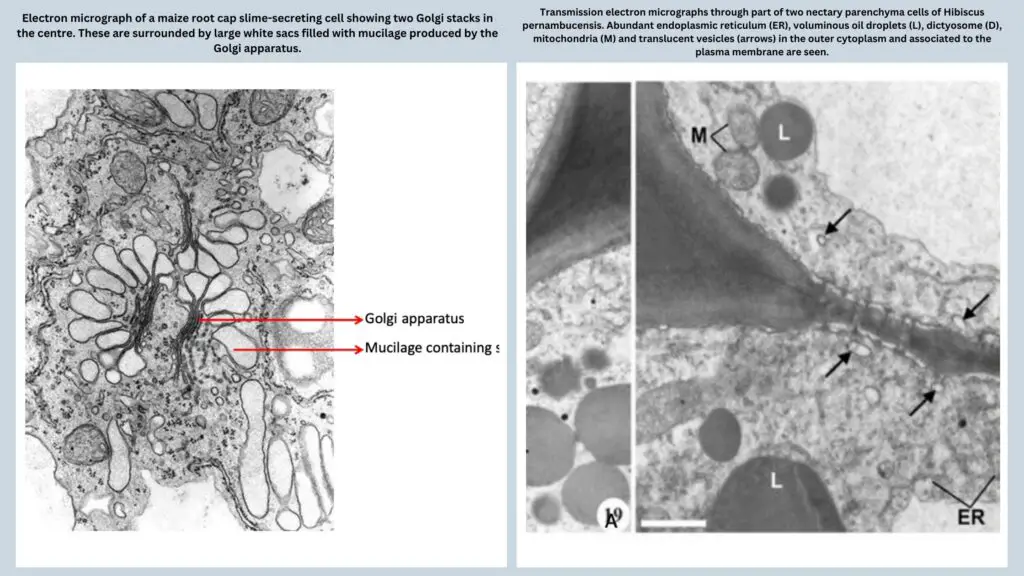
- Mucilage secretion is facilitated by Golgi bodies, which release mucilage through exocytosis. In certain taxa, mucilage cells may also contain bundles of needle-like crystals known as raphides.
- Tannin Idioblasts:
- Tannin idioblasts are parenchyma cells that contain tannins, a common secondary metabolite.
- These cells form interconnected systems and may associate with vascular bundles, appearing in families such as Ericaceae, Myrtaceae, Fabaceae, Crassulaceae, Rosaceae, and Vitaceae.
- Tannins within these cells oxidize to form phlobaphenes, which can be observed under a microscope as brown or reddish-brown pigments.
- The synthesis of tannins occurs in the rough endoplasmic reticulum, and these compounds are sequestered within the vacuoles of the cells.
- Tannins serve as significant deterrents against herbivory in angiosperms.
Secretory cavities and ducts
Secretory cavities and ducts are specialized structures in plants that serve as sites for the secretion of various substances into intercellular spaces. These secretory structures may exhibit a more or less isodiametric form, resembling glands, or may be considerably elongated, resembling ducts. Based on their development, these cavities and ducts can be categorized into three primary types: schizogenous, lysigenous, and schizolysigenous.
- Schizogenous Cavities and Ducts
- Schizogenous cavities and ducts form through the separation of cells at the middle lamella, primarily due to the mechanisms associated with phragmoplasts and cell plates during cell division.
- As new cell plates form, they connect with existing walls without fully penetrating them. Consequently, as turgor pressure builds within the cell, swelling occurs, leading to tearing at the points of weakness between the new cell plates and the old walls. This results in the formation of intercellular spaces.
- Such spaces may ultimately coalesce to form larger cavities filled with gases or liquid secretions.
- Examples of plants exhibiting schizogenous cavities include resin ducts found in Coniferae, secretory ducts in Asteraceae and Umbelliferae, as well as secretory cavities in Eucalyptus and Lysimachia.
- Resin Ducts
- Resin ducts are among the most well-studied types of secretory ducts in conifers, characterized by a schizogenous development pattern.
- They are distributed throughout vascular and ground tissues across all plant organs.
- The secretion of resin occurs schizogenously in most plant tissues, with an exception noted in the bud scales of Pinus pinaster, where a schizolysigenous pattern has been observed.
- Studies utilizing electron microscopy have revealed that resin ducts contain a higher number of undifferentiated plastids than the surrounding cortical cells, and these plastids are enveloped by endoplasmic reticulum, which may facilitate the transport of resins within the ducts.
- Lysigenous Cavities and Ducts
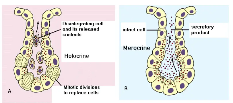
- Lysigenous cavities and ducts arise from the complete breakdown of cells (holocrine secretion).
- Notable examples of lysigenous cavities can be found in aquatic plants, certain monocotyledon roots, and the primary resin ducts of Mangifera indica.
- Kino Veins
- Kino veins frequently develop in the wood of Eucalyptus as a response to wounding or fungal infections.
- They represent a specialized form of traumatic duct that emerges from traumatic parenchyma produced by the cambium shortly after the stimulus for vein formation is introduced.
- Kino veins vary significantly in size, ranging from isolated veins to dense masses that encircle the tree.
- These veins contain polyphenols, including tannins, and their formation in both xylem and phloem is initiated through the lysigenous breakdown of parenchyma bands by the vascular cambium.
- The hormone ethylene, released by microbes or host plants, serves as a stimulus for kino vein formation post-injury, facilitating the release of polyphenols into the duct lumen.
- Schizolysigenous Cavities and Ducts
- In some cases, schizogenous processes may precede cellular autolysis, resulting in the formation of schizolysigenous cavities.
- The epithelial cells lining the cavity undergo autolysis, thereby enlarging the space.
- Examples of such cavities can be found in the leaves and fruits of citrus species, notably in the pericarp of oranges and lemons.
- Schizolysigenous oil glands also occur in the floral parts of clove (Eugenia caryophyllata), serving as a source of clove oil.
- Gum Production and Gummosis
- The traumatic formations in secondary phloem and xylem of conifers and woody angiosperms yield resin, gum-resin, and gum ducts.
- These secretory structures play a significant role in chemical defense mechanisms, promoting wound healing and deterring herbivores.
- Gummosis, often observed in plants such as Acacia and Citrus, results from responses to fungal and viral diseases. For instance, the “brown rot” gummosis in citrus trees, caused by Phytophthora citrophthora, leads to the development of gum ducts through schizogenous processes in the cambium.
- Laticifers
- Laticifers represent a heterogeneous group of specialized secretory cells, derived from parenchymatous cells, exhibiting distinct metabolic and structural characteristics.
- Typically, secretory activities and products are localized within the laticifer system, without programmed release to surrounding tissues or the external environment.
- A central vacuole within these cells often contains an emulsion or suspension of latex, which forms an anastomosing system closely associated with vascular bundles, particularly in secondary phloem.
- The cytoplasm of laticifers may undergo specialization or degeneration over time, indicating the adaptability of these structures to varying physiological conditions.
- Types of Laticifers
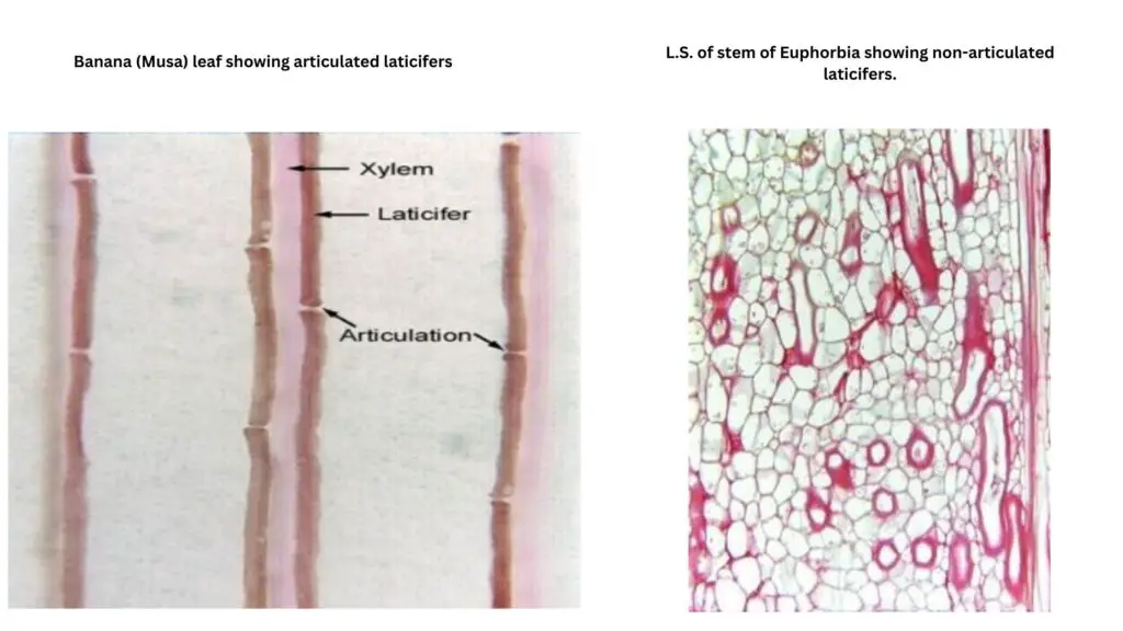
- Articulated Laticifers:
- These laticifers are compound in origin and consist of longitudinal cell files, with end walls that may break down partially or completely.
- Notable examples include Hevea brasiliensis (the primary source of rubber) and various species in families such as Asteraceae and Euphorbiaceae.
- Nonarticulated Laticifers:
- These are multinucleate structures that originate from single cells and develop symplatically, resulting in tube-like formations.
- Nonarticulated laticifers are common in families such as Moraceae and Apocynaceae.
- Articulated Laticifers:
- Latex Appearance and Composition
- Latex, the fluid secreted by laticifers, can vary in appearance and composition:
- It may be clear and colorless (e.g., Nerium oleander), milky (e.g., Euphorbia), or yellow-brown (e.g., Cannabis).
- The composition of latex includes organic acids, carbohydrates, mucilage, sterols, and fats, with common components being terpenoids and rubber (cis-1,4-polyisoprene).
- Additionally, latex may contain various alkaloids, glycosides, sugars, tannins, and proteins, along with specific enzymes, such as proteolytic enzymes like papain in Carica papaya.
- Latex, the fluid secreted by laticifers, can vary in appearance and composition:
Mechanisms of Secretion
The mechanisms of secretion in plants involve various methods through which secretory substances are eliminated from protoplasts. Understanding these mechanisms is crucial for comprehending how plants manage their internal processes and interactions with the environment. The primary types of secretion mechanisms include holocrine, merocrine, granulocrine, and eccrine secretion.
- Holocrine Secretion:
- In holocrine secretion, substances are released due to the complete disintegration of the cell.
- The entire contents of the cell become part of the secretion, which is liberated when the cell undergoes lysis.
- This process may involve extensive alterations in cell organelles, leading to a total breakdown of the cellular structure.
- Merocrine Secretion:
- Merocrine secretion occurs when the protoplast releases substances through its intact plasmalemma or tonoplast, allowing for a more controlled release of secretory products.
- This type of secretion can be further classified into two categories: granulocrine secretion and eccrine secretion.
- Granulocrine Secretion:
- In granulocrine secretion, secretory substances accumulate in vesicles formed by the endoplasmic reticulum or dictyosomes, or both.
- These vesicles then fuse with the plasma membrane, and the secretory substances are eliminated through a process known as exocytosis.
- The synthesis of these secretory products can occur either within the vesicles via synthetases located in the vesicle membrane or in the cytoplasm, followed by transport into the vesicles through ATP-driven molecular pumps.
- As the secretory products accumulate, the vesicles move toward the plasmalemma for secretion. Upon fusion, the vesicle membranes integrate into the plasmalemma, and the contents of the vesicles are released into the external environment.
- Notably, when granulocrine secretion is rapid, there is a significant flow of membrane materials, necessitating membrane recycling. This recycling is facilitated by coated vesicles, which transport the membrane components back into the cytoplasm for reuse.
- Eccrine Secretion:
- Eccrine secretion differs slightly in that the secretory substances exit the cytoplasm through the plasma membrane as individual molecules or ions.
- This process can be passive or active; passive secretion occurs due to concentration gradients driven by osmosis, while active secretion requires metabolic energy.
- In the active process, membrane-bound molecular pumps recognize and bind to the molecules to be secreted, forcing them across the plasma membrane or tonoplast.
- An example of eccrine secretion in action is the exudation of water through hydathodes, which occurs due to pressure within the conducting elements.
Economic Importance of Plant Secretions
The economic importance of plant secretions is vast, with numerous secretions serving critical roles in various industries. Among these, latex and its derivatives, particularly rubber, stand out as some of the most significant contributors to the global economy.
- Natural Rubber:
- Natural rubber is produced by over 2,500 plant species; however, the rubber content in most of these species is insufficient for commercial extraction.
- The primary source of natural rubber is the rubber tree, Hevea brasiliensis, which is extensively cultivated for this purpose.
- Rubber production in Hevea can be enhanced through the application of hormones, and its yield can also be influenced by various minerals in the soil.
- Besides Hevea brasiliensis, Guayule (Parthenium argentatum), a member of the Asteraceae family, produces rubber but lacks laticifers.
- Gutta-Percha:
- Gutta-percha is derived from the coagulated latex of species in the genus Palaquium.
- This substance is utilized in several applications, including the manufacture of golf balls and submarine cables, where its water-resistant properties are particularly valuable.
- Chicle:
- The latex from other members of the Sapotaceae family, such as Achras sapota, is the original source of chicle, which is used to produce chewing gum.
- Opium:
- Opium and its derivatives, obtained from specific plant species, are widely used in medicine. Their applications range from pain relief to the treatment of various health conditions, underscoring the pharmaceutical significance of these plant secretions.
- Plant Oils:
- In addition to rubber and its derivatives, many plant oils have substantial economic value.
- Olive Oil: This oil is extensively used in cooking and food preparation, noted for its health benefits and flavor.
- Safflower Oil: Increasingly important due to its unsaturated fat content, safflower oil is utilized in the production of margarine and various cooking oils, appealing to health-conscious consumers.
- Palm Oil: Commonly used in the manufacture of soap and candles, palm oil has become a staple in the cosmetics and food industries.
- https://devamatha.ac.in/ckfinder/userfiles/files/BOTANY%20(SF)_pptx.pdf
- https://www.cambridge.org/core/books/abs/an-introduction-to-plant-structure-and-development/secretion-in-plants/4BE4336968729FC7BBCA306D4EBFB495
- https://www.studocu.com/in/document/the-maharaja-sayajirao-university-of-baroda/plant-physiology/unit-2-secretory-systems/43114013
- Text Highlighting: Select any text in the post content to highlight it
- Text Annotation: Select text and add comments with annotations
- Comment Management: Edit or delete your own comments
- Highlight Management: Remove your own highlights
How to use: Simply select any text in the post content above, and you'll see annotation options. Login here or create an account to get started.