Rhizopus species, such as Rhizopus microsporus, are frequently associated with mucormycosis, a fatal fungal infection that affects immunocompromised patients. Mucormycosis necessitates the development of new therapeutic modalities due to its escalating prevalence, unacceptably high mortality rate, and the extreme morbidity of its highly disfiguring surgical treatment. Creative Biolabs has the knowledge and experience in the pharmaceutical industry to assist you with your antifungal drug discovery initiatives against a variety of fungi, including Rhizopus microsporus.
Rhizopus microsporus (R. microsporus) belongs to the class Zygomycetes and the order Mucorales, which contains the genus Rhizopus. This species is a widespread soil fungus that can induce mucormycosis in immunocompromised patients and rice seedling blight. In addition, R. microsporus is one of the very few fungi that host the -proteobacteria Burkholderia rhizoxinica and Burkholderia endofungorum as bacterial endosymbionts. Mycotoxin rhizoxin, a toxin that inhibits mitosis in eukaryotic cells by binding to -tubulin, is secreted by the endobacteria that have colonised the host plant. Due to an amino acid exchange in its tubulin protein, Rhizopus itself is resistant to the toxin.
Habitat of Rhizopus microsporus
Rhizopus microsporus is a species of filamentous fungus commonly known as black bread mold. It is typically found in various habitats that provide favorable conditions for its growth. Here are some common habitats where Rhizopus microsporus can be found:
- Soil: Rhizopus microsporus is commonly found in soil rich in organic matter. It thrives in environments where decaying plant material is present, such as compost piles, forest floors, and agricultural fields.
- Bread and other food items: As a bread mold, Rhizopus microsporus can be found on stale or moldy bread and other carbohydrate-rich food items. It can quickly colonize these surfaces, especially if they are moist and have favorable temperatures.
- Fruits and vegetables: Rhizopus microsporus can infect various fruits and vegetables, causing them to rot. It is often responsible for the black mold commonly seen on fruits like strawberries, tomatoes, and grapes.
- Damp and humid environments: This fungus prefers damp and humid conditions for its growth. It can thrive in environments with high moisture levels, such as bathrooms, basements, and areas with water damage or leaks.
- Plants and plant debris: Rhizopus microsporus can be found on the surfaces of living plants or plant debris. It can cause disease in plants, especially when there are wounds or damage that provide entry points for the fungus.
Overall, Rhizopus microsporus is a widespread fungus that can be found in diverse habitats, particularly those that offer organic matter, moisture, and suitable temperatures for its growth.
Morphology of Rhizopus microsporus
Rhizopus microsporus is a filamentous fungus that exhibits characteristic morphological features. Here are the key aspects of its morphology:
- Hyphae: Rhizopus microsporus consists of long, branched, and septate hyphae. Hyphae are the thread-like structures that make up the body (mycelium) of the fungus. They have cross-walls or septa that divide the hyphae into individual cells.
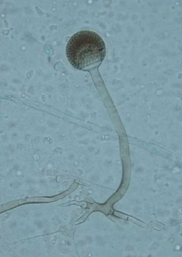
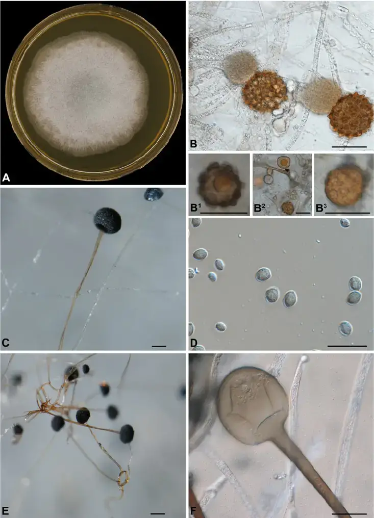
- Sporangia: This species produces specialized structures called sporangia. Sporangia are sac-like structures that are attached to the tips of aerial hyphae. They are usually spherical or ellipsoidal in shape and have a dark-colored outer covering. Sporangia contain numerous spores.
- Sporangiophores: Sporangiophores are specialized hyphae that bear sporangia. They emerge from the substrate or mycelium and extend upwards, giving rise to sporangia at their tips. Sporangiophores are usually erect and can branch to produce multiple sporangia.
- Spores: Rhizopus microsporus produces a large number of asexual spores called sporangiospores within the sporangia. These spores are typically dark-colored and appear as powdery masses when mature. They are released from the sporangia and can disperse through the air or other means.
- Rhizoids: Rhizopus microsporus possesses specialized structures called rhizoids. Rhizoids are root-like structures that anchor the fungus to its substrate and aid in nutrient absorption. They are non-septate and typically colorless.
- Stolons: Stolons are horizontal hyphae that grow along the surface of the substrate. They serve as a means of vegetative propagation and can give rise to new sporangiophores and mycelium.

Life Cycle of Rhizopus microsporus
The life cycle of Rhizopus microsporus involves both sexual and asexual reproduction. Here is an overview of its life cycle:
- Spore Germination: The life cycle begins when the spores of Rhizopus microsporus land on a suitable substrate in a favorable environment. The spores are typically dispersed by air or other means. When conditions are favorable, the spores germinate, and each spore gives rise to a hypha.
- Vegetative Growth: The germinated spore grows into a vegetative mycelium. The mycelium consists of a network of hyphae that spread and colonize the substrate, extracting nutrients for growth and development. The hyphae secrete enzymes to break down complex organic matter into simpler compounds that can be absorbed.
- Asexual Reproduction – Sporangiospores: Under favorable conditions, specialized hyphae called sporangiophores develop from the mycelium. Sporangia, which are sac-like structures, form at the tips of sporangiophores. Within the sporangia, haploid nuclei divide and differentiate into a large number of asexual spores called sporangiospores. The sporangiospores are released from the sporangium and can disperse to new locations, allowing the fungus to spread and colonize new substrates.
- Asexual Reproduction – Germination of Sporangiospores: When sporangiospores land on a suitable substrate, they can germinate under favorable conditions. Each sporangiospore undergoes germination, forming a new hypha, which grows into a mycelium, continuing the vegetative growth phase.
- Sexual Reproduction – Plasmogamy: Under certain environmental conditions, sexual reproduction can occur in Rhizopus microsporus. Different mating types of the fungus come into contact, and the cytoplasm of the hyphae fuses in a process called plasmogamy. Plasmogamy results in the formation of a multinucleate structure called a zygospore.
- Sexual Reproduction – Zygospore Formation: The multinucleate zygospore develops a thick, dark-walled protective coat. Inside the zygospore, the nuclei undergo karyogamy, which is the fusion of nuclei, resulting in the formation of a diploid nucleus.
- Sexual Reproduction – Meiosis: Following karyogamy, meiosis occurs within the zygospore, leading to the formation of haploid nuclei. These haploid nuclei develop into individual haploid spores within the zygospore.
- Sexual Reproduction – Zygospore Germination: The zygospore, containing the haploid spores, can remain dormant until conditions become favorable. When conditions are suitable, the zygospore germinates, and each haploid spore gives rise to a new hypha, initiating the vegetative growth phase and completing the life cycle.
Cultural Characterisitcs of Rhizopus microsporus
Cultural characteristics of Rhizopus microsporus refer to the observable traits and features of the fungus when grown in laboratory or artificial culture conditions. Here are some common cultural characteristics of Rhizopus microsporus:
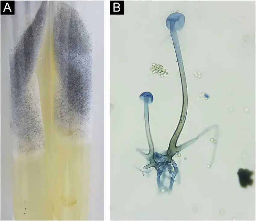
- Colony Appearance: When grown on appropriate culture media, Rhizopus microsporus colonies typically appear as rapidly growing, woolly or cottony masses. The colonies often exhibit a white to grayish coloration, which may darken to a brown or black color as they mature.
- Growth Rate: Rhizopus microsporus is known for its rapid growth rate in suitable conditions. It can quickly colonize the culture media, forming extensive mycelial networks and spreading over the surface.
- Aerial Hyphae and Sporangiophores: As the colony grows, Rhizopus microsporus produces aerial hyphae that extend vertically from the substrate. These hyphae give rise to specialized structures called sporangiophores. The sporangiophores bear the characteristic sporangia at their tips.
- Sporangia: Rhizopus microsporus produces sporangia, which are spherical or ellipsoidal sac-like structures. The sporangia are typically dark-colored, ranging from dark brown to black. They can be observed as distinctive structures on the sporangiophores or scattered throughout the colony.
- Sporulation: Rhizopus microsporus is a prolific sporulator, meaning it produces a large number of spores. The sporangia contain numerous asexual spores called sporangiospores. These spores are released from the sporangia and can be dispersed to new locations.
- Optimal Temperature: Rhizopus microsporus thrives at temperatures between 20°C to 30°C (68°F to 86°F). It grows well under mesophilic conditions, which are moderate temperature ranges.
- Media Preferences: Rhizopus microsporus can grow on various culture media. It shows preference for media rich in organic matter, such as nutrient agar or potato dextrose agar. These media provide the necessary nutrients for the fungus to grow and reproduce.


It’s important to note that cultural characteristics may vary depending on the strain or isolate of Rhizopus microsporus and the specific growth conditions provided. These observations are commonly used in laboratory settings to identify and study the fungus.
Culture media used for the growth of Rhizopus microsporus
| Culture Medium | Description |
|---|---|
| Sabouraud dextrose agar (SDA) | A general-purpose fungal growth medium |
| Potato dextrose agar (PDA) | A more complex medium that is often used for the growth of filamentous fungi |
| Malt extract agar (MEA) | A rich medium that is often used for the growth of yeasts |
| Yeast extract mannitol agar (YEMA) | A medium that is specifically designed for the growth of dermatophytes, such as Rhizopus microsporus |
| Czapek yeast extract agar (CYA) | A complex medium that is often used for the growth of aerobic fungi |
| Nutrient broth (NB) | A liquid medium that is often used for the growth of fungi |
Rhizopus microsporus can be cultivated and studied in the laboratory using various culture media that provide the necessary nutrients for its growth. Here are some commonly used culture media for the cultivation of Rhizopus microsporus:
- Potato Dextrose Agar (PDA): PDA is a widely used general-purpose medium for the cultivation of many fungi, including Rhizopus microsporus. It contains mashed potatoes and dextrose (glucose) as carbon sources, along with agar to solidify the medium.
- Sabouraud Dextrose Agar (SDA): SDA is another common medium used for the cultivation of fungi, including Rhizopus microsporus. It consists of dextrose, peptone, and agar. It has a lower pH compared to PDA, which inhibits the growth of bacteria and promotes fungal growth.
- Malt Extract Agar (MEA): MEA is a nutrient-rich medium that contains malt extract, peptone, and agar. It provides a favorable environment for the growth of Rhizopus microsporus and other fungi.
- Czapek-Dox Agar: Czapek-Dox Agar is a selective medium commonly used for filamentous fungi. It contains sodium nitrate, potassium chloride, magnesium sulfate, and other salts, along with glucose as a carbon source. This medium provides specific nutritional requirements for the growth of Rhizopus microsporus.
- Yeast extract peptone dextrose agar (YEPD): YEPD is a rich medium that is often used to grow fungi. It contains yeast extract, peptone, glucose, agar, and a pH indicator.
- Synthetic Media: In addition to the above-mentioned media, synthetic media can be formulated with specific nutrient compositions tailored to the requirements of Rhizopus microsporus. These media often contain specific sources of carbon, nitrogen, vitamins, and minerals to support the growth of the fungus.
Pathogenesis of Rhizopus microsporus
- Rhizopus microsporus is a necrotroph where a symbiotic relationship exists between the fungus and the endobacteria it harbours (Paraburkholderia rhizoxinica).
- Endobacteria secrete rhizoxin, a toxin that inhibits cell mitosis and vegetative production, in order to destroy the living cells of their host.
- Due to an amino acid substitution in the -tubulin protein, R. microsporus has developed resistance to the toxin.
- By grazing on the decaying matter, the necrosis of the plant tissue provides nutrients to both the fungus and the bacteria.
- In all known cases, the virulence factor is biosynthesized by the pathogenic fungus. In this case of symbiosis between R. microsporus and B. rhizoxinica, the population of the hosted bacteria produces the agent that causes rice seedling blight.
- In analogy with Koch’s postulates, the discovery that rhizoxin-producing strains of R. microsporus contained symbionts demonstrates that the bacteria produce toxins.
- Removal of the symbionts from the host reduced rhizoxin production, after which the symbionts were grown in pure culture. Lastly, the reintroduction of the bacteria cultivated in pure culture into the host plant restored rhizoxin production.
- Maintaining symbiosis is essential for sporulation to occur. To accomplish symbiosis, endofungal bacteria possess a type III secretion system (T3SS).
- Mutants deficient in the T3SS mechanism have decreased intracellular survival and do not produce spores.This T3SS is a pathogenicity factor necessary for the pathogen to cause disease.
Infections caused by Rhizopus microsporus
Rhizopus microsporus is known to cause various types of infections, particularly in individuals with compromised immune systems or underlying health conditions. The most notable infection caused by this fungus is called rhinocerebral mucormycosis, which primarily affects the nasal passages, sinuses, and can spread to the brain. Here are some infections associated with Rhizopus microsporus:
- Rhinocerebral Mucormycosis: This is the most common and severe infection caused by Rhizopus microsporus. It typically occurs in individuals with uncontrolled diabetes, immunocompromised individuals (such as those with organ transplants or undergoing chemotherapy), or those with iron overload conditions. The infection begins in the nasal passages and sinuses, and if left untreated, can invade the blood vessels and spread to the brain and other organs. Symptoms include facial pain, nasal congestion, black necrotic lesions on the palate or nasal mucosa, fever, headache, and neurological symptoms.
- Pulmonary Mucormycosis: Rhizopus microsporus can also cause pulmonary mucormycosis, which is an infection of the lungs. It occurs when spores of the fungus are inhaled and establish an infection in the lung tissue. This type of infection is more common in individuals with weakened immune systems, such as those with hematological malignancies, solid organ transplants, or undergoing immunosuppressive therapy. Symptoms include cough, chest pain, fever, shortness of breath, and sometimes hemoptysis (coughing up blood).
- Cutaneous Mucormycosis: Cutaneous mucormycosis is a rare but possible infection caused by Rhizopus microsporus. It occurs when the fungus enters the skin through a wound or burn. The infection can lead to necrotic skin lesions, tissue destruction, and potential spread to deeper tissues. Immunocompromised individuals and those with poorly controlled diabetes are more susceptible to this type of infection.
- Intestinal mucormycosis: This is an infection of the intestines that can result in abdominal pain, diarrhoea, and vomiting. It is most prevalent in people who have had surgery or whose immune systems are compromised.
- Systemic mucormycosis: This is an uncommon but severe infection that can affect any organ in the body. It is most frequently observed in immunocompromised individuals.
Diagnosis of Rhizopus microsporus infection
The diagnosis of a Rhizopus microsporus infection, particularly rhinocerebral mucormycosis, typically involves a combination of clinical evaluation, imaging studies, and laboratory tests. Here are the common diagnostic approaches used:
- Clinical Evaluation: A thorough clinical evaluation is conducted to assess the symptoms, medical history, and risk factors of the individual. Rhizopus microsporus infections, such as rhinocerebral mucormycosis, often occur in immunocompromised individuals or those with underlying health conditions like uncontrolled diabetes or iron overload. The characteristic symptoms, such as facial pain, necrotic lesions, and involvement of nasal passages, provide initial clues for diagnosis.
- Imaging Studies: Imaging studies, such as computed tomography (CT) or magnetic resonance imaging (MRI), are often performed to visualize the affected areas. These imaging techniques can reveal the extent of tissue involvement, identify necrotic areas, and assess the spread of the infection to adjacent structures, such as the sinuses or brain.
- Biopsy and Histopathological Examination: A biopsy may be performed to obtain a tissue sample for laboratory analysis. During the biopsy, a small portion of the affected tissue is removed, and it is examined under a microscope. Histopathological examination of the tissue can reveal characteristic features of mucormycosis, such as invasion of blood vessels, tissue necrosis, and the presence of fungal hyphae. Rhizopus microsporus can be identified based on the morphology and characteristics of the fungal hyphae.
- Microbiological Culture: Microbiological culture of the collected tissue or specimens can be performed to isolate and identify the causative fungus. Fungal cultures are usually done on specific media, such as Sabouraud dextrose agar (SDA) or potato dextrose agar (PDA). Growth of Rhizopus microsporus colonies, along with its characteristic features, can help confirm the diagnosis.
- Molecular Tests: Molecular techniques, such as polymerase chain reaction (PCR), may be employed to detect the genetic material of Rhizopus microsporus. PCR-based methods can provide rapid and specific identification of the fungus, aiding in early diagnosis and appropriate management.
Treatment of Rhizopus microsporus infection
The treatment of Rhizopus microsporus infections, particularly invasive mucormycosis caused by the fungus, typically involves a combination of antifungal therapy and surgical intervention. Prompt and aggressive treatment is essential to improve outcomes. Here are the main approaches used:
- Antifungal Therapy: The primary class of antifungal medications used to treat Rhizopus microsporus infections is called polyenes. Lipid formulations of amphotericin B, such as liposomal amphotericin B, are commonly used as the first-line treatment. These medications are administered intravenously at high doses to achieve therapeutic concentrations in the infected tissues. Prolonged treatment courses are often necessary, and the duration depends on the extent of the infection and individual response.
- Surgical Intervention: Surgical intervention is an important aspect of the management of Rhizopus microsporus infections. The goal of surgery is to remove infected and necrotic tissues as much as possible. Surgical debridement or resection may be performed to reduce the fungal burden and improve the effectiveness of antifungal therapy. In cases of rhinocerebral mucormycosis, surgical intervention may involve removal of necrotic nasal tissues, sinuses, and, in severe cases, potentially the involvement of the orbit or brain.
- Control of Predisposing Factors: Treating and controlling the underlying predisposing factors that contribute to the susceptibility to mucormycosis is crucial. This may involve optimizing blood glucose control in diabetes, correcting immune deficiencies, discontinuing immunosuppressive medications (if feasible), and managing other coexisting medical conditions.
- Supportive Care: Supportive care measures, such as intravenous fluid resuscitation, pain management, and nutritional support, play a crucial role in the overall management of individuals with Rhizopus microsporus infections. Close monitoring of vital signs, oxygenation, and organ function is essential.
Prevention and control of Rhizopus microsporus infection
Prevention and control of Rhizopus microsporus infection, particularly in individuals at high risk, focus on reducing exposure to the fungus and managing predisposing factors. Here are some preventive measures that can help minimize the risk of infection:
- Good Hygiene Practices: Practicing good hygiene is essential in preventing fungal infections. This includes regular handwashing with soap and water, especially before handling food or touching the face. Maintaining cleanliness in living areas, healthcare settings, and food preparation areas can help minimize the risk of exposure to fungal spores.
- Controlling Underlying Health Conditions: Proper management of underlying health conditions is crucial to reduce the risk of Rhizopus microsporus infections. This may involve controlling blood sugar levels in individuals with diabetes, optimizing immune function in immunocompromised individuals, and managing iron overload conditions. Regular medical follow-up, adherence to prescribed medications, and lifestyle modifications are important for disease control.
- Environmental Control: Avoiding exposure to environments where the fungus thrives can be helpful. This includes minimizing exposure to organic matter, such as decaying vegetation, compost, or other potential sources of fungal spores. Individuals with compromised immune systems should avoid working with soil or handling organic material without appropriate protective measures.
- Proper Wound Care: Prompt and proper wound care is important to minimize the risk of cutaneous mucormycosis. Keeping wounds clean, covered, and properly dressed can help prevent fungal entry and infection. Seek medical attention for any persistent or worsening wound infections.
- Personal Protective Equipment (PPE): Healthcare workers and individuals working in environments with a high risk of exposure to Rhizopus microsporus should use appropriate personal protective equipment, including gloves, masks, and gowns, as recommended by infection control guidelines.
- Education and Awareness: Raising awareness about Rhizopus microsporus infections, their risk factors, and preventive measures is important. Educating healthcare professionals, high-risk individuals, and their caregivers about the signs and symptoms of infection, as well as preventive strategies, can help in early recognition and prompt management.
FAQ
What is Rhizopus microsporus?
Rhizopus microsporus is a filamentous fungus belonging to the order Mucorales. It is commonly found in soil, decaying plant matter, and food.
What infections can Rhizopus microsporus cause?
Rhizopus microsporus can cause invasive mucormycosis, particularly rhinocerebral mucormycosis, pulmonary mucormycosis, and cutaneous mucormycosis.
Who is at risk of Rhizopus microsporus infections?
Individuals with weakened immune systems, such as those with uncontrolled diabetes, organ transplant recipients, cancer patients undergoing chemotherapy, and individuals with iron overload conditions, are at higher risk.
How is Rhizopus microsporus infection diagnosed?
Diagnosis involves a combination of clinical evaluation, imaging studies (CT or MRI), histopathological examination of tissue samples, microbiological culture, and molecular tests.
What is the treatment for Rhizopus microsporus infection?
Treatment typically involves a combination of antifungal therapy (such as liposomal amphotericin B) and surgical intervention to remove infected tissues. Control of predisposing factors is also important.
Can Rhizopus microsporus infections be prevented?
While complete prevention may not be possible, practicing good hygiene, controlling underlying health conditions, proper wound care, environmental control, and using appropriate personal protective equipment can help minimize the risk.
Can Rhizopus microsporus infections spread from person to person?
No, Rhizopus microsporus infections are not known to spread from person to person. They occur due to environmental exposure to fungal spores.
Is Rhizopus microsporus a common cause of fungal infections?
Rhizopus microsporus is one of the most common causative agents of mucormycosis, accounting for a significant number of invasive fungal infections.
Can Rhizopus microsporus infect animals or pets?
While Rhizopus microsporus primarily affects humans, it can occasionally cause infections in animals, particularly in those with compromised immune systems.
Are there any vaccines available for Rhizopus microsporus infections?
Currently, there are no vaccines specifically available for Rhizopus microsporus infections. Prevention mainly relies on risk reduction strategies and proper management of underlying health conditions.
References
- https://www.sciencedirect.com/topics/medicine-and-dentistry/rhizopus-microsporus
- https://www.creative-biolabs.com/drug-discovery/therapeutics/rhizopus-microsporus.htm
- Text Highlighting: Select any text in the post content to highlight it
- Text Annotation: Select text and add comments with annotations
- Comment Management: Edit or delete your own comments
- Highlight Management: Remove your own highlights
How to use: Simply select any text in the post content above, and you'll see annotation options. Login here or create an account to get started.