

Membrane lipids are the lipids present in the cell membrane and membranes surrounding different cell organelles. They form the main lipid portion of these membranes. Two different regions are present in most of these molecules, one is hydrophilic and another is hydrophobic. The hydrophilic part has attraction towards water. Hydrophobic part, however, remains away from water and associates with other hydrophobic regions. Because both the regions are present in the same molecule, such lipids are referred to as amphipathic lipids. In aqueous condition, this property allows the molecules to arrange themselves into a lipid bilayer, with hydrophilic regions facing water and hydrophobic regions remaining inside.
The major membrane lipids include phospholipids, glycolipids, and sterols. Among these, phospholipids form the major part and contain a polar head group with nonpolar hydrocarbon tails. Cholesterol is an important sterol of animal cell membranes. Storage lipids are different from these structural lipids. Triacylglycerols, for example, are mainly hydrophobic and are stored in lipid droplets for energy reserve. They are not used for formation of the basic membrane bilayer. Some membrane lipids also take part in cell signaling and other functions associated with membrane.
Characteristics of Membrane Lipids
The following are some of the important characteristics of membrane lipids–
- Membrane lipids are amphipathic molecules having a polar hydrophilic region and non-polar hydrophobic region.
- Most of the membrane lipids are arranged in the form of a lipid bilayer. The hydrophobic tails remain towards inside while the polar heads are exposed to the aqueous environment.
- The lipid bilayer has self-sealing property. Small breaks formed in the membrane are closed by rearrangement of the lipid molecules.
- Membrane lipids are highly dynamic and are not fixed at one place. They can move laterally within the same layer and can also rotate.
- Movement of lipid from one membrane leaflet to another is referred to as flip-flop. This movement is very slow in most phospholipids and is also carried out with the help of specific membrane proteins.
- The two leaflets of membrane have different lipid composition. This property is known as lipid asymmetry.
- Membrane fluidity depends upon temperature and the nature of fatty acid chains. Short and unsaturated fatty acid chains generally make the membrane more fluid.
- Cholesterol is present between phospholipid molecules of animal membranes. It regulates the fluidity, packing and permeability of membrane.
- There are different types of membrane lipids such as glycerophospholipids, sphingolipids and sterols. Their amount varies in different cells, organelles and also between the membrane leaflets.
- The distribution of lipids over the membrane is also not uniform. Some lipids become concentrated in particular small regions, forming lipid domains.
- The hydrophobic inner part of lipid bilayer acts as a permeability barrier, restricting the free movement of many charged and water-soluble substances.
- Membrane lipids also interact with membrane proteins and affect their activity and location. Some lipids are involved in cell signaling also.
Why Do Membrane Lipids Form a Bilayer?
The following are the reasons for the formation of membrane lipid bilayer-
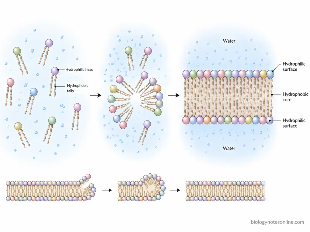
- Amphipathic nature- Membrane lipids are amphipathic molecules having a hydrophilic polar head and hydrophobic hydrocarbon tails. These two regions have different nature. When placed in water, the lipid molecules arrange according to these hydrophilic and hydrophobic regions.
- Interaction with water- The polar heads interact with the surrounding water and remain towards the aqueous region. The non-polar fatty acid tails do not show favourable interaction with water. Thus, these tails remain away from water.
- Hydrophobic effect– When hydrophobic tails remain exposed to water, the surrounding water molecules become more ordered. The lipid tails therefore come together and their contact with water is reduced. This is an important force responsible for the spontaneous arrangement of membrane lipids.
- Formation of two layers- Membrane phospholipids generally have one polar head and two hydrocarbon tails. Due to this approximately cylindrical structure, these lipids can pack in the form of a bilayer rather than small spherical micelles. The hydrophobic tails are present towards inside and polar heads are exposed to water on both sides.
- Non-covalent interactions- The hydrocarbon tails interact with the nearby tails by van der Waals interactions. Polar head groups can interact with water and other polar groups by electrostatic interactions and hydrogen bonding. These interactions are individually weak. But together they help in maintaining the membrane structure.
- Energetically favourable arrangement– In the bilayer, most of the hydrophobic parts remain protected from water while the hydrophilic heads remain hydrated. Thus, when membrane lipids are present in an aqueous environment, the bilayer can be formed spontaneously. Covalent bonds between the individual lipid molecules are not required for this arrangement.
- Self-sealing property- An open edge of lipid bilayer exposes its hydrophobic tails directly to water. This condition is unfavourable. The lipid molecules rearrange, due to which the exposed edge tends to close. It also allows bilayer sheets to form closed compartments and small membrane disruptions can be resealed.
The formed lipid bilayer has a hydrophobic interior and hydrophilic surfaces on its two sides. It forms a stable barrier between the two aqueous regions. The lipid molecules are still able to move within the membrane. Therefore, the amphipathic nature and molecular shape of membrane lipids are mainly involved in bilayer formation.
Major Types of Membrane Lipids
Membrane lipids are amphipathic molecules, having both the hydrophilic and hydrophobic regions. These molecules form the major part of the lipid bilayer of biological membranes. The membrane does not contain all lipids in equal amount. Their distribution also differs between cells, cell organelles and between the two layers of the same membrane.
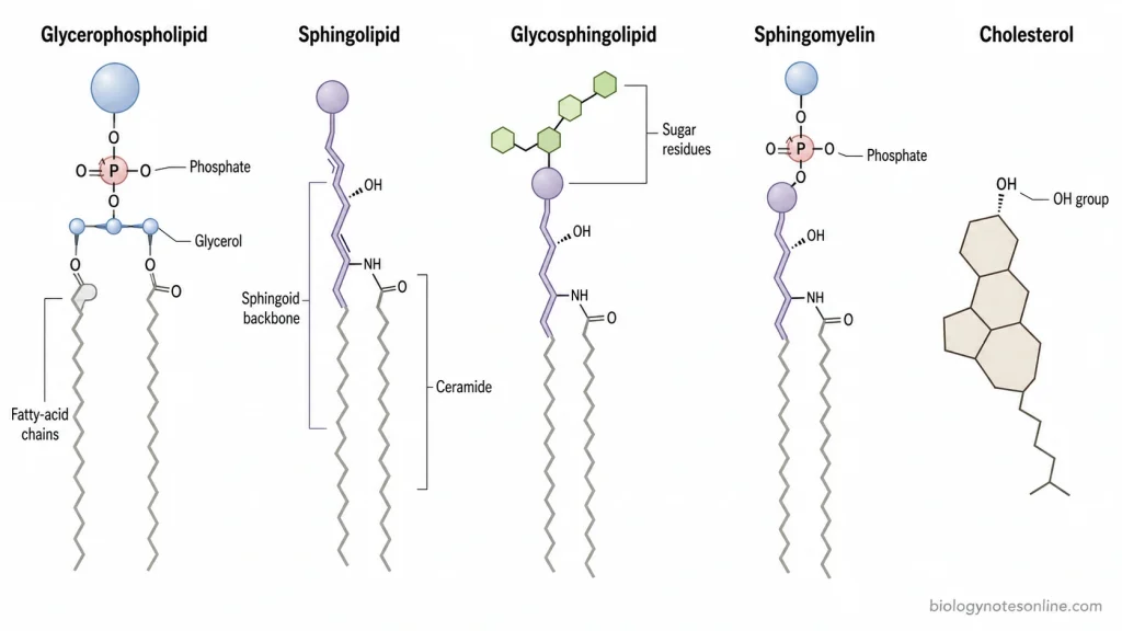
The major types of membrane lipids include:
- Glycerophospholipids
- Sphingolipids
- Sterols
1. Glycerophospholipids
Glycerophospholipids are the major phospholipid group found in many biological membranes. It is made up of glycerol to which two fatty acid chains are attached along with a phosphate-containing polar head group. The fatty acids make the hydrophobic region while the phosphate head forms the hydrophilic region. These two regions allow the phospholipids to arrange themselves into lipid bilayers.
Some of the important glycerophospholipids are-
Phosphatidylcholine (PC)- It contains choline as its polar head group. This is one of the abundant phospholipids of eukaryotic cell membranes.
Phosphatidylethanolamine (PE)- Another common phospholipid, having ethanolamine in its head group. In many animal plasma membranes, most of it is present towards the inner leaflet.
Phosphatidylserine (PS) contains a negatively charged serine head group. It is largely maintained on the cytoplasmic side of animal plasma membrane.
Phosphatidylinositol (PI)- It is present in smaller quantity in the membrane. Its phosphorylated forms are used in different cell signalling processes.
Cardiolipin is a characteristic glycerophospholipid of mitochondria, and is particularly enriched in the inner mitochondrial membrane of mammals.
2. Sphingolipids
Sphingolipids are another group of membrane lipids, but instead of glycerol these possess a sphingoid backbone. A fatty acid combines with the sphingoid base forming ceramide. This ceramide acts as the basic structure from which different sphingolipids are formed.
They are common structural components of eukaryotic plasma membranes. Some products formed from sphingolipids also have functions in cellular signalling.
The important sphingolipids include-
Sphingomyelin- It is commonly present in animal cell membranes. Sphingomyelin is a phosphosphingolipid having phosphocholine or another related phosphorus-containing group attached to the ceramide.
Glycosphingolipids– These contain sugar residues instead of a phosphate-containing head group. One or more sugars can be attached with the ceramide. Cerebrosides and gangliosides are the examples. Their carbohydrate portions are generally directed towards the non-cytosolic side of plasma membrane, thus remaining exposed towards the extracellular region where they participate in membrane organization and cellular interactions.
3. Sterols
Sterols are different from the phospholipids in their basic structure. They have four fused hydrocarbon rings and a small polar hydroxyl group. The sterol molecules are located between other lipid molecules of the membrane and affect their packing, membrane permeability and fluidity.
Cholesterol- It is the major sterol of animal cell membranes. At high temperature, movement of the lipid chains is restricted by cholesterol. At lower temperature, it prevents the membrane lipids from packing very closely.
Plant membranes have several sterols rather than cholesterol being the major one. β-sitosterol, stigmasterol and campesterol are some of the important plant sterols. In fungi, the major membrane sterol is ergosterol.
How Lipid Composition Determines Membrane Properties
The properties of a biological membrane are determined to a large extent by the type of lipids present and their relative amount. Lipids having different fatty acid chains and head groups do not pack in the same manner. Hence changes in lipid composition affect membrane fluidity, permeability, thickness and even the bending of membrane.
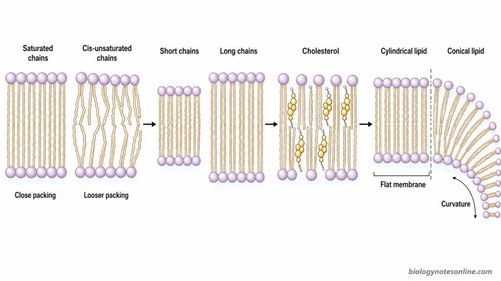
Some of the important effects of lipid composition are-
Fatty acid saturation and membrane fluidity– Membranes containing more cis-unsaturated fatty acids have bends in their hydrocarbon chains, because of which the lipid molecules cannot pack very closely. These membranes generally remain more disordered. On the other hand, saturated fatty acid chains pack closely with each other and increase lipid ordering. Shorter and unsaturated chains also have lower phase-transition temperature compared to long, saturated chains.
Effect of cholesterol- Cholesterol is fitted between the phospholipid molecules of membrane. In fluid membranes, it restricts the movement of hydrocarbon chains and increases their ordering, making the membrane less permeable. At lower temperature, cholesterol prevents regular close packing of phospholipids and their easy conversion into a solid-like state. It also increases membrane thickness and mechanical stiffness in many lipid bilayers.
Chain length, thickness and permeability
The length of the fatty acid chains also affects the hydrophobic region of membrane. Increase in acyl chain length can increase the hydrophobic thickness, and the movement of small polar solutes across such membrane becomes lower. Cholesterol also produces a more tightly packed membrane, hence permeability is reduced.
Polar head groups and membrane curvature- All the membrane lipids do not have the same molecular shape. Phosphatidylcholine (PC) and phosphatidylserine (PS) have a more cylindrical shape and normally favour flat bilayers. Phosphatidylethanolamine (PE) has a comparatively smaller head group, giving it a conical shape which favours negative membrane curvature. Lipids having a larger head region can favour curvature in the opposite direction. Therefore, change in the relative amount of these lipids can affect bending and shape of the membrane.
Lipid asymmetry- The two layers of plasma membrane also differ in their lipid composition. In animal plasma membrane, PS and PE are concentrated mainly towards the inner leaflet, whereas PC and sphingomyelin occur mainly towards the outer leaflet. This unequal distribution gives different properties to both sides of membrane and also contributes to membrane curvature. Exposure of PS towards the external surface can have important physiological functions.
Formation of membrane domains
Sphingolipids generally contain more saturated chains and show close packing with cholesterol. Because of these interactions, sphingolipid and cholesterol-rich regions can become more ordered than the surrounding membrane containing more unsaturated phospholipids. Such differences in lateral lipid organization can form small membrane domains with different physical properties.
How Are Lipids Organized Within the Membrane?
Membrane lipids are arranged mainly in the form of a lipid bilayer, with the polar region remaining in contact with water and hydrocarbon chains towards the interior. The lipids are not equally distributed on both sides of membrane. They also move within the membrane and some lipids remain associated in particular regions.
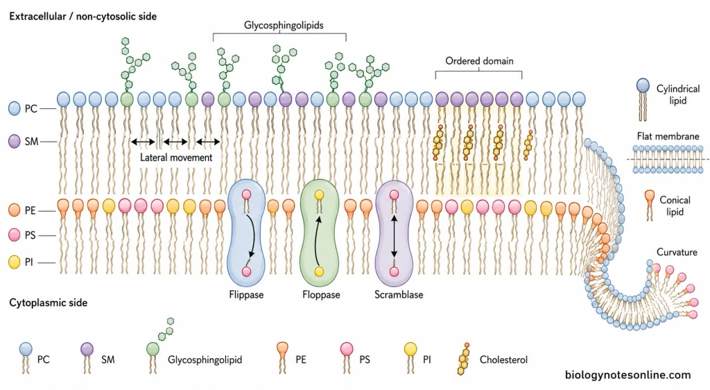
Lipid bilayer
The hydrophilic heads of membrane lipids are directed towards the aqueous region. Their hydrophobic fatty acid chains remain away from water and face each other in the interior. In this arrangement, two layers of lipids are formed, which is referred to as lipid bilayer.
The outer surface of bilayer is therefore formed by the polar head groups while the middle portion contains hydrocarbon chains. Water is present on both sides but does not enter freely through this hydrophobic region.
Lateral movement
Lipid molecules are not fixed at one particular position. They can move from one place to another within the same leaflet and also rotate. Such lateral movement occurs rapidly in the membrane.
Flip-flop or transverse movement
Movement from one membrane layer to the other is called flip-flop or transverse movement. This movement occurs very slowly for most phospholipids because the polar head has to cross the hydrophobic region of membrane.
Some membrane proteins are involved in movement of lipids between the two leaflets. Flippases move particular lipids mainly towards the cytoplasmic leaflet, whereas floppases transfer lipids in the opposite direction. Scramblases move lipids between both sides and can disturb the normal lipid distribution.
Lipid asymmetry
The two layers of plasma membrane have different lipid composition. In animal plasma membrane, phosphatidylcholine (PC) and sphingomyelin (SM) are present mainly towards the outer leaflet. Phosphatidylethanolamine (PE), phosphatidylserine (PS) and much of phosphatidylinositol (PI) occur towards the cytoplasmic side.
Glycosphingolipids are mainly found on the non-cytosolic side. In plasma membrane, their carbohydrate portion is exposed towards the outside of cell.
Lateral organization
All the lipids present in one leaflet are also not uniformly mixed. Some lipids remain more closely associated with each other and form regions having a different lipid composition from surrounding membrane.
Lipid rafts
Lipid rafts are such small membrane regions rich in cholesterol and sphingolipids along with some membrane proteins. Sphingolipids having saturated hydrocarbon chains pack more closely with cholesterol. These regions are more ordered compared to the surrounding lipid membrane and their composition can change.
How Are Membrane Lipids Synthesized and Distributed?
Most of the membrane lipids are synthesized in the endoplasmic reticulum (ER) and then transported to other cell membranes. The Golgi apparatus is also involved, mainly in formation of sphingomyelin and glycolipids. The following are the major steps involved-
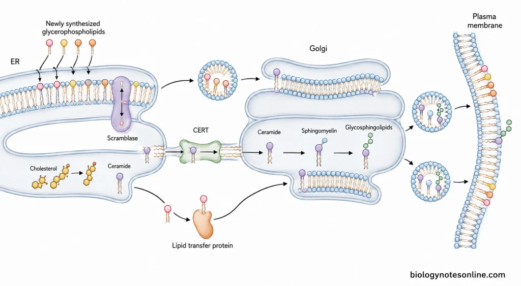
- Synthesis of glycerophospholipids in ER- The major site of glycerophospholipid synthesis is the cytosolic surface of ER. Fatty acids are first attached with glycerol-3-phosphate forming phosphatidic acid. Different polar head groups are then added and phosphatidylcholine, phosphatidylserine, phosphatidylethanolamine and phosphatidylinositol are formed.
- Distribution between ER membrane layers- Newly formed phospholipids are first added towards the cytosolic leaflet. Some of these lipids are then transferred to the opposite leaflet by scramblase, so that both sides of ER membrane can grow. During this process, phospholipids become distributed between the two layers.
- Formation of cholesterol and ceramide- The ER is also the major site for synthesis of cholesterol and ceramide. Ceramide is the basic molecule from which many sphingolipids are later produced.
- Transport from ER to Golgi- Lipids are moved from ER by vesicular as well as non-vesicular transport. Ceramide transfer protein (CERT) carries ceramide from ER to Golgi, where further reactions take place. Other lipid transfer proteins can also transfer lipids between closely located organelle membranes.
- Modification in Golgi apparatus- In this step, ceramide is converted into sphingomyelin and glycosphingolipids. Sugar groups are added stepwise during formation of the complex glycosphingolipids.
- Transport to different cell membranes- Lipids formed or modified in ER and Golgi are distributed to plasma membrane and different cell organelles. Vesicles carry many of these lipids through the secretory pathway, while some lipids are transferred directly with the help of lipid transfer proteins.
- Formation of membrane lipid asymmetry- The lipids reaching a membrane are not kept equally on its two sides. Flippases move particular phospholipids towards the cytoplasmic leaflet while floppase activity can move lipids towards the opposite side. Scramblases can transfer lipids in both directions. In this way, the characteristic lipid distribution of membrane is maintained.
Why Do Different Membranes Have Different Lipid Compositions?
The lipid composition is not same in all the biological membranes. It changes according to the type of organism, cell organelle and the function carried out by the membrane. Therefore, the lipid composition of animal plasma membrane cannot be used to describe all other membranes.
Plasma Membrane vs Intracellular Membranes
- Animal plasma membrane contains a higher amount of cholesterol and sphingolipids. These lipids make the membrane more closely packed and are important for maintaining its barrier property.
- The membrane of endoplasmic reticulum (ER) has much lower cholesterol than plasma membrane. It has a comparatively loose lipid arrangement and remains more flexible. The ER is also the major region where many membrane lipids and proteins are synthesized.
- Golgi membrane, mitochondrial membrane and membranes of other cell organelles also differ in their lipids. Different organelles perform different reactions and their membrane composition changes accordingly.
Animal, Plant, and Fungal Membrane Sterols
- In animals, cholesterol is the major membrane sterol. Its amount is particularly high in the plasma membrane.
- Plant membranes contain several types of sterols. β-sitosterol, stigmasterol and campesterol are some of the important sterols present in plants, instead of cholesterol being the main sterol.
- Ergosterol is the major membrane sterol in fungi. It is involved in maintaining the membrane fluidity and permeability, similar to the function performed by cholesterol in animal membranes.
Bacterial Membrane Lipids
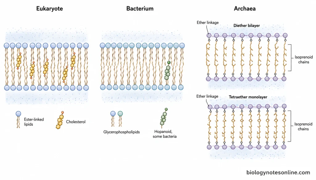
- Bacterial membranes generally contain mainly glycerophospholipids. In Escherichia coli, phosphatidylethanolamine, phosphatidylglycerol and cardiolipin are the major membrane lipids. The lipid composition however is not same in all bacterial species.
- Most bacteria do not contain sterols in their membrane. Some groups are exceptions, and some bacteria also contain hopanoids which affect membrane fluidity and permeability.
- The amount and type of bacterial membrane lipids may change with temperature and other environmental conditions. In this way, the required physical condition of membrane is maintained.
Archaeal Ether Lipids and Tetraether Membranes
- Archaeal membrane lipids are quite different from those of bacteria and eukaryotes. The hydrocarbon region is made up of isoprenoid chains which are attached to glycerol-1-phosphate by ether bonds. Bacteria and eukaryotes generally have fatty acids attached through ester bonds.
- Many archaea contain diether lipids which arrange to form a usual lipid bilayer. Therefore, all archaeal membranes are not formed as tetraether monolayers.
- In some archaea, tetraether lipids extend across the complete membrane and form a monolayer. This type of membrane is especially found in several thermophilic and thermoacidophilic archaea and provides greater stability under high temperature and other harsh conditions.
Functions of Membrane Lipids
Membrane lipids perform different structural and cellular functions. The following are some of the important functions-
- Formation of lipid bilayer- The major function is formation of basic membrane structure. The hydrophobic chains remain towards inside while the polar heads face the aqueous region.
- Permeability barrier- Lipid bilayer prevents free passage of ions and many water-soluble substances across the membrane.
- Membrane fluidity- Fatty acid chain length, degree of saturation and cholesterol content control the fluid nature of membrane.
- Cell signalling- Some membrane lipids act as signalling molecules or form signalling products. PIP₂, DAG and IP₃ are important examples.
- Membrane protein function- The surrounding lipids are required for proper positioning and activity of many membrane proteins.
- Membrane curvature- Different lipid shapes help in bending of membrane during vesicle formation, fusion and fission.
- Membrane domains- Cholesterol and sphingolipids can remain together forming ordered regions called lipid rafts.
- Cell recognition- Glycolipids have carbohydrate groups towards the outer surface of plasma membrane. These are used in cell recognition and interaction.
Membrane Lipids at a Glance
The major membrane lipids and their important characteristics are given below-
| Lipid type | Representative examples | Membrane location | Key structural feature | Major function |
|---|---|---|---|---|
| Glycerophospholipids | PC, PE, PS, PI | Found in most cellular membranes. In animal plasma membrane, PC is more towards outer leaflet while PE, PS and PI occur mainly towards inner side. | Glycerol with two fatty acid chains and phosphate-containing head group. | These are the major bilayer-forming lipids. Also involved in membrane barrier and signalling. |
| Sphingolipids | Sphingomyelin, glycosphingolipids | Mainly abundant in animal plasma membrane, especially towards the outer leaflet. | Ceramide forms their basic structure. Sugar or phosphocholine can be present as head group. | Membrane organization, cell recognition and formation of cholesterol-rich membrane regions. |
| Sterols | Cholesterol, β-sitosterol, stigmasterol, campesterol, ergosterol | Cholesterol in animal membranes, phytosterols in plants and ergosterol in fungal membrane. | A small polar hydroxyl group with rigid fused hydrocarbon rings. | Mainly involved in controlling membrane fluidity, packing and permeability. |
| Cardiolipin | Cardiolipin (CL) | Mainly inner mitochondrial membrane and also present in many bacterial membranes. | It is a dimeric phospholipid having four acyl chains. | Important for organization and functioning of energy-producing membrane protein complexes. |
| Bacterial membrane lipids | PE, PG, cardiolipin | Cytoplasmic membrane of bacteria. The exact composition differs among bacterial groups. | Mostly glycerol-based phospholipids with ester-linked fatty acids. | Formation of bacterial membrane and maintaining its required physical properties. |
| Archaeal ether lipids | Archaeol, caldarchaeol | Cytoplasmic membrane of archaea. | Isoprenoid chains are attached to glycerol-1-phosphate through ether bonds. Diether or tetraether forms occur. | Diether lipids form bilayers while tetraether lipids can form a membrane monolayer, providing membrane stability. |
References
- Alberts, B., Johnson, A., Lewis, J., Raff, M., Roberts, K., & Walter, P. (2002). Molecular biology of the cell (4th ed.). Garland Science. https://www.ncbi.nlm.nih.gov/books/NBK21054/
- Andersen, J. P., Vestergaard, A. L., Mikkelsen, S. A., Mogensen, L. S., Chalat, M., & Molday, R. S. (2016). P4-ATPases as phospholipid flippases—Structure, function, and enigmas. Frontiers in Physiology, 7, 275. https://doi.org/10.3389/fphys.2016.00275
- Blom, T., Somerharju, P., & Ikonen, E. (2011). Synthesis and biosynthetic trafficking of membrane lipids. Cold Spring Harbor Perspectives in Biology, 3(8), a004713. https://doi.org/10.1101/cshperspect.a004713
- Casares, D., & Escribá, P. V. (2019). Membrane lipid composition: Effect on membrane and organelle structure, function and compartmentalization and therapeutic avenues. International Journal of Molecular Sciences, 20(9), 2167. https://doi.org/10.3390/ijms20092167
- Chong, P. L.-G., Chang, A., Yu, A., & Mammedova, A. (2022). Vesicular and planar membranes of archaea lipids: Unusual physical properties and biomedical applications. International Journal of Molecular Sciences, 23(14), 7616. https://doi.org/10.3390/ijms23147616
- Cockcroft, S., & Raghu, P. (2018). Phospholipid transport protein function at organelle contact sites. Current Opinion in Cell Biology, 53, 52–60. https://doi.org/10.1016/j.ceb.2018.04.011
- Cooper, G. M. (2000). The cell: A molecular approach (2nd ed.). Sinauer Associates. https://www.ncbi.nlm.nih.gov/books/NBK9839/
- Dufourc, E. J. (2008). Sterols and membrane dynamics. Journal of Chemical Biology, 1(1–4), 63–77. https://doi.org/10.1007/s12154-008-0010-6
- Escribá, P. V., González-Ros, J. M., Goñi, F. M., Kinnunen, P. K. J., Vigh, L., Sánchez-Magraner, L., Fernández, A. M., Busquets, X., Horváth, I., & Barceló-Coblijn, G. (2008). Membranes: A meeting point for lipids, proteins and therapies. Journal of Cellular and Molecular Medicine, 12(3), 829–875. https://doi.org/10.1111/j.1582-4934.2008.00281.x
- Fadeel, B., & Xue, D. (2009). The ins and outs of phospholipid asymmetry in the plasma membrane: Roles in health and disease. Critical Reviews in Biochemistry and Molecular Biology, 44(5), 264–277. https://doi.org/10.1080/10409230903193307
- Frallicciardi, J., Melcr, J., Siginou, P., Marrink, S. J., & Poolman, B. (2022). Membrane thickness, lipid phase and sterol type are determining factors in the permeability of membranes to small solutes. Nature Communications, 13(1), 1605. https://doi.org/10.1038/s41467-022-29272-x
- Harayama, T., & Antonny, B. (2023). Beyond fluidity: The role of lipid unsaturation in membrane function. Cold Spring Harbor Perspectives in Biology, 15(7), a041409. https://doi.org/10.1101/cshperspect.a041409
- Harayama, T., & Riezman, H. (2018). Understanding the diversity of membrane lipid composition. Nature Reviews Molecular Cell Biology, 19, 281–296. https://doi.org/10.1038/nrm.2017.138
- Houtkooper, R. H., & Vaz, F. M. (2008). Cardiolipin, the heart of mitochondrial metabolism. Cellular and Molecular Life Sciences, 65(16), 2493–2506. https://doi.org/10.1007/s00018-008-8030-5
- Huang, Z., & London, E. (2016). Cholesterol lipids and cholesterol-containing lipid rafts in bacteria. Chemistry and Physics of Lipids, 199, 11–16. https://doi.org/10.1016/j.chemphyslip.2016.03.002
- Jain, S., Caforio, A., & Driessen, A. J. M. (2014). Biosynthesis of archaeal membrane ether lipids. Frontiers in Microbiology, 5, 641. https://doi.org/10.3389/fmicb.2014.00641
- Kraft, M. L. (2017). Sphingolipid organization in the plasma membrane and the mechanisms that influence it. Frontiers in Cell and Developmental Biology, 4, 154. https://doi.org/10.3389/fcell.2016.00154
- McMahon, H. T., & Boucrot, E. (2015). Membrane curvature at a glance. Journal of Cell Science, 128(6), 1065–1070. https://doi.org/10.1242/jcs.114454
- Mouritsen, O. G. (2011). Lipids, curvature, and nano-medicine. European Journal of Lipid Science and Technology, 113(10), 1174–1187. https://doi.org/10.1002/ejlt.201100050
- Pereira de Sa, N., & Del Poeta, M. (2022). Sterylglucosides in fungi. Journal of Fungi, 8(11), 1130. https://doi.org/10.3390/jof8111130
- Ramstedt, B., & Slotte, J. P. (2006). Sphingolipids and the formation of sterol-enriched ordered membrane domains. Biochimica et Biophysica Acta (BBA) – Biomembranes, 1758(12), 1945–1956. https://doi.org/10.1016/j.bbamem.2006.05.020
- Rao, R. P., & Acharya, J. K. (2008). Sphingolipids and membrane biology as determined from genetic models. Prostaglandins & Other Lipid Mediators, 85(1–2), 1–16. https://doi.org/10.1016/j.prostaglandins.2007.10.002
- Ryan, E., Joyce, S. A., & Clarke, D. J. (2023). Membrane lipids from gut microbiome-associated bacteria as structural and signalling molecules. Microbiology, 169(3), 001315. https://doi.org/10.1099/mic.0.001315
- Sarmento, M. J., Llorente, A., Petan, T., Khnykin, D., Popa, I., Nikolac Perkovic, M., Konjevod, M., & Jaganjac, M. (2023). The expanding organelle lipidomes: Current knowledge and challenges. Cellular and Molecular Life Sciences, 80(8), 237. https://doi.org/10.1007/s00018-023-04889-3
- Scott, H. L., Kennison, K. B., Enoki, T. A., Doktorova, M., Kinnun, J. J., Heberle, F. A., & Katsaras, J. (2021). Model membrane systems used to study plasma membrane lipid asymmetry. Symmetry, 13(8), 1356. https://doi.org/10.3390/sym13081356
- Subczynski, W. K., Pasenkiewicz-Gierula, M., Widomska, J., Mainali, L., & Raguz, M. (2017). High cholesterol/low cholesterol: Effects in biological membranes: A review. Cell Biochemistry and Biophysics, 75(3–4), 369–385. https://doi.org/10.1007/s12013-017-0792-7
- Sunshine, H., & Iruela-Arispe, M. L. (2017). Membrane lipids and cell signaling. Current Opinion in Lipidology, 28(5), 408–413. https://doi.org/10.1097/MOL.0000000000000443
- van Meer, G., Voelker, D. R., & Feigenson, G. W. (2008). Membrane lipids: Where they are and how they behave. Nature Reviews Molecular Cell Biology, 9(2), 112–124. https://doi.org/10.1038/nrm2330
- Vogel, P., Persson, S., Moreno-Pescador, G., & Noack, L. C. (2025). Sterols in plant biology—Advances in studying membrane dynamics. The Cell Surface, 13, 100147. https://doi.org/10.1016/j.tcsw.2025.100147
- Willdigg, J. R., & Helmann, J. D. (2021). Mini review: Bacterial membrane composition and its modulation in response to stress. Frontiers in Molecular Biosciences, 8, 634438. https://doi.org/10.3389/fmolb.2021.634438
- Xu, F., Rychnovsky, S. D., Belani, J. D., Hobbs, H. H., Cohen, J. C., & Rawson, R. B. (2005). Dual roles for cholesterol in mammalian cells. Proceedings of the National Academy of Sciences of the United States of America, 102(41), 14551–14556. https://doi.org/10.1073/pnas.0503590102