What is Neuroglia?
- Neuroglia, often referred to as glial cells, play a vital role in the central and peripheral nervous systems. Although commonly described as the “glue” of the nervous system, their functions extend far beyond mere support. Neuroglial cells are classified as non-neuronal cells that do not generate electrical impulses but are essential for maintaining the overall homeostasis of neural tissues.
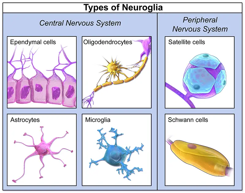
- Neuroglia are categorized into various types based on their origin and function. In the central nervous system (CNS), this includes astrocytes, oligodendrocytes, microglia, and ependymal cells. Each type has distinct roles, such as supporting neurons, insulating axons, and responding to injury or infection. For instance, astrocytes contribute to the structural integrity of the CNS, while oligodendrocytes are responsible for the formation of myelin sheaths, which enhance signal transmission.
- The overarching function of neuroglia encompasses a range of homeostatic processes. These cells are crucial for maintaining the chemical environment around neurons, ensuring appropriate concentrations of ions, neurotransmitters, and metabolic substrates. For example, astrocytes regulate potassium levels and transport nutrients like glucose and lactate to neurons, supporting their energy needs.
- Moreover, neuroglial cells facilitate communication within the nervous system. They play a key role in synaptic plasticity, influencing how signals are transmitted between neurons. This modulation of neurotransmission underscores the complexity of neuroglial functions, which were historically underestimated. Recent research indicates that glial cells actively participate in the signaling processes, affecting neuronal communication and overall brain function.
- Neuroglia also engage in various defensive roles. Microglia, the resident immune cells of the CNS, monitor the environment for pathogens and respond to injuries. They are involved in clearing debris and dead cells, thus maintaining a healthy neural landscape. Additionally, during pathological conditions, glial cells can become reactive, initiating inflammatory responses to protect the nervous tissue.
- The heterogeneity of neuroglial cells reflects their evolutionary specialization, enabling them to perform a diverse array of functions. From supporting neuronal development and shaping the cytoarchitecture of the nervous system to participating in immune responses, neuroglia are indispensable to the proper functioning of both the CNS and peripheral nervous system (PNS).
Definition of Neuroglia
Neuroglia, or glial cells, are non-neuronal cells in the nervous system that provide support, insulation, and protection for neurons. They play crucial roles in maintaining homeostasis, facilitating communication, and responding to injury or disease within the central and peripheral nervous systems.
Classification of Neuroglia
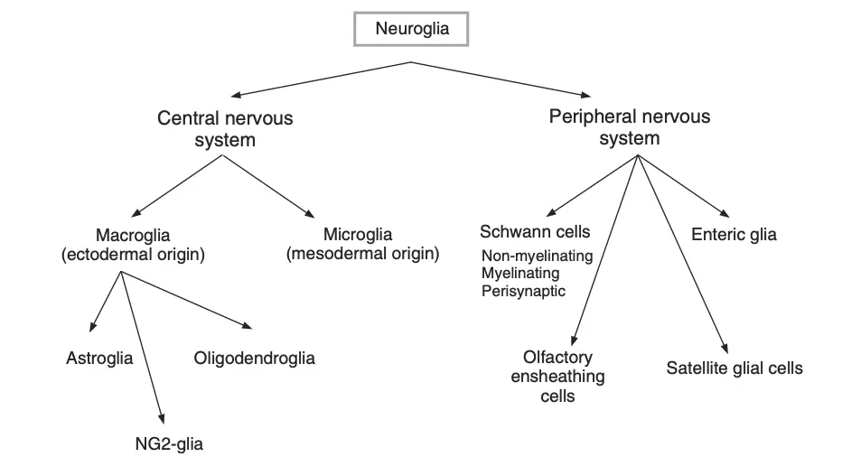
Neuroglia, or glial cells, are essential components of the nervous system, classified into two main categories based on their origin and function: microglia and macroglia. Each category contains various cell types, each serving unique and vital roles within the central nervous system (CNS) and peripheral nervous system (PNS).

- Microglia
- These are small, phagocytic cells that act as the immune defenders of the nervous system.
- Microglia continuously monitor the environment, engulfing pathogens and debris to maintain neural health.
- Their flexible shape allows them to adapt and change, particularly when responding to foreign bodies, and they are distributed throughout the brain and spinal cord.
- Macroglia
- This group comprises larger glial cells and is further divided into several types based on their specific functions and locations.
- Astrocytes
- Star-shaped cells that provide structural support and fill the spaces between neurons.
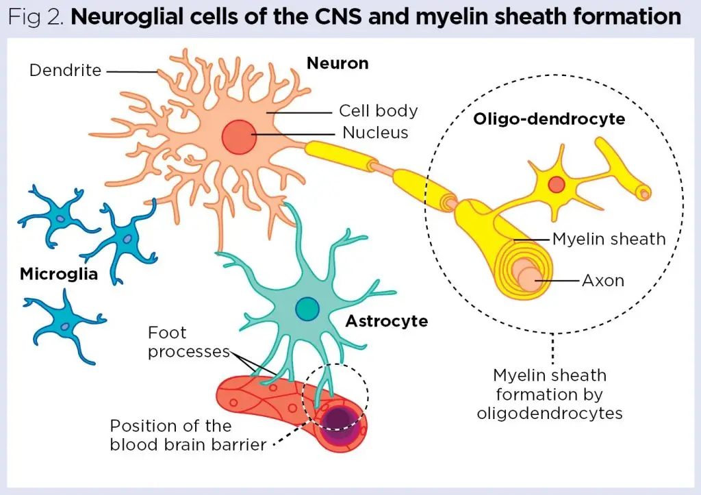
- They form the blood-brain barrier through their end-feet, protecting the brain from harmful substances while facilitating nutrient exchange between neurons and blood vessels.
- Oligodendrocytes
- Present in the CNS, these cells are responsible for producing the myelin sheath that insulates neuronal axons, enhancing the speed of electrical signal transmission.
- Ependymal Cells
- These cells line the cavities of the CNS and are subdivided into three types:
- Ependymocytes promote the movement of substances between neurons and cerebrospinal fluid (CSF).
- Tanycytes respond to hormonal changes in the blood, influencing various physiological functions.
- Choroidal Epithelial Cells regulate the chemical composition of the CSF.
- These cells line the cavities of the CNS and are subdivided into three types:
- Radial Glial Cells
- They serve as temporary scaffolding for new neurons during development, guiding their migration and positioning within the CNS.
- Schwann Cells
- Found in the PNS, these cells myelinate peripheral axons similarly to oligodendrocytes in the CNS.
- They also possess phagocytic capabilities, helping to clear debris in the peripheral nervous system.
- Satellite Cells
- Located around the sensory and autonomic nerve cells in the PNS, these cells maintain a stable chemical environment for neurons, supporting their function.
- Enteric Glial Cells
- Present in the gastrointestinal tract, these cells are involved in digestive processes and overall homeostasis of the enteric nervous system.

How many glial cells are in the brain?
The quantification of glial cells in the human brain remains an area of active research, with estimates suggesting significant numbers that emphasize their importance in neural function. Historically, it was believed that glial cells outnumbered neurons by a factor of ten; however, contemporary studies indicate a more balanced ratio between these cell types.
- Total Cell Counts in the Human Brain
- Recent techniques, such as isotropic fractionation, have revealed that the human brain contains approximately 86 billion neurons and 84 billion non-neuronal cells, which include glial cells.
- Therefore, the overall count of glial cells is nearly equal to that of neurons in the adult human brain, challenging previous assumptions.
- Variability Across Brain Regions
- The distribution of glial cells varies significantly across different regions of the brain:
a. Cerebellum: This region has the lowest glia-to-neuron ratio, with estimates of 70 billion neuronal nuclei and 16 billion non-neuronal nuclei, resulting in a ratio of 0.22.
b. Cerebral Cortex: In contrast, the cortex exhibits a higher ratio of 3.76, consisting of around 60 billion non-neuronal cells alongside 16 billion neurons.
c. Basal Ganglia: This area has an even higher ratio of approximately 11.3, with 0.69 billion neurons and 7.73 billion non-neuronal cells.
- The distribution of glial cells varies significantly across different regions of the brain:
- Comparative Ratios in Other Species
- Across various species, the glial cell to neuron ratio tends to increase with brain size:
a. In rodents, the ratio is about 0.3–0.4.
b. In cats, it is 1.1; in horses, it is 1.2; and in rhesus monkeys, it varies between 0.5 and 1.0.
c. Humans exhibit a ratio of approximately 1.5–1.7, whereas elephants and certain whale species can reach ratios as high as 4–6.
- Across various species, the glial cell to neuron ratio tends to increase with brain size:
- Diversity of Glial Cells
- While the total numbers provide insight into glial abundance, they do not reflect the diversity within the glial population.
- Stereological studies indicate the following approximate distribution among glial subtypes in the human brain:
a. Astrocytes: About 20% of the total glial population.
b. Oligodendrocytes: Roughly 75%.
c. Microglia: Approximately 5%.
- Methodological Challenges
- The estimation of glial cell numbers is fraught with challenges, including variability in methodologies and potential loss of nuclei during processing.
- Additionally, previous studies using two-dimensional counting may not provide a comprehensive view of glial diversity, as they lacked specific staining techniques to accurately identify cell types.

Embryogenesis and development of neuroglia in mammals
The development of neuroglial cells during embryogenesis in mammals is a complex and finely orchestrated process. Neuroglia, comprising various supportive cells, arise from neural progenitors that play critical roles in the formation and maintenance of the nervous system. Understanding the lineage and differentiation of these cells sheds light on their essential functions in both development and adulthood.
- Origin of Neuroglial Cells
- All neural cells, including neurons and macroglial cells, originate from the neuroepithelium, which forms the neural tube.
- Neuroepithelial cells are pluripotent and can differentiate into either neurons or glial cells, functioning as true neural progenitors.
- These progenitors give rise to neuronal precursors (neuroblasts) and glial precursors (glioblasts), which further differentiate into their respective cell types.
- Radial Glial Cells
- Early in development, neuroepithelial cells differentiate into radial glial cells, which are pivotal in neurogenesis.
- Radial glial cells extend processes from the ventricular zone to the pia mater, providing structural scaffolding that guides migrating neurons to their final destinations.
- These cells generate various cell types, including neurons, astrocytes, and some oligodendrocytes, with the majority of oligodendrocytes originating from distinct precursors.
- Astrocyte Development
- Astrocytes can arise from both radial glial cells and specific glial precursors, with the ratio of origin varying by region within the central nervous system (CNS).
- In the perinatal cortex, astrocytes exhibit proliferative capabilities, with new astroglial cells arising from the symmetric division of differentiated astrocytes.
- Some astrocytes derive from oligodendrocyte precursor cells (OPCs), migrating to various sites in the brain.
- Oligodendrocyte Lineage
- Oligodendrocytes develop from committed glial precursors through several characterized intermediate stages.
- These cells are essential for myelination in the CNS and are derived from specific progenitor populations located in the brain and spinal cord.
- Microglial Development
- Microglial cells originate from myelomonocytic progenitors that develop from mesodermal sources.
- These progenitors migrate into the neural tube early in embryonic development, evolving into embryonic microglia with distinct morphological features.
- Microglia serve critical roles during development, including phagocytosing apoptotic neurons and mediating immune responses within the CNS.
- Neurogenesis in Adulthood
- Neurogenesis persists in specific regions of the adult brain, such as the hippocampus and the subventricular zone, where astrocytes have been identified as neural stem cells.
- The production of new neurons contributes to the brain’s plasticity and ability to adapt to new experiences throughout life.
- Peripheral Glia and Schwann Cells
- Peripheral glial cells, including Schwann cells, arise from neural crest cells and are crucial for peripheral nerve function.
- Schwann cell precursors differentiate into either myelinating or non-myelinating Schwann cells, depending on their interactions with axons.
Functions of Neuroglia
The key functions of neuroglia include:
- Support and Structure
- Neuroglia provide structural support to neurons, maintaining the architecture of the nervous system.
- Nutrient Supply
- Glial cells help supply nutrients to neurons, ensuring they receive the necessary resources for energy metabolism and function.
- Myelination
- Oligodendrocytes (in the CNS) and Schwann cells (in the PNS) produce myelin, a fatty substance that insulates axons, facilitating faster signal transmission.
- Homeostasis
- Neuroglia maintain the extracellular environment, regulating ion concentrations and removing excess neurotransmitters, thus contributing to homeostasis.
- Immune Response
- Microglia act as the immune cells of the CNS, responding to injury and infection by phagocytosing debris and secreting inflammatory mediators.
- Neurotransmitter Regulation
- Astrocytes uptake and recycle neurotransmitters, such as glutamate, which prevents excitotoxicity and modulates synaptic activity.
- Scar Formation
- Following injury, astrocytes can proliferate and form glial scars, which can help protect the damaged area but may also inhibit regeneration.
- Guidance During Development
- During embryonic development, radial glial cells guide the migration of neurons to their final positions, influencing the formation of neural circuits.
- Signal Modulation
- Glial cells can influence neuronal signaling through the release of gliotransmitters, affecting synaptic transmission and plasticity.
- Regulation of Blood Flow
- Astrocytes regulate cerebral blood flow by signaling to blood vessels, ensuring adequate oxygen and nutrient delivery in response to neuronal activity.
- Purves D, Augustine GJ, Fitzpatrick D, et al., editors. Neuroscience. 2nd edition. Sunderland (MA): Sinauer Associates; 2001. Neuroglial Cells. Available from: https://www.ncbi.nlm.nih.gov/books/NBK10869/
- https://opentextbc.ca/biology/chapter/16-1-neurons-and-glial-cells/
- https://www.lecturio.com/concepts/histology-of-the-nervous-system/
- https://www.verywellhealth.com/what-are-glial-cells-and-what-do-they-do-4159734
- https://www.urmc.rochester.edu/neuroscience/_shared/ResearchProjects/403/c03-AV.pdf
- https://www.toppr.com/guides/biology/cell-the-unit-of-life/neuroglia-definition-functions-types/
- https://qbi.uq.edu.au/brain-basics/brain/brain-physiology/types-glia
- Text Highlighting: Select any text in the post content to highlight it
- Text Annotation: Select text and add comments with annotations
- Comment Management: Edit or delete your own comments
- Highlight Management: Remove your own highlights
How to use: Simply select any text in the post content above, and you'll see annotation options. Login here or create an account to get started.