Fish Integument (Skin)
- The integument, or skin, of a fish serves as a crucial protective barrier that shields the body from injuries and infections. However, beyond protection, the fish integument fulfills several other important roles, including respiration, excretion, and osmoregulation. Through these functions, fish are able to maintain internal balance while interacting with their aquatic environment. The integument is not just a physical barrier; it is also the main interface between the fish and its surroundings.
- Specialized structures, such as phosphorescent organs, poison glands, and electric organs, are often embedded within the fish’s skin. These adaptations are evolutionary responses to specific environmental challenges, enhancing the fish’s ability to survive, communicate, or defend itself. Phosphorescent organs, for example, enable deep-sea fish to generate light, while poison glands provide a chemical defense mechanism against predators. Electric organs help some species to navigate or stun prey.
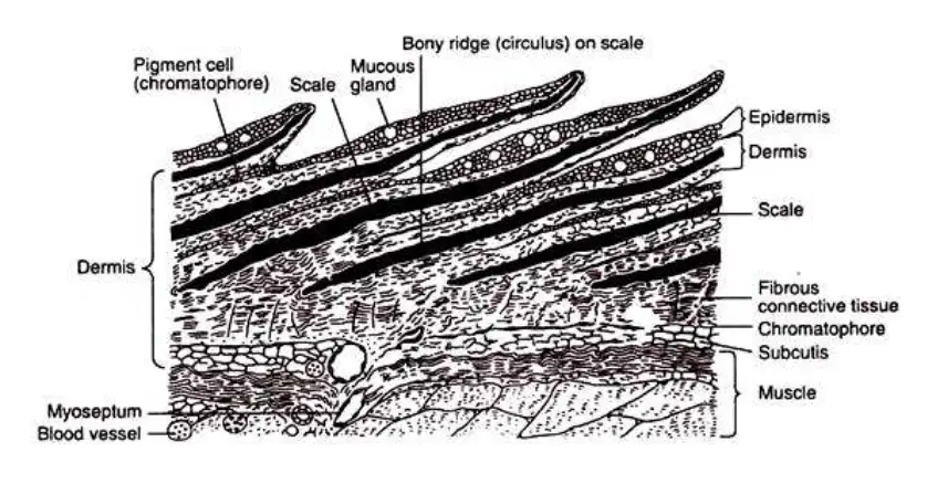
- The fish skin is divided into distinct layers: the epidermis, dermis, and subcutis. The outermost layer, the epidermis, consists of living cells, which is unique compared to the dead, keratinized outer layers found in terrestrial animals. Beneath the epidermis lies the dermis, which contains connective tissue, blood vessels, and nerves. This layer provides structural support and helps in thermoregulation. Finally, the subcutis, the innermost layer, consists of loose connective tissue and fat, aiding in insulation and buoyancy.
Epidermis And Dermis
Epidermis
The epidermis is the outermost layer of the skin, derived from the ectoderm, and consists of multiple layers of epithelial cells. These layers vary in thickness depending on the species, age, size, and reproductive stage of the organism. The cells in the epidermis are arranged in specific layers, each contributing to its overall function. Below are detailed points explaining its structure and functions:
- Basal Layer:
The basal layer is the innermost layer of the epidermis and serves as the foundation for the other layers. It is composed of columnar or cuboidal cells, which constantly divide to produce new cells. These newly formed cells gradually migrate upward to replenish the layers above, maintaining the integrity of the epidermis. This regenerative process is vital for the continual replacement of surface cells that are lost due to damage or natural wear. - Middle Layer:
Situated above the basal layer, the middle layer consists of more mature epithelial cells that have moved upward from the basal layer. These cells begin to flatten and prepare for their eventual transition to the surface. Their primary role in this layer is to provide structural stability to the epidermis and to act as an intermediary zone between the proliferative basal layer and the surface layer. As they move up, these cells also contribute to the formation of various secretions, such as mucus in fish, which aids in protection and lubrication. - Superficial Layer:
The superficial layer forms the outermost boundary of the epidermis and is made up of flattened epithelial cells. These cells, having reached the end of their life cycle, form a protective barrier that shields the underlying tissues from physical damage, infections, and dehydration. In many species, particularly aquatic organisms like fish, the superficial layer also secretes mucus, which plays a crucial role in reducing friction, protecting against parasites, and facilitating efficient gas exchange. - Thickness Variation:
The thickness of the epidermis is not constant and can vary significantly depending on several factors:- Species: Different species have epidermal layers of varying thickness to adapt to their specific environmental conditions.
- Age: In older individuals, the epidermis may become thicker or thinner, depending on the balance between cell production and loss.
- Size: Larger individuals may have thicker epidermal layers to accommodate the increased surface area and physical stress.
- Reproductive Cycle: During certain stages of reproduction, such as spawning, some species exhibit changes in the thickness of their epidermis, likely as an adaptation to environmental or physiological demands.
- Function of Epithelial Cells:
The epithelial cells within the epidermis serve multiple functions, including:- Protection: They create a barrier to prevent harmful substances, pathogens, and mechanical damage from reaching the deeper tissues.
- Regeneration: By continuously producing new cells in the basal layer, the epidermis can repair itself and maintain its protective function.
- Secretion: In certain species, particularly aquatic ones, epithelial cells produce mucus that serves multiple purposes, including lubrication, protection, and involvement in respiration or osmoregulation.

Epithelial cells
Epithelial cells play a crucial role in the structural and functional integrity of various organisms, particularly in fish. These cells form protective barriers, facilitate absorption and secretion, and participate in sensory functions. Understanding their morphology and functions is essential for students and educators in the biological sciences.
- Morphology of Epithelial Cells:
- The deepest layers of epithelial cells are predominantly composed of columnar or cuboidal cells, characterized by round or elongated nuclei. These basal cells are arranged in a single row, forming the basal layer. They undergo mitotic division to generate new cells, thereby replenishing the outermost layer as it becomes worn or damaged.
- The middle layer consists of polygonal cells, each featuring a round nucleus. This layer serves as an intermediate stage in the epithelial structure.
- The outermost layer, known as the superficial layer, may exhibit a variety of shapes, including rectangular, columnar, or cuboidal. The nuclei of these cells are typically located at the basal region. In many fish species, these outer cells possess secretory capabilities, containing vesicles that release their contents externally. This secretion forms an extracellular cuticular coat, often referred to as the cuticle or glycocalyx.
- Types of Epithelial Cells:
- Mucous Cells: These are numerous in the epidermis of fish and function as unicellular glands that open onto the skin surface via minute pores. Their shape may vary, including flask-like, goblet-like, or tubular forms extending into the dermis. The primary secretion, a slippery mucus containing a lipoprotein known as mucin, reduces friction during swimming. Additionally, the regular secretion of mucus serves as a protective mechanism, washing away microorganisms and irritants, thereby preventing potential infections. In some species, this mucus facilitates chemical communication, while in others, it aids in nesting behavior for egg-laying, such as seen in Macropodus, Gasterosteus, and Betta. Moreover, in certain species like Protopterus and Lepidosiren, the mucus forms a protective cocoon during periods of aestivation.
- Club Cells: Recognized as “alarm substance cells,” these larger cells can be either uninucleate or multinucleate. They produce pheromones that trigger alarm responses in other fish within the school when detected by their olfactory organs. Club cells are strategically positioned in the middle layer of the epidermis and do not open to the epithelial surface. Their abundance varies across different fish species.
- Poison Glands: Some epidermal glandular cells undergo modifications to become poison glands, which secrete toxic substances for defense against predators and for offensive purposes. These glands are typically located at the base of specific structures, such as spines and stingers, allowing for the injection of venom when the structures penetrate a target. Notable examples include the stingray, which possesses venomous spines, and the chimaeras, which have venom glands associated with dorsal fin spines. In the Scorpaenidae family (scorpion fish), poison glands are situated in the grooves of various fins, while sturgeon exhibit poison glands on the sides of their caudal peduncle.
- Photophores: Found predominantly in marine fishes, these specialized multicellular glands arise from the basal layer of the epidermis. They are absent in freshwater species. Photophores are embedded in the dermis and are responsible for bioluminescence, allowing deep-sea fishes to produce light.
- Sacciform Cells: These sac-like unicellular glands are scattered throughout the epidermis of various fish species. They contain a basal nucleus and release proteinaceous substances. These cells open to the exterior through small pores, and their secretions are vital for maintaining skin moisture and facilitating cutaneous respiration.
- Other Cell Types: In addition to the aforementioned cells, the epidermis of marine fish contains ionocytes, also known as chloride cells, which serve excretory functions. Various types of immune cells, including lymphocytes and granulocytes, are present in the intercellular spaces. Lymphocytes are typically interspersed among the basal layer cells, while chromatophores, which contain pigment granules, contribute to color variation in fish skin.
- Sensory Receptors: The fish epidermis is rich in sensory receptors, including taste buds, ampullary organs, epithelial sensory cells, and free nerve endings. These receptors are distributed throughout the body surface and play vital roles in environmental sensing and interaction.
Dermis
The dermis is a vital layer of skin located beneath the epidermis, primarily composed of connective tissue. This layer provides structural support, nourishment, and various functionalities essential for the health and integrity of the skin. Understanding its composition and roles is crucial for both students and educators in the biological sciences.
- Structure of the Dermis:
- The dermis is characterized by two distinct layers: the upper stratum spongiosum and the lower stratum compactum.
- Stratum Spongiosum: This layer is relatively thin and consists of loose connective tissue. The loose structure allows for flexibility and the accommodation of various cellular components, which include fibroblasts, macrophages, and mast cells. This layer also contains blood vessels that play a critical role in thermoregulation and nutrient delivery.
- Stratum Compactum: In contrast, this layer is denser and thicker, composed of tightly packed connective tissue fibers. The density of this layer provides mechanical strength and resilience, enabling the dermis to withstand various stresses and strains.
- Vascularization:
- The dermis is richly supplied with blood vessels. This vascular network serves multiple functions:
- It provides essential nutrients to the avascular epidermis, which lacks its own blood supply.
- The blood vessels assist in thermoregulation by facilitating heat exchange; for instance, vasodilation increases blood flow to the skin, aiding in cooling, while vasoconstriction reduces blood flow to retain heat.
- The dermis is richly supplied with blood vessels. This vascular network serves multiple functions:
- Nerve Supply:
- Besides blood vessels, the dermis is also innervated with a variety of nerve endings. These sensory neurons are crucial for the perception of touch, temperature, pain, and pressure.
- Specialized structures, such as Meissner’s corpuscles and Pacinian corpuscles, are embedded within the dermis, allowing for enhanced tactile sensation and pressure detection, respectively.
- Functions of the Dermis:
- Support and Protection: The dermis provides structural support to the epidermis and serves as a protective barrier against mechanical injury, pathogens, and harmful substances.
- Homeostasis: Through its vascular and nerve supply, the dermis plays a significant role in maintaining homeostasis, particularly in temperature regulation and fluid balance.
- Immune Response: The presence of immune cells within the dermis contributes to the skin’s defense mechanisms. These cells can identify and respond to foreign pathogens, thus playing a crucial role in immune surveillance.
- Hair and Gland Support: The dermis houses hair follicles and various glands (such as sebaceous and sweat glands), which are essential for thermoregulation, lubrication, and excretion.
- Interaction with the Epidermis:
- The dermis and epidermis are interconnected through a basement membrane, which provides structural integrity and facilitates nutrient exchange.
- The health of the epidermis is reliant on the dermis, as the latter supplies necessary nutrients and supports the overall skin architecture.
Functions of the integument
The integument, or skin, serves as a multifunctional organ system in various organisms, particularly in fish. It provides a barrier against environmental challenges and plays a critical role in maintaining homeostasis. Understanding the various functions of the integument is essential for students and educators in the biological sciences.
- Protection and Support:
- The integument acts as a protective barrier, safeguarding underlying soft tissues from abrasion, pathogens, and external threats. It is equipped with mucous glands that secrete a copious amount of mucus, creating a thick, slimy layer over the body. This mucosal layer is instrumental in defending against parasites, fungi, and bacteria.
- Mucous also aids in the lubrication of the fish’s body, which reduces friction during swimming. This reduction in resistance allows fish to navigate through water more efficiently, enhancing their speed and agility.
- Wound Healing:
- The secretion of mucous contributes to the repair and healing of wounds. The mucosal barrier helps to protect damaged areas, while promoting a conducive environment for tissue regeneration.
- Nest Preparation:
- Certain fish species, such as Betta, Gasterosteus, and Macropodus, utilize the mucous they produce for constructing nests. This behavior not only supports reproductive activities but also enhances the survival of eggs by providing a protective environment.
- Sensory Reception:
- The integument is sensitive to external stimuli, such as temperature variations, chemical changes in water quality, and other environmental factors. This sensory capability allows fish to respond effectively to their surroundings, facilitating survival.
- Regulation of Water and Ion Exchange:
- The integument is involved in regulating the exchange of water and ions between the body fluids and the external environment. This function is crucial for maintaining osmotic balance, particularly in aquatic environments where salinity levels may vary.
- Thermoregulation:
- The integument contributes to thermoregulation by facilitating heat exchange. The vascular network within the dermis helps to regulate the fish’s body temperature in response to environmental changes.
- Cutaneous Respiration:
- In some fish species, such as Anguilla (eel) and Periophthalmus (mudskipper), the integument serves as an accessory respiratory organ. In these cases, the dermis becomes highly vascularized, allowing for gas exchange directly through the skin.
- Structural Derivatives:
- Various structures, including scales, plates, and spines, are derivatives of the integument. These features provide additional protection to the fish’s body, serving as physical barriers against predators and environmental hazards.
- Defense Mechanisms:
- The integument also includes specialized modifications such as poison glands found in scorpion fish and toad fish. These glands are adaptations of mucous glands and function in both offense and defense, allowing these species to deter predators.
- Bioluminescence:
- In deep-sea fish, such as lantern fish, epidermal glands are modified into photophores, which produce light. This bioluminescence serves various purposes, including communication and camouflage in dark environments.
- Coloration:
- The presence of chromatophores within the dermis contributes to the coloration of fish. These pigment-containing cells allow fish to adapt their coloration for camouflage, signaling, or communication with conspecifics.
- Alarm Substances:
- Club cells located in the epidermis secrete pheromones, which act as “alarm substances.” These chemicals serve to warn other fish of potential danger or the presence of predators, enhancing group survival through coordinated responses.
Fish Scales
Fish scales are integral components of the integumentary system in many fish species, serving a variety of essential functions. These structures form an exoskeleton that contributes to the overall protection and functionality of the fish. Understanding the characteristics and roles of fish scales can enhance the knowledge of students and educators in the biological sciences.
- Composition and Development:
- Fish scales are derived from mesenchymal cells within the dermis, a layer of connective tissue beneath the epidermis. This developmental origin allows scales to serve multiple purposes beyond mere protection.
- Types of Scales:
- There are various types of scales found in fish, including:
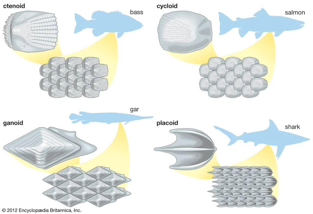
- Cycloid Scales: These are smooth and rounded, typically found in many teleost fish, contributing to a streamlined body shape.
- Ctenoid Scales: These have tiny spines or projections along their edges, providing additional texture and protection.
- Ganoid Scales: Present in some primitive fish like sturgeons, these scales are thick and bony, offering substantial protection.
- Placoid Scales: Found in cartilaginous fish such as sharks and rays, these scales resemble tiny teeth and contribute to hydrodynamics and protection.
- There are various types of scales found in fish, including:
- Protection:
- The primary function of fish scales is to provide a protective barrier against environmental hazards. They shield the underlying tissues from physical abrasions, pathogens, and parasites.
- The scales also minimize water resistance as the fish swims, aiding in more efficient movement through aquatic environments.
- Adaptations in Scale Presence:
- While many fish are covered in scales, some species, such as freshwater catfish, are described as “naked,” lacking scales altogether. This adaptation may be beneficial for specific environmental conditions or behaviors.
- Certain fish species exhibit an intermediate condition, being generally naked but possessing scales in restricted areas. For example, the paddlefish (Polyodon) has scales localized to its throat, pectoral region, and the base of its tail, demonstrating an evolutionary variation in scale distribution.
- Modification of Scales:
- In various fish species, scales can be modified into other structures:
- Teeth: Some fish have evolved to have scale-like teeth, which serve feeding purposes and enhance predation.
- Bony Armor Plates: In species like seahorses, scales may be transformed into protective bony plates that provide defense against predators.
- Spiny Stings: In stingrays, modified scales form spines that can deliver venomous stings, serving as a defense mechanism against threats.
- In various fish species, scales can be modified into other structures:
- Size and Visibility:
- In certain fish, such as the freshwater eel (Anguilla), the scales are so small and deeply embedded in the skin that the fish appears to be naked. This adaptation might assist in camouflage or aid in specific swimming behaviors.
- Hydrodynamic Function:
- Scales play a critical role in enhancing the hydrodynamic properties of fish. Their arrangement and structure help reduce drag in water, allowing for more efficient movement and agility in swimming.
- Thermoregulation and Osmoregulation:
- Scales can assist in thermoregulation by providing a barrier that helps maintain body temperature. Additionally, they contribute to osmoregulation, helping fish maintain the balance of water and electrolytes in their bodies, especially in varying salinity levels.
Type of scales in fishes
Fish scales are diverse structures that serve vital functions in the protection and adaptation of fish species. The classification of fish scales is based on their structural characteristics and shapes, which are often specific to different groups of fish. Understanding the various types of scales provides insights into their biological roles and evolutionary significance.
- Classification of Scales:
- Fish scales can be broadly categorized into two main types based on their origin and structure:
- Placoid Scales: These scales are primarily found in Elasmobranchii, such as sharks and rays. They are considered dermal denticles and have a unique structure.
- Non-Placoid Scales: These develop from the dermis and encompass several subtypes:
- Cosmoid Scales: Present in extinct crossopterygii and some living species like Latimeria.
- Ganoid Scales: Found in fish like gars and sturgeons.
- Cycloid Scales: Common in soft-rayed fishes such as Burbot.
- Ctenoid Scales: Characteristic of spiny-rayed bony fishes, belonging to the order Acanthopterygii.
- Fish scales can be broadly categorized into two main types based on their origin and structure:
- Placoid Scales:
- Structure: Placoid scales consist of two primary components:
- Upper Part: Known as the ectodermal cap or spine, this layer is coated with a hard, transparent substance called vitrodentine, similar to enamel in human teeth. Beneath this is dentine, which surrounds a pulp cavity.
- Lower Part: The basal plate is disc-like and embedded in the dermis, with the spine projecting outward through the epidermis. The basal plate features an aperture that allows blood vessels and nerves to enter the pulp cavity.
- Function: Placoid scales serve not only as a protective layer but can also be modified into various structures, including teeth, spines, and saw-like features in specific species.
- Structure: Placoid scales consist of two primary components:
- Cosmoid Scales:
- Structure: These scales are plate-like and consist of three layers:
- An outer layer of vitreodentine, which is thin and enamel-like.
- A middle layer composed of a non-cellular material called cosmine, characterized by branching tubules.
- An innermost layer made of a vascularized perforated bony substance known as isopedine.
- Growth: Cosmoid scales increase in size from the edges as new layers of isopedine are added.
- Structure: These scales are plate-like and consist of three layers:
- Ganoid Scales:
- Structure: Ganoid scales are thick and heavy, characterized by an outer layer of ganoine (a type of inorganic substance), a middle layer similar to cosmine, and an innermost bony layer of isopedine.
- Arrangement: These scales fit edge-to-edge, covering the body, and can be modified into large bony scutes in species like Acipenser.
- Growth: They also grow at the edges, continuously adding new layers of isopedine.
- Bony Ridge Scales:
- Characteristics: Found in bony fishes (Osteichthyes), these scales are semitransparent and thin due to the absence of dense enamel and dentine.
- Structure: The surface of these scales features alternating bony ridges and grooves arranged in concentric rings. The inner layer consists of fibrous connective tissue.
- Development: Bony ridge scales originate from accumulations of dermal cells, with the formation of a focus that guides their growth. As they develop, the ridges or circuli emerge at the scale’s surface.
- Cycloid Scales:
- Structure: Cycloid scales are thin and rounded, typically lacking teeth on their edges, and are characterized by concentric growth rings.
- Function: These scales contribute to the hydrodynamic efficiency of the fish by reducing drag and improving swimming performance. They often overlap slightly, providing effective coverage.
- Ctenoid Scales:
- Characteristics: Ctenoid scales have small, tooth-like projections along their posterior edges, enhancing their protective capabilities.
- Arrangement: They overlap in a manner where the posterior edge of one scale covers the anterior edge of the next, creating a flexible armor.
- Function: The unique structure of ctenoid scales aids in minimizing water resistance while allowing for greater mobility.

Uses of scales
Fish scales serve multiple purposes beyond their primary function of providing protection and supporting the skin. Their structural and biological characteristics render them valuable in various scientific and ecological contexts. Understanding the uses of fish scales contributes to the study of ichthyology, ecology, and conservation efforts.
- Identification and Classification:
- Scales are instrumental in the identification and classification of different fish species. Their unique shapes, sizes, and structures allow researchers to differentiate between species and understand evolutionary relationships. For example, the presence or absence of specific types of scales can indicate the evolutionary lineage of a fish.
- Age Determination and Growth Assessment:
- Scales can be used to calculate the age of fish, providing insights into their growth rates. This process involves measuring the spacing in the annual rings of the scales, akin to counting tree rings. Each ring represents a year of growth, allowing scientists to estimate the age and assess growth patterns over time.
- Spawning History:
- In certain species, such as the Atlantic salmon, scales exhibit distinct spawning marks. These marks indicate the number of spawning events and the timing of the first spawning. By analyzing these marks, researchers can gather data on reproductive cycles and population dynamics, which are crucial for effective fishery management.
- Paleoichthyology:
- Scales provide significant information about extinct fish species. Fossilized scales can help scientists reconstruct past environments and ecological conditions, offering insights into the evolutionary history of fish. This knowledge aids in understanding how ancient species adapted to their habitats and how they relate to modern fish.
- Ecological Insights:
- Scales can also inform researchers about the dietary habits of piscivorous animals, including predators that consume fish. By studying the scales found in the stomach contents of these predators, scientists can infer the feeding habits and preferences of both the predator and prey, contributing to the understanding of food webs and ecosystem dynamics.
Modifications of scales
Fish scales exhibit remarkable adaptability, evolving into various modifications that enhance survival in specific ecological niches. These modifications serve diverse functions, from protection to predation, demonstrating the evolutionary significance of scales beyond their traditional role.
- Transformation into Teeth:
- In species such as sharks, placoid scales are adapted into teeth, providing efficient means for capturing prey. This modification enhances the predator’s ability to grasp and consume various marine organisms. A notable example is the sawfish (Pristis), where the teeth resemble modified scales, contributing to their distinctive saw-like appearance used for hunting.
- Scute-like Armor Plates:
- The sturgeon fish displays an adaptation where rows of ganoid scales are enlarged into scute-like armor plates. This modification not only provides robust protection against predators but also offers a streamlined body shape that aids in navigating through various aquatic environments.
- Cutting Blades in Surgeonfish:
- The surgeonfish (Acanthurus) features sharp, cutting blades formed from modified scales at the base of the tail. These structures serve dual purposes: they provide defense against predators and can be utilized in combat with other fish, contributing to social hierarchies within their habitats.
- Enlarged Spines in Pufferfish:
- In species such as the pufferfish (Teratodon) and the porcupinefish (Diodon), scales undergo significant enlargement and modification into spines. This adaptation serves as an effective defense mechanism, making these fish less palatable to potential predators and providing a formidable barrier against attacks.
- Bony Rings in Seahorses and Pipefish:
- Seahorses and pipefish have developed protective bony rings surrounding their bodies, formed from modified scales. This adaptation not only provides physical protection but also contributes to their unique morphology, enabling them to blend into their environments effectively.
- Rigid Protective Structures in Cofferfish:
- The cofferfish (Ostracion) is encased in a rigid protective box formed by articulating bony plates, which are also derived from scales. This structural adaptation offers substantial protection from predators while allowing the fish to maintain its distinctive, compressed body shape.
Pigmentation & colouration in fishes
Pigmentation and coloration in fish are complex phenomena resulting from the presence and arrangement of various pigments within specialized cells known as chromatophores and iridocytes. These features not only contribute to the aesthetic diversity of fish species but also play crucial roles in their survival, communication, and environmental adaptation.
- Pigment Sources and Functions:
- The coloration in fish arises from several types of pigments located in the integument, including chromatophores and iridocytes. Chromatophores are embedded in the dermis and come in several types: erythrophores (red/orange), xanthophores (yellow), and melanophores (black). They contain various pigments such as carotenoids (yellow and red), melanin (black), purines (white or silvery), and flavins (yellow).
- The distribution of these chromatophores varies by species and contributes to the diverse color patterns observed in fish. For example, brightly colored species like Carassius (goldfish) and Colisa exhibit vivid patterns that enhance their visibility in aquatic environments.
- Iridocytes and Reflective Properties:
- Iridocytes, also referred to as “mirror cells,” possess significant reflective capabilities due to the presence of guanine crystals. These cells contribute to the silvery or iridescent appearance of many fish, particularly on the ventral side, where they help in camouflage against predators from below.
- Color Change Mechanisms:
- Fish can alter their coloration in response to environmental changes, primarily through two mechanisms: temporary and semipermanent changes.
- Temporary Change: This occurs through the rearrangement of pigment granules within chromatophores, enabling rapid adaptation to shifting surroundings.
- Semipermanent Change: This involves a gradual increase or decrease in the number of chromatophores, influenced by factors such as habitat, species, age, sex, and overall health. For example, a fish transitioning to a darker environment may slowly adjust its coloration to blend in more effectively.
- Neural and Hormonal Control of Pigmentation:
- The control of pigment movement within chromatophores involves both neural and hormonal mechanisms. Nerves innervate chromatophores in various fish species, where chemical messengers called neurohumors facilitate pigment dispersion or concentration. Two types of nerve fibers are involved: one promotes dispersion, while the other induces concentration.
- For instance, in Phoxinus phoxinus, stimulation of nerves leads to melanin aggregation, demonstrating the rapid response capability of neural control.
- Hormonal Regulation:
- The pituitary gland significantly influences pigment granule migration through the secretion of melanophore-stimulating hormone (MSH). When the pituitary gland is removed (hypophysectomy), fish typically exhibit lighter coloration due to decreased melanin concentration. Conversely, injection of pituitary extracts can temporarily restore darker coloration.
- Other hormones, such as adrenaline and thyroxine, can also affect pigmentation, with studies showing that treatments with adrenaline promote pigment concentration in species like the eel (Anguilla anguilla).
- Classification by Neural Control:
- Fish can be categorized based on the degree of neural control over their melanophores:
- Aneuronic: Melanophores lack nerve innervation (e.g., skates, dogfish).
- Mononeuronic: Chromatophores are innervated by a single nerve fiber that primarily promotes dispersion (e.g., Mustelus squalus).
- Dineuronic: Chromatophores are connected to both dispersing and concentrating fibers, enabling more complex color changes (e.g., teleosts).
- Fish can be categorized based on the degree of neural control over their melanophores:

Significance of pigmentation & colouration in fishes
Pigmentation and coloration in fishes are essential adaptations that serve multiple functions critical to their survival and ecological interactions. These adaptations enable fishes to thrive in diverse environments, providing mechanisms for protection, communication, and prey capture.
- Protection through Camouflage:
- Many fish species utilize pigmentation to blend seamlessly with their surroundings, facilitating camouflage. This ability to change color allows them to conceal themselves from predators and ambush prey effectively.
- Various forms of camouflage exist, including:
- Protective Resemblance: Certain species mimic inanimate objects or living organisms in their environment. For example, the juvenile Platax orbicularis resembles a leaf, making it less conspicuous to predators.
- Disruptive Coloration: This strategy involves patterns that break up the outline of a fish, making it harder to detect against the backdrop of the aquatic habitat.
- Countershading: Fish are darker on their dorsal side and lighter on their ventral side, which helps to reduce visibility when viewed from above or below.
- Mirror-Siding: Some fish possess reflective scales that mimic the surrounding water surface, further enhancing their invisibility.
- Transparency: Certain species, like some juvenile fishes, exhibit transparency, allowing them to evade detection.
- Behavioral Adaptations for Concealment and Disguise:
- The ability to adapt coloration in response to environmental cues is crucial for predator evasion. For instance, the Hippocampus bargibanti (seahorse) takes on the coloration of the coral it attaches to, thereby enhancing its concealment while simultaneously providing a means to ambush unsuspecting prey.
- Communication and Social Interaction:
- Beyond protection, coloration plays a pivotal role in communication among fishes. Brightly colored individuals can attract mates or signal territorial claims to other fish. The vivid colors may indicate health, genetic fitness, or readiness to breed, thus facilitating reproductive success.
- Advertising Presence:
- Pigmentation can also serve as a means of advertisement, helping fish identify one another. This visual recognition is vital in maintaining social structures within schools or populations. For instance, some species use specific color patterns to recognize individuals, fostering group cohesion and cooperative behaviors.
- Influence on Ecosystem Dynamics:
- The significance of pigmentation and coloration extends to ecological interactions beyond individual species. For example, the presence of brightly colored reef fish can indicate healthy coral ecosystems, while changes in coloration patterns may reflect environmental stress or habitat degradation.
- Handbook of Fish Biology and Fisheries Volume 1 Fish Biology Edited by Paul J.B. Hart
Department of Biology University of Leicester and John D. Reynolds School of Biological
Sciences University of East Anglia, Wiley Online Library. - A Textbook of Fish Biology and Fisheries by S.S. Khanna and H.R. Singh Published by
Narendera Publishing House. - Fish pigmentation and coloration: Molecular mechanisms and aquaculture perspectives:
Mingkun Luo, Guoqing Lu, Haoran Yin, Lanmei Wang, Malambugi Atuganile and Zaijie Dong:
Reviews in Aquaculture, 2021:1-18. - Fish & Fisheries Digest Part-2: The Integument & Colouration by S.K.Gupta
- Text Highlighting: Select any text in the post content to highlight it
- Text Annotation: Select text and add comments with annotations
- Comment Management: Edit or delete your own comments
- Highlight Management: Remove your own highlights
How to use: Simply select any text in the post content above, and you'll see annotation options. Login here or create an account to get started.