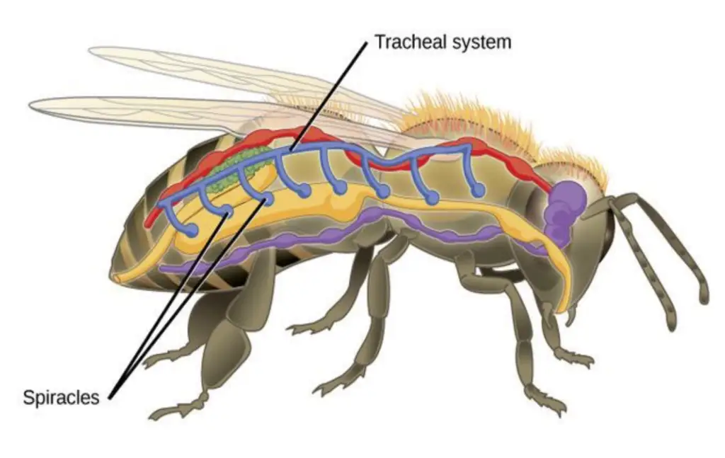
The respiratory system of insects is a sophisticated network designed for efficient gaseous exchange. This system relies on a series of internal tubes known as tracheae, which permeate the insect’s body, extending to all tissues, including muscle fibers. Unlike in vertebrates, where oxygen is transported via blood, the tracheal system allows oxygen to reach its utilization sites directly, thereby eliminating the need for blood in gas transport.
Tracheae open to the external environment through openings called spiracles. These spiracles are typically equipped with mechanisms that minimize water loss, a critical adaptation given the high surface area exposed to the environment. Spiracles can open or close in response to changes in internal oxygen levels or the accumulation of carbon dioxide in the tissues. Specifically, they tend to open when there is a low concentration of oxygen or a high concentration of carbon dioxide, ensuring that gas exchange remains efficient.
While diffusion suffices for meeting the oxygen demands of many smaller and less active insects, larger and more active species necessitate a more robust system. In these cases, the tracheal system becomes essential, as it allows for rapid movement of air throughout the body. This enhanced capability is crucial during periods of increased metabolic activity, such as flight or intense locomotion.
In summary, the insect respiratory system exemplifies a highly adapted means of gaseous exchange. Through the intricate design of tracheae and spiracles, insects can efficiently manage their respiratory needs while minimizing water loss, showcasing the evolutionary success of this group in diverse environments.

The tracheal system
The tracheal system of insects is a highly specialized and efficient network designed for respiratory function. This system facilitates the exchange of gases, supplying oxygen directly to tissues while expelling carbon dioxide. Its intricate structure comprises tracheae, tracheoles, and air-sacs, each serving specific functions essential for maintaining the insect’s metabolic needs.
- Tracheae: These are the larger tubes that begin at the spiracles and extend inward. The tracheae typically branch into finer tubes, with the smallest being approximately 2 microns in diameter. Originating from ectodermal tissue, tracheae possess a cuticular lining that is shed during molting. The inner surface features a spiral thickening known as taenidia, which prevents the tracheae from collapsing under low internal pressure. The structure of the tracheae consists of an intima made of cuticulin, lined with a protein and chitin layer externally.
- Air-sacs: In certain areas, tracheae expand to form air-sacs, which have thin walls and lack well-developed taenidia. These air-sacs can collapse under pressure but play a crucial role in ventilating the tracheal system. They contribute to respiration by allowing for a more effective exchange of gases, especially during periods of high metabolic demand.
- Tracheoles: These are the smaller, finer branches of the tracheal system that extend from the tracheae. Tracheoles typically measure about 1 micron in diameter at their proximal end and taper down to approximately 0.1 microns distally. They are formed from epidermal cells known as tracheoblasts and retain their cuticular lining during molting. Tracheoles closely associate with various tissues, particularly muscle fibers, where they may indent the plasma membrane but generally do not penetrate deeply into the cells. They either end blindly or may form connections with other tracheoles.
- Distribution of the Tracheal System: The tracheal system originates at the spiracles, which serve as external openings. In many Apterygota, tracheae from each spiracle may form unconnected tufts. However, in most insects, neighboring tracheae anastomose to form longitudinal trunks that run along the body. Typically, there are lateral trunks on either side, with dorsal and ventral trunks present as well. These longitudinal tracheae are interconnected by transverse commissures, allowing for efficient gas distribution to various body segments.
- Specific Supply to Organs: The arrangement of the tracheal system varies across insect species. Generally, the heart and dorsal muscles receive branches from the dorsal trunks, while the alimentary canal, gonads, legs, and wings are supplied by the lateral trunks. The ventral trunks or transverse commissures supply the central nervous system. The head receives oxygen through spiracle 1 via two main tracheal branches on each side, which provide a dorsal branch to the antennae, eyes, and brain, as well as a ventral branch to the mouthparts and associated muscles. In some species, constrictions in connecting tubes help isolate the head’s tracheal system, ensuring a sufficient oxygen supply to the brain and sensory organs. Similar isolation occurs in the pterothorax of certain insects.
Spiracles
Spiracles are vital external openings that serve as the entry points for air into the insect tracheal system. Located laterally, usually on the pleura, these structures play a crucial role in facilitating gaseous exchange while minimizing water loss. The configuration and functionality of spiracles vary among different insect groups, reflecting their adaptation to diverse environments.
- Structure of Spiracles: In their simplest form, spiracles function as direct openings leading to the tracheae. However, in most insects, the spiracle opens into a cavity known as the atrium, from which the tracheae branch off. Together, the opening and atrium comprise what is referred to as the spiracle. The atrial walls often contain hair-like structures that filter out dust particles, while some insects have a sieve plate with small pores for additional filtration.
- Number and Distribution: Insects typically possess a maximum of ten pairs of spiracles, comprising two thoracic and eight abdominal spiracles. Based on the number and distribution of functional spiracles, respiratory systems can be classified as follows:
- Polypneustic: Characterized by at least eight functional spiracles on each side.
- Holopneustic: Ten spiracles, such as in bibionid larvae (one mesothoracic, one metathoracic, and eight abdominal).
- Peripneustic: Nine spiracles (one mesothoracic and eight abdominal), as seen in cecidomyid larvae.
- Hemipneustic: Eight spiracles (one mesothoracic and seven abdominal), exemplified by mycetophilid larvae.
- Oligopneustic: Characterized by one or two functional spiracles on each side.
- Amphipneustic: Two spiracles (one mesothoracic and one post-abdominal), as in psychodid larvae.
- Metapneustic: One spiracle (post-abdominal), as observed in culicid larvae.
- Propneustic: One spiracle (mesothoracic), as found in dipterous pupae.
- Apneustic: Refers to the absence of functional spiracles, as seen in chironomid larvae. It is important to note that apneustic insects still possess a tracheal system; however, it does not connect to the external environment.
- Polypneustic: Characterized by at least eight functional spiracles on each side.
- Closing Mechanism: Many terrestrial insects have developed sophisticated closing mechanisms for their spiracles, which are crucial for conserving water. These mechanisms may involve one or two movable valves located at the spiracular opening, or they may function internally to constrict the atrium, thereby restricting airflow to the tracheae. For instance, in grasshoppers, spiracle 2 is equipped with two semicircular valves that close through muscular action. The valves, unsclerotized except at the hinge, are thickened at the base to accommodate muscle insertion. This muscle facilitates closure by pulling down the valves, while the surrounding cuticle’s elasticity usually allows the spiracle to open.
- Control of Opening and Closing: Spiracles are generally kept open only for the briefest time necessary for effective respiration, minimizing water loss from the tracheal system. The closure of spiracles results from the sustained contraction of closer muscles, whereas opening occurs through the elastic recoil of the surrounding cuticle when these muscles relax. Control of this process is governed by the central nervous system, which may also respond to local chemical stimuli.
Classification of tracheal system
The classification of the tracheal system in insects is based on the number and arrangement of functional spiracles, which are crucial for gaseous exchange. Most insects typically possess a maximum of ten pairs of spiracles. However, variations exist in the functionality and arrangement of these spiracles among different species. The following classification outlines the primary categories based on the number and functionality of spiracles:
- Holopneustic: This is a primitive type characterized by the presence of two pairs of thoracic spiracles and eight pairs of abdominal spiracles, all of which are functional. The arrangement is as follows:
- Structure: 1 (mesothoracic) + 1 (metathoracic) + 8 (abdominal).
- Examples: Commonly observed in insects such as dragonflies, grasshoppers, and cockroaches.
- Hemipneustic: In this classification, one or more pairs of spiracles are non-functional. It can be further divided into the following subcategories:
- Peripneustic: The metathoracic spiracle is closed, leading to the structure of 1 + 0 + 8.
- Examples: Larvae of Lepidoptera, Hymenoptera, and Coleoptera.
- Amphipneustic: Only the mesothoracic and the last pair of abdominal spiracles are functional, structured as 1 + 0 + 1.
- Examples: Larvae of cyclorrhaphan Diptera.
- Propneustic: This type has only the mesothoracic spiracles open, resulting in a structure of 1 + 0 + 0.
- Examples: Mosquito pupae.
- Metapneustic: In this case, only the last pair of abdominal spiracles remains functional, leading to a structure of 0 + 0 + 1.
- Examples: Mosquito larvae.
- Apneustic: This classification denotes the absence of functional spiracles altogether.
- Examples: Found in organisms like mayfly larvae and the nymphs of Odonata.
- Peripneustic: The metathoracic spiracle is closed, leading to the structure of 1 + 0 + 8.
- Hypopneustic: Insects classified as hypopneustic may have one or two pairs of spiracles completely absent or non-functional.
- Examples: Observed in some species of Siphunculata and Mallophaga.
- Hyperpneustic: This classification is characterized by the presence of more than ten pairs of spiracles.
- Examples: Notable in the species of Japyx (dipluran).
Respiration in aquatic insects
Aquatic insects have adapted to their environments with various specialized methods to obtain oxygen, either from the air or directly from water. Their respiratory adaptations are diverse, enabling them to thrive in habitats ranging from stagnant ponds to fast-flowing streams. Below is an explanation of how aquatic insects manage respiration, highlighting the key modifications they utilize.
- Tracheal Gills:
- Found in the immature stages of insects like stoneflies, mayflies, and some Odonata (dragonflies and damselflies).
- These gills consist of evaginated trachea or tracheoles that extend outward to absorb dissolved oxygen from water.
- The tracheal gills are typically located on the sides of the abdomen or thorax, allowing efficient gas exchange.
- Rectal Gills:
- Specifically found in the nymphs of Odonata.
- The rectum contains specialized gills with tracheoles. Water is taken into the rectal chamber, where oxygen is extracted, and the water is then expelled.
- This process not only allows for respiration but also enables the insect to propel itself through water by forcefully ejecting water from the rectum.
- Spiracular Gills:
- Found in aquatic Diptera, especially in those inhabiting streams that may dry up periodically.
- These gills can function in both water and air. When submerged, they act as gills; when the water evaporates, part of the gill breaks off, allowing air to enter the spiracle for aerial respiration.
- Respiratory Tubes:
- Seen in insects like Nepidae (water scorpions).
- These insects possess siphon-like respiratory tubes, which are extended to the water’s surface, allowing them to breathe air while submerged.
- Post-Abdominal Siphon:
- Found in mosquito larvae.
- This sharp, posterior siphon pierces plant tissues to access oxygen trapped within the plant’s internal air spaces, or it can be extended to the water’s surface to draw in air.
- Air Bubble Respiration:
- Common in aquatic Hemiptera and Coleoptera.
- These insects trap a bubble of air at the water’s surface and carry it below as a portable air supply.
- Oxygen from the surrounding water can diffuse into the air bubble, providing the insect with a sustained source of breathable oxygen while submerged.
- Plastron Respiration:
- A specialized adaptation seen in some aquatic insects.
- A plastron consists of a thin layer of air held by hydrofuge (water-repelling) hairs on the insect’s body.
- This bubble does not collapse and can last for extended periods—up to four months—allowing the insect to remain underwater for prolonged durations without needing to resurface for air.
Other types of respiration
Insects, while primarily relying on the tracheal system for respiration, exhibit various specialized methods of respiration depending on their environment. These alternative forms of respiration are adaptations that allow different species to thrive in unique conditions. Here is a breakdown of these other types of respiration found in insects:
- Cutaneous Respiration:
- Insects like Protura, Collembola, and endoparasites utilize respiration through their body wall.
- Oxygen diffuses directly through the skin without the use of a tracheal system.
- This method is common in smaller, soft-bodied insects or those living in moist environments.
- Tracheal Gills / Abdominal Gills:
- Found in larvae of Trichoptera and nymphs of Ephemeroptera.
- These are outgrowths of the trachea that form gills, located on the lateral sides of the body.
- The gills absorb dissolved oxygen from water, and their shapes can vary, appearing lamellate (plate-like) or filamentous.
- Spiracular Gills:
- Seen in aquatic pupae.
- The spiracle or its atrium extends into a long filament.
- This adaptation allows for both aquatic and aerial respiration, enabling insects to live in air, moist environments, or fully submerged in water.
- Blood Gills:
- These are tubular or digitiform (finger-like) structures found at the anal end in larvae of Trichoptera.
- In Chironomid larvae, two pairs of these gills are present on the penultimate segment along with four shorter anal gills.
- These structures are called blood gills as they contain blood, although they may sometimes house trachea.
- Their primary function is the absorption of water and inorganic ions rather than respiration.
- Rectal Gills:
- Found in dragonfly nymphs (naids).
- The rectum is modified into a barrel-like chamber, with the rectal wall forming basal thick pads and distal gill filaments richly supplied with tracheoles.
- This allows the nymphs to extract oxygen directly from water through the rectum.
- Air Sacs:
- Present in winged insects, these are dilated parts of the trachea that form thin-walled air sacs.
- Unlike the rest of the tracheal system, air sacs do not contain taenidia (the spiral thickening that supports tracheal tubes).
- They appear as glistening sac-like structures and function as storage reservoirs for air.
- These sacs also help in reducing body weight and regulating internal pressure during respiration.
- Plastron Respiration:
- Common in aquatic beetles, plastron respiration involves a special type of air store in the form of a thin film, held in place by hydrofuge (water-repelling) hairs or scales.
- The air volume remains constant and can act as a physical gill.
- If the surrounding water contains enough dissolved oxygen, this plastron can directly facilitate gas exchange with the environment.
Functions of Insect Respiratory System
Below is an overview of the major functions of the insect respiratory system:
- Oxygen Supply:
- The primary function of the respiratory system is to supply oxygen to all tissues and cells.
- The tracheal system delivers oxygen directly to cells through a network of branching tubes that extend throughout the body, ensuring an efficient and rapid oxygen delivery.
- Carbon Dioxide Removal:
- Just as oxygen must be delivered, carbon dioxide—a metabolic waste product—must be removed.
- The tracheal system helps expel carbon dioxide from tissues through the spiracles, preventing the build-up of toxic gases.
- Minimization of Water Loss:
- The spiracles, small openings on the insect’s body, can be closed when not in use to minimize water loss, which is crucial for insects living in arid environments.
- The closing mechanism of the spiracles helps insects conserve moisture while still facilitating respiration.
- Air Storage and Regulation:
- Some insects, particularly those that fly, have specialized structures called air sacs that store air and assist with respiration.
- These air sacs can expand and contract with body movements, helping to regulate the volume of air in the tracheal system, especially during active periods like flight.
- Adaptation to Aquatic Environments:
- In aquatic insects, the respiratory system has special adaptations such as tracheal gills, spiracular gills, and siphons that allow them to extract oxygen from water or air.
- These modifications enable insects to thrive in various aquatic habitats, ensuring they can maintain adequate oxygen levels even when submerged.
- Support for High Metabolic Activity:
- Many insects, especially flying species like bees and butterflies, have high metabolic rates that require large amounts of oxygen.
- The tracheal system efficiently meets these demands by rapidly delivering oxygen to muscles and tissues, supporting sustained activity.
- Regulation of Gas Exchange:
- The opening and closing of spiracles are controlled by the insect’s central nervous system, which responds to internal oxygen and carbon dioxide levels.
- This regulation ensures that spiracles remain open for the shortest time possible, reducing exposure to environmental threats like dehydration or harmful gases.
- Facilitating Flight:
- The tracheal system works in coordination with the insect’s muscular system to enhance respiration during flight.
- During flight, movements of the thorax help pump air through the tracheal tubes, ensuring a continuous supply of oxygen to flight muscles.
- Adaptation to Low Oxygen Environments:
- Insects that live in environments with low oxygen availability, such as in soil or decaying matter, have specialized adaptations like spiracular control or air-bubble trapping to extract oxygen from their surroundings.
- http://courseware.cutm.ac.in/wp-content/uploads/2020/06/Insect-Respiratory-System.pdf
- https://www.ndsu.edu/pubweb/~rider/Pentatomoidea/Teaching%20Structure/Lecture%20Notes/Week%2012a%20Respiratory%20System.pdf
- https://www.ncbi.nlm.nih.gov/pmc/articles/PMC1570919/
- http://www.rnlkwc.ac.in/pdf/study-material/zoology/Respiratory.pdf
- https://faculty.ksu.edu.sa/sites/default/files/respiration_in_insect_1.pdf