Human teeth play a crucial role in the process of digestion, enabling the breakdown of food into smaller, manageable pieces before swallowing. Unlike some animals that can consume food in one large gulp, humans rely on their teeth to perform various functions essential for effective mastication. This article provides an overview of the structure, types, and developmental stages of human teeth, emphasizing their significance in both digestion and speech.
Teeth are composed of a hard, calcified substance primarily made up of proteins, such as collagen, and minerals, particularly calcium. This unique composition makes teeth one of the strongest structures in the human body. The different types of teeth, each adapted for specific functions, include incisors, canines, premolars, and molars. These teeth are activated by powerful jaw muscles, while saliva produced by the salivary glands facilitates lubrication, aiding in the swallowing process.
The arrangement and quantity of teeth in vertebrates are categorized using a dental formula, expressed as fractions that detail the number and types of teeth present. For adults, this formula indicates a total of 32 permanent teeth, which include eight incisors, four canines, eight premolars, and twelve molars—this total encompasses the four wisdom teeth. The incisors are primarily responsible for cutting food, canines for tearing, premolars for shearing, and molars for grinding and crushing.
Infants are born without teeth, relying exclusively on milk for nutrition. As they grow, primary teeth, also known as milk teeth, begin to emerge, typically around six months of age. This set consists of 20 teeth: eight incisors, four canines, and eight molars, with the lower incisors being the first to erupt. Most children will have all their primary teeth by the age of three. The transition from primary to permanent teeth generally begins around the age of six and continues until the individual reaches about 24 to 25 years of age, at which point the full set of permanent teeth is established.
Human teeth exhibit three notable characteristics: they are heterodont, meaning they come in various shapes and types; thecodont, which indicates they are securely embedded in the jawbone’s socket; and diphyodont, signifying that humans develop only two sets of teeth in their lifetime. The first set, the deciduous teeth, is gradually replaced by permanent teeth in a well-defined sequence.
The development and eruption of teeth follow a consistent pattern. As the child ages, the shedding of primary teeth and the emergence of permanent teeth occur systematically. Wisdom teeth, or third molars, typically appear between the ages of 17 and 21, completing the transition to the adult dental structure.
Primary and Permanent Dentition
The human dentition is categorized into two main types: primary dentition and permanent dentition. Understanding the structure and timing of these two stages is crucial for grasping the developmental phases of human teeth.

- Primary Dentition: This set consists of 20 teeth, with 10 located in each dental arch.
- Each quadrant contains five teeth: two incisors (one central and one lateral), one canine, and two molars.
- These teeth are designated by letters A through E, with the central incisor being A and the second molar being E.
- The eruption of primary teeth typically begins around six months of age, providing infants with the ability to chew and aid in speech development.
- Primary teeth play a significant role in guiding the growth of the jaw and the alignment of the permanent teeth that will follow.
- Permanent Dentition: The permanent set comprises 32 teeth, with 16 in each arch.
- In each quadrant, there are eight teeth: two incisors (central and lateral), one canine, two premolars, and three molars.
- These teeth are numbered from 1 to 8, starting with the central incisor as tooth 1 and ending with the third molar (wisdom tooth) as tooth 8.
- The transition from primary to permanent teeth generally begins at around six years of age, as the permanent teeth start to erupt and replace the primary ones.
- By the age of approximately 13 years, most permanent teeth have erupted; however, the third molars typically emerge later, usually between the ages of 17 and 21.

Structure of Human Teeth
Human teeth exhibit a complex structure that is essential for their functions in mastication and speech. Understanding this anatomy is crucial for students and educators alike, as it lays the foundation for dental health and oral biology.

- Main Parts of a Tooth:
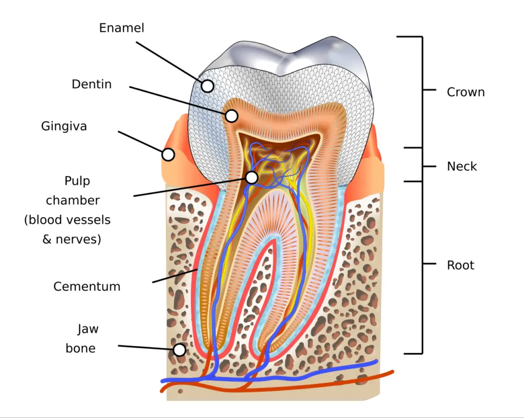
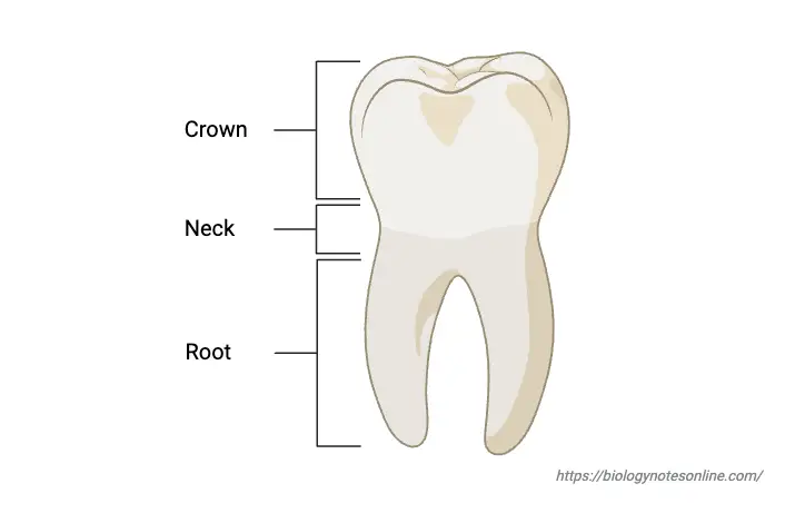
- A tooth consists of three primary sections: the crown, the root, and the neck (or cervix).
- The crown is the uppermost part, visible above the gum line, and is responsible for the initial engagement with food during chewing.
- The root is the lower portion, anchored securely within the jawbone’s socket (alveolus), playing a vital role in stabilizing the tooth.
- The neck serves as the transitional area between the crown and the root, surrounded by gum tissue.
- A tooth consists of three primary sections: the crown, the root, and the neck (or cervix).
- Composition of the Tooth:
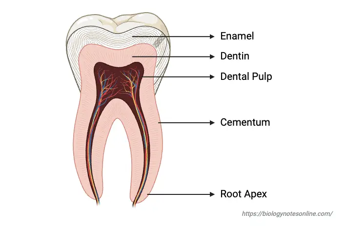
- Teeth are primarily composed of four main components: enamel, dentine, pulp cavity, and cementum.
- Enamel: This is the hardest substance in the human body, covering the outer surface of the crown. It is highly mineralized and provides a durable layer that withstands the forces of chewing and protects the underlying tissues from thermal and chemical damage. However, enamel is vulnerable to decay from dental caries and wear over time.
- Dentine: Situated beneath the enamel and cementum, dentine is a hard, living tissue that constitutes the majority of the tooth structure. It contains microscopic tubules that run from the pulp cavity to the outer layers, allowing for the sensation of pressure and pain. This vital tissue also plays a role in supporting the tooth’s integrity.
- Pulp Cavity: The pulp cavity is the central, hollow region of the tooth filled with dental pulp, a connective tissue rich in blood vessels and nerves. This living component is essential for nourishing the tooth and enabling sensory functions. The health of the pulp is critical; infection can lead to severe complications.
- Cementum: This is a specialized bone-like tissue that covers the root of the tooth, anchoring it within the alveolar bone. Cementum facilitates the attachment of the periodontal fibers, which hold the tooth in place. It is softer than enamel and sensitive, becoming exposed during certain dental conditions.
- Teeth are primarily composed of four main components: enamel, dentine, pulp cavity, and cementum.
- Tooth Surfaces and Junctions:
- The boundary where the anatomic crown meets the root is known as the cemento-enamel junction (CEJ). This area is critical for the integrity of the tooth as it delineates the transition from enamel to cementum.
- The dentino-enamel junction (DEJ) is where the dentine and enamel intersect, serving as a significant structural interface within the tooth.
- Roots and Root Variability:
- The number of roots varies among different types of teeth. For example, incisors and canines typically possess a single root, while premolars may have one or occasionally two roots. Molars generally have multiple roots, ranging from two to four, facilitating their function in grinding food.
- Developmental Aspects:
- Teeth develop from both the dermis and epidermis during embryonic growth, resulting in the unique combination of hard tissues that make up the dental structure. This dual origin underscores the complexity of tooth anatomy and its functional adaptation for dietary needs.

Types of Teeth
Teeth are categorized into distinct types, each serving specialized functions during mastication and digestion. Understanding the different types of teeth enhances comprehension of their roles in both oral health and overall well-being.

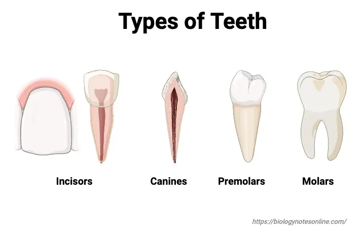
- Incisors: Incisors are located at the front of the mouth and are essential for cutting food.
- Humans have a total of eight incisors, divided into four in the upper jaw (maxillary) and four in the lower jaw (mandibular).
- These teeth feature sharp edges that facilitate the incising of food, making them particularly useful for biting into softer foods.
- The central incisors are positioned nearest the midline, with lateral incisors situated between the central incisors and the canines.
- Due to their exposed position, incisors are vulnerable to trauma, especially in children, where approximately one in ten experience dental injuries that can affect both function and aesthetics.
- Canines: Also known as cuspids, canines are located at the corners of the dental arches.
- Humans possess four canines, with two in the upper jaw and two in the lower jaw.
- These teeth have a sharp, triangular projection on the incisal edge, enabling them to grip and tear food, particularly tougher items like meat.
- Canines have long, stable roots, allowing them to withstand greater forces compared to incisors.
- In some cases, canines may become impacted, particularly in adolescents with dental crowding, necessitating surgical intervention to aid eruption.
- Premolars: Referred to as bicuspids, premolars are situated behind the canines and play a dual role in the chewing process.
- In permanent dentition, humans typically have eight premolars—two on each side of both jaws.
- These teeth have a flat surface with ridges that facilitate the crushing and grinding of food.
- Notably, premolars are absent in primary dentition, which means they develop only after the primary teeth have been shed.
- Dentists may extract premolars to alleviate crowding before orthodontic treatments.
- Molars: Molars are the largest and strongest teeth, crucial for effective grinding and chewing.
- Adults generally have twelve molars—six in the upper jaw and six in the lower jaw, with four of these being wisdom teeth (third molars) that typically erupt between ages 17 and 25.
- The surface of molars is large and flat, designed for grinding food into smaller pieces.
- The number of cusps on molars can vary from three to five, contributing to their ability to handle various food textures.
- Due to their anatomy, molars are particularly susceptible to dental caries, as their deep grooves and wider points of contact with adjacent teeth make them difficult to clean.
Functions of Teeth
Teeth serve multiple essential functions that contribute significantly to both physiological processes and overall facial aesthetics. Understanding these roles is crucial for students and educators, as it highlights the importance of dental health and its impact on daily life.
- Mastication of Food: The primary function of teeth is to facilitate the mechanical breakdown of food. This process, known as mastication, is vital for digestion.
- Incisors: These are the front teeth with sharp edges designed for cutting food into smaller pieces. Their shape enables efficient biting, particularly for softer foods.
- Canines: Positioned next to the incisors, canines have pointed tips that assist in tearing food. Their strength is particularly beneficial for gripping and ripping through tougher substances.
- Premolars: These teeth play a dual role in mastication. They have a flat surface that allows them to tear and crush food, making them integral to the initial stages of grinding before it reaches the molars.
- Molars: Located at the back of the mouth, molars are equipped with a broad, flat surface ideal for grinding and crushing food into finer particles. This function is essential for effective digestion, as it increases the surface area of food, allowing for more efficient enzymatic action during digestion.
- Clear Pronunciation: Beyond their role in digestion, teeth are crucial for speech. They contribute to articulating sounds and forming words. The precise positioning of the tongue against the teeth allows for the correct pronunciation of various phonemes, underscoring the connection between dental health and effective communication.
- Shaping the Face: Teeth also play a significant role in maintaining facial structure. They contribute to the overall shape and contour of the face, influencing one’s appearance. Well-aligned teeth help support the lips and cheeks, providing a balanced and aesthetically pleasing facial profile. Conversely, tooth loss or misalignment can lead to changes in facial structure, impacting self-esteem and social interactions.
- Jágr, Michal & Eckhardt, Adam & Pataridis, Statis & Broukal, Zdenek & Duskova, Jana & Miksík, Ivan. (2014). Proteomics of Human Teeth and Saliva. Physiological research / Academia Scientiarum Bohemoslovaca. 63 Suppl 1. S141-54. 10.33549/physiolres.932702.
- https://moodle.beverleyhigh.net/mod/resource/view.php?id=6106&forceview=1
- https://www.thoughtco.com/human-teeth-and-evolution-1224798
- https://onlinesciencenotes.com/human-teeth-types-dental-formula-structure-composition-and-functions/
- https://byjus.com/biology/types-of-teeth-in-humans/
- https://teachmeanatomy.info/head/other/child-adult-dentition/
- https://en.wikipedia.org/wiki/Human_tooth
- https://www.hovedentalclinic.co.uk/blog/types-of-teeth/