What is cleavage?
- Cleavage can define as the division of zygote or fertilized egg into smaller cells, which are called blastomeres.
- It’s a repeated mitotic division that happens very quickly after fertilization, no growth occurs between divisions.
- During cleavage, the overall size of embryo not increase; instead, the large egg cytoplasm is split into many smaller parts.
- Each blastomere formed contains a portion of cytoplasm and a nucleus – they together form a structure called morula (like a berry shape).
- Pattern of cleavage vary by species; can be holoblastic (complete) or meroblastic (incomplete) depending on amount of yolk present in egg.
- In holoblastic cleavage, whole egg divides totally (example – Amphioxus, Frog).
- In meroblastic cleavage, only part of egg undergo division, mainly the active cytoplasmic region, seen in Birds / Reptiles.
- Rate and plane of cleavage are influenced by factors like yolk quantity, distribution, and genetic control etc.
- After several cleavages, blastula stage is reached where cavity (blastocoel) appear inside mass of cells.
Characteristics of cleavage
- Cleavage is termed as rapid and multiple mitotic division of fertilized egg, which turns zygote into many small cells called blastomeres.
- No increase of embryo size occur, even after many divisions, only number of cells become more and more.
- The daughter cells get smaller each time, while total volume of embryo almost remain same.
- The cell cycle during this period is very short – mostly contain S and M phase, without proper G1/G2 growth phase.
- Whole embryo shape stays maintained during cleavage, though inside so much division happens.
- Number of blastomeres often follow geometric progression like 2, 4, 8, 16, 32 etc., that sequence goes fast.
- Cytoplasm portion divided and shared among blastomeres, so regulatory molecules get distributed in new pattern.
- Ratio of nucleus to cytoplasm continuously increase — that’s quite significant for next developmental step.
- After many divisions, a solid mass called morula form, later it turns into blastula or blastocyst stage.
- Cleavage type depend mainly on yolk amount and how it distributed inside egg; it may be holoblastic (complete) or meroblastic (incomplete).
- In eggs having less yolk, like amphibians, full cleavage seen. But in birds or reptiles, cleavage only partial due to heavy yolk portion.
- Division begin soon after fertilization and it end when nucleus–cytoplasm balance achieved again.
- In most species, except mammals, maternal proteins/mRNAs already stored inside oocyte guide the cleavage direction and rate.
- Early cleavages often synchronous but later they become irregular and asynchronous — typical pattern of developing embryo.
- This entire process, though seem mechanical, actually prepare for next stages – gastrulation and cell differentiation, etc.
Step by step process of Cleavage
- Cleavage is initiated soon after fertilization, and the zygote is driven into mitotic cycles by activation of MPF (Mitosis Promoting Factor).
- The fertilized egg is subdivided, and the first mitotic spindle is formed so that karyokinesis is carried out, producing two nuclei.
- Cytokinesis is accomplished by formation of a contractile ring of actin microfilaments, a cleavage furrow is formed and the cell is pinched into two daughter blastomeres.
- Successive rounds of mitosis continue, so the embryo progresses through 2 → 4 → 8 → 16 cell stages, each division producing more blastomeres while total embryo volume remains essentially constant.
- The cell cycle is simplified to alternate S (DNA synthesis) and M (mitosis) phases only, G₁ and G₂ being omitted, and thus divisions are very rapid.
- With each division the blastomeres become progressively smaller, and the nuclear : cytoplasmic ratio is increased continuously, which will act as a timer for later events.
- Karyokinesis (nuclear division) and cytokinesis (cytoplasmic division) are coordinated each cycle, but they are driven by different machineries — spindle for the former, actin–myosin ring for the latter.
- As cleavages continue, spatial arrangement of cleavage planes (meridional, equatorial, latitudinal etc.) determine the relative positions of blastomeres and embryo symmetry.
- When cell number is high a compact mass called morula is formed (in mammals compaction at ~8-cell stage promotes this), cells are closely appressed to one another.
- A cavity, the blastocoel, is then formed within the morula and the structure is termed blastula (or blastocyst in mammals) with an outer trophoblast-like layer and an inner cell mass in some groups.
- Maternal mRNAs and proteins govern early cleavage, and gradually they are degraded while zygotic transcription is prepared, this transition culminates at the MBT (mid-blastula transition).
- Cleavage is said to end when a balance between nuclear and cytoplasmic material is approached and when zygotic genome activation (MBT) allows more conventional cell cycles (G₁, S, G₂, M) to be resumed.
- After MBT, cell divisions slow and become asynchronous, and morphogenetic processes (like gastrulation) are permitted because cells now have size, polarity and new gene expression programs.
Types of cleavages
The cleavage pattern in embryos mainly determined by yolk quantity and how that yolk is distributed by the cytoplasm.Also, timing and direction of mitotic spindle (inside egg) play important role. Yolk usually slow / restrict the process of complete division.
Broadly it divided into Holoblastic (complete) and Meroblastic (incomplete) type.
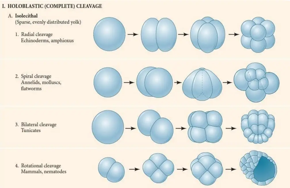
I. Holoblastic (Complete) Cleavage
Occurs when cleavage furrow cut across the whole egg. Found in eggs having very less / moderate yolk like Mammals, Sea urchins, Amphibians etc.
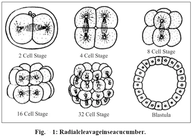
a. Radial Holoblastic Cleavage –
- Typical for deuterostomes like Echinoderms, Hemichordates, Amphioxus.
- Division planes are parallel or at right angle (90°) to polar axis.
- Blastomeres placed one above another forming radial symmetry.
- Usually results in indeterminate type development; each cell can form full embryo if separated.
- In Frog, though radial type, 3rd cleavage is displaced toward animal pole making micromeres and macromeres unequal.
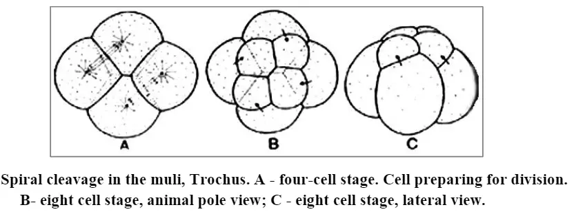
b. Spiral Holoblastic Cleavage –
- Common in Protostomes (Lophotrochozoa) like Annelids, Molluscs, Flatworms.
- Cleavage planes set at oblique angle, not 90°, so cells arranged spirally.
- Upper tier cells lie over junctions of lower ones; rotation alternate clockwise and anticlockwise.
- Gives determinate (mosaic) type development; cell fate fixed early.
- Produce small micromeres and large macromeres sometimes.
c. Rotational Holoblastic Cleavage –
- Seen in Mammals (like Human, Mouse) and Nematodes (C. elegans).
- First division meridional, next one rotated—one divides meridional, other equatorial (90° rotation).
- Division asynchronous; embryos may have 3, 5, 7 cell stages.
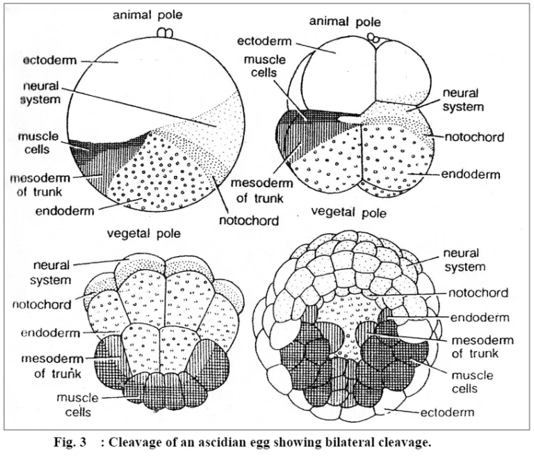
d. Bilateral Holoblastic Cleavage –
- Found in Tunicates and Amphioxus.
- First cleavage split zygote into two mirror halves – right and left.
- All next cleavages follow this bilateral axis.
e. Biradial Cleavage –
- Happens in Ctenophores.
- First three cleavage planes not perfectly perpendicular, give mixed radial-bilateral pattern.
- Usually form 8 blastomeres, 4 large and 4 small ones.
II. Meroblastic (Incomplete) Cleavage
Occurs when yolk amount is high; furrow can’t penetrate through whole egg. Only active cytoplasm divides.
a. Discoidal Meroblastic Cleavage –
- Seen in Birds, Reptiles, Fishes, Monotremes.
- Yolk heavy and concentrated at vegetal pole (telolecithal egg).
- Cleavage confined to a disc-like region at animal pole (called blastodisc).
- Division form a multi-layered blastoderm over the yolk mass.
b. Superficial Meroblastic Cleavage –
- Found in Insects (like Drosophila).
- Yolk in center (centrolecithal).
- First, many nuclear divisions occur without cell wall formation (karyokinesis only).
- Later, nuclei migrate to periphery, where cytoplasmic division (cytokinesis) finally takes place.
The yolk thus decide whether cleavage will be full or partial, fast or slow, equal or unequal. Whatever type it takes, final result always – formation of blastula ready for gastrulation and further embryonic morphogenesis.


Difference between Holoblastic (complete) cleavage and Meroblastic (incomplete) cleavage
| Feature | Holoblastic (Complete) Cleavage | Meroblastic (Incomplete) Cleavage |
|---|---|---|
| 1. Definition / Extent | Cleavage furrow passes through whole egg, dividing it fully into separate blastomeres. | Cleavage furrow limited to active cytoplasmic region only; yolky portion stay undivided. |
| 2. Yolk Concentration | Found in eggs with very little / moderately distributed yolk — isolecithal or mesolecithal type. | Seen in eggs containing large yolk quantity – telolecithal or centrolecithal type. |
| 3. Resulting Blastomeres | All cytoplasm gets partitioned into cells which may be equal or unequal size (depending on yolk). | Only yolk-free cytoplasm divide; gives a thin cellular disc (blastodisc) over yolk mass. |
| 4. Examples | Amphibians (Frog), Mammals, Sea urchins, Annelids, Molluscs, Nematodes. | Birds, Reptiles, Fishes, Insects (like Drosophila). |
| 5. Subtypes / Patterns | Radial, Spiral, Rotational, Bilateral, Biradial types are common. | Discoidal (in birds/reptiles) and Superficial (in insects). |
| 6. Influence of Yolk | Because yolk less or moderate, cleavage furrow can travel entire zygote, even if slow at vegetal pole. | High yolk density block cleavage furrow; so division happen only at cytoplasm layer. |
| 7. Special Note (Balfour’s Law) | Cleavage rate faster when yolk amount less, so holoblastic eggs divide more quickly. | Cleavage rate slower as yolk amount increase; heavy yolk restrict cytoplasmic movement. |
| 8. Final Structure | Produce complete set of small blastomeres → forming morula then blastula. | Produce partial cellular layer (blastoderm) above yolk → leads to blastodisc stage. |
Thus, main deciding factor is yolk – more yolk cause incomplete cleavage, less yolk allow full division. Both however reach same destination – multicellular early embryo, ready for gastrulation and morphogenetic changes.
Laws of cleavage
Balfour’s Law – It state that rate of cleavage become slower when yolk amount is high, since yolk kind of hinder / restrict the protoplasm division. In eggs with more yolk, cleavage occur very slow or irregular (like at the vegetal pole of Rana tigrina). Less yolky parts divide faster, that’s why animal pole shows more quick divisions.
Sachs’ Law (or Sack’s law) – It describe how cleavage planes arranged geometrically. The division usually form equal daughter cells, and the next plane come almost at right angle (90°) to the previous one. Sometimes small deviation seen, but general rule stays like that.
Hertwig’s Law – According to this, the nucleus stays near the center of the active cytoplasm, and the mitotic spindle orient itself along longest axis of protoplasmic mass. Then, the cleavage plane cut that mass nearly perpendicular to this axis. So, the position of nucleus / spindle basically decide where the cell split.
Pflüger’s Law – This one tells that spindle elongates in direction where there is least resistance (less yolk, more soft area). Because of that, spindle find its path through easier cytoplasmic zone, especially in uneven yolk distribution eggs.
These four laws together explain the main pattern how cleavage behaves—about rate, direction, position and geometry—all depend upon yolk content and spindle orientation etc., which give predictable blastomere arrangement.
The planes of cleavage
1. Meridional Plane – This plane of cleavage pass along animal–vegetal axis of the egg, cutting both poles. It divide egg into two equal symmetrical halves. The first cleavage in many forms like Frog, Sea urchin, and mammals is meridional. It’s a normal type of vertical division where plane go through center part of zygote.
2. Vertical Plane – The vertical plane also goes from animal pole to vegetal pole but not always through the center. Because of that, cells form may become unequal in size. In biradial cleavage (like in Ctenophora), this one become third plane. It’s still upright but slightly shifted, giving asymmetrical arrangement sometimes.
3. Equatorial Plane – This plane run at right angle (90°) to main axis, passing through the equator of egg. It cut the zygote horizontally into upper and lower parts. In radial cleavage (ex: Echinoderm eggs), the third cleavage is equatorial. It happen when cytoplasmic furrow moves orthogonal to long axis, creating smaller, even blastomeres.
4. Latitudinal Plane (Transverse / Horizontal) – Almost same like equatorial but occur either above or below the equator, so not exactly middle. In Amphioxus, the third cleavage is latitudinal and occur just above equator, giving rise to small micromeres and big macromeres. Also, in Frog, the same happen—plane slightly above equator, leading unequal size cells.
The orientation of these planes depend on how mitotic spindle align and cytoplasmic forces act. As stated in Hertwig’s law, division plane cut protoplasm at right angle to its long axis. Sachs’ law also tell that each new plane come almost at 90° to previous one, forming regular cleavage geometry.
Sequence of these planes define types of cleavage – In Radial, all planes stay parallel / perpendicular to polar axis. In Spiral, they become oblique (not 90°) forming spiral blastomeres. In Rotational cleavage (mammals, nematodes), first division is meridional, then one blastomere divide meridional and another equatorial (rotated 90°). In Bilateral type (tunicates), first plane divide zygote into right-left halves and next planes stay symmetrical to that axis.
Therefore, orientation and timing of each cleavage plane basically govern how blastomeres positioned, that’s why final embryo structure depend strongly on these specific cleavage directions.
Chemical Changes during Cleavage
- During cleavage, rapid chemical events are observed inside the zygote, and intense DNA replication is driven by maternal reserves.
- DNA synthesis is increased markedly, nuclei are multiplied quickly while cell size is not enlarged, so the cytoplasmic : nuclear ratio is reduced progressively.
- Nucleotides and raw material are supplied from the egg cytoplasm, including mitochondrial DNA and yolk platelets( and this supply is pivotal).
- Early transcription is usually repressed and divisions are carried out by maternal mRNA and proteins that were stored in the oocyte.
- The mid-blastula transition (MBT) is the time when zygotic transcription is activated, in Xenopus laevis this occurs after about 12 divisions, and G1/G2 phases are added then.
- RNA synthesis (mRNA, tRNA) is increased later in cleavage, but early stages are mostly independent of new zygotic transcripts.
- Protein synthesis is up-regulated during cleavage and mitotic cycles are driven by MPF (Mitosis Promoting Factor), which is cyclic in activity.
- Cyclin B accumulates during S phase and is degraded at mitotic exit, this periodic degradation control the timing of successive mitoses.
- The CDK component of MPF phosphorylations are carried out on targets like histones, nuclear lamins, and myosin regulators leading to chromatin condensation and nuclear envelope breakdown.
- Cyclin B mRNA is maternally stored in the cytoplasm, and if its translation is blocked the embryo will not enter mitosis (translation must occur).
- In mammals, large fraction of maternal polyA⁺ RNA and many maternal proteins are degraded early (for example ~75% of maternal polyA⁺ RNA and ~50% proteins are lost by the 2-cell stage in mouse).
- Protein degradation pathways such as ubiquitin–proteasome and macroautophagy are involved in removing maternal components; they act continuously.
- Chromatin remodeling is undertaken after fertilization: paternal protamine-packaged DNA is decondensed and repackaged with maternal histones, and DNA breaks are repaired during this process.
- The paternal genome is actively demethylated while the maternal genome is passively demethylated over cleavages, producing overall hypomethylation except at imprinted loci.
- ATP-dependent chromatin remodelers are recruited to reposition nucleosomes, allowing transcription factors to access promoters when zygotic activation begins.
- Overall, cleavage chemistry is run largely by a preloaded maternal machinery (MPF oscillator, stored mRNAs/proteins), while a timed degradation and remodeling sequence prepares the embryo so it can prevail (prevail = mistakenly used here for “prevent”/protect) to switch to zygotic control at MBT.
Significance of cleavage
- During cleavage, the single zygote is divided into many smaller nucleated cells called blastomeres, which together form the early embryo body.
- It works as a cell multiplication process without increase in the total size / volume of embryo, so the organism remain same size though cell number rise rapidly.
- The process supply basic cellular units, needed later for formation of tissues and organs, basically the raw material for organogenesis.
- Cytoplasm of zygote is partitioned among cells, that distribution helps in spreading regulatory molecules (cytoplasmic determinants) so that every blastomere receive different sets controlling gene expression & fate.
- Cleavage leads to morula stage (a compact cell ball) and then to blastula (in mammals – blastocyst), forming the first real embryonic structure.
- Inside blastula a cavity called blastocoel is formed, which act as fluid space where presumptive organ areas can move during gastrulation, allowing correct body plan development.
- The process prepare the embryo for mid-blastula transition (MBT), when cleavage speed reduces and zygotic genome starts transcription — at this point control shift from maternal to embryonic.
- As division continue, the nuclear : cytoplasmic ratio increase continuously, that ratio believed to help timing activation of many new embryonic genes.
- It sets up the platform for gastrulation and organogenesis, ensuring cell number, polarity, and orientation are properly arranged before morphogenetic movements begin.
- In deuterostomes (like Mammals, Echinoderms), cleavage is regulative, meaning separated early blastomeres can still develop into full individuals – showing their totipotency, which also explain identical twinning.
- In ART (Assisted Reproductive Technology), morphology of cleavage-stage embryos (about Day–3 post fertilization) is used to judge embryo quality and implantation potential; such evaluation is key for embryo transfer success.
- Thus, the main significance of cleavage lies in transforming a single cell into structured multicell system, organizing cytoplasmic content, establishing developmental cues, and setting the embryo’s self-governing genetic stage — it’s small but mighty start of life (literally).
Examples of Cleavage in Different Chordates
- Primitive Chordates – Amphioxus / Tunicates
- Cleavage is holoblastic (complete) because eggs contain little yolk (isolecithal).
- In Amphioxus, divisions are equal and radial, each new plane nearly 90° to the last.
- 1st cleavage meridional; 2nd also meridional but perpendicular; 3rd latitudinal—slightly above equator—produces 4 small micromeres (animal side) and 4 large macromeres (vegetal side).
- Later cleavages give a hollow sphere—coeloblastula, with internal blastocoel.
- Tunicates show bilateral holoblastic cleavage, first plane split zygote into left-right halves (symmetry established).
- Second plane a bit shifted posteriorly, creating 2 large anterior + 2 small posterior blastomeres; pattern becomes slightly asymmetrical though overall bilateral persists.
- Amphibians – (Frog)
- Egg is mesolecithal, yolk moderate & uneven → cleavage displaced radial holoblastic (unequal).
- The first two cleavages are meridional, giving 4 nearly equal blastomeres.
- Third plane is latitudinal but displaced toward animal pole, producing 4 small micromeres (top) and 4 large macromeres (bottom).
- Because of Balfour’s law, yolk at vegetal pole slow down division.
- Cleavage remains asynchronous — micromeres divide faster than macromeres — forming amphiblastula, with blastocoel shifted toward animal pole.
- Reptiles / Birds / Non-Teleost Fish
- Their eggs are telolecithal, very dense yolk → cleavage meroblastic (incomplete).
- Pattern is discoidal, division restricted only to small blastodisc (cytoplasmic cap) at animal pole.
- In Chick, cleavage start by meridional furrow in blastodisc but never cut through yolk.
- Multiple furrows make multilayered blastoderm resting above yolk, separated by sub-germinal cavity.
- This structure acts as embryonic disc where later gastrulation will begin.
- Mammals – (Placental / Marsupials)
- Egg is isolecithal (almost alecithal) → cleavage holoblastic but with unique rotational pattern.
- 1st cleavage meridional (normal). 2nd cleavage: one blastomere divide meridional, the other equatorial—rotated 90°, that’s why called rotational.
- Cleavage is slow (every 12–24 hr) and asynchronous.
- At 8-cell stage, compaction occur—blastomeres flatten tightly forming morula.
- Later, cells differentiate: outer ones → trophectoderm (placental), inner mass → ICM (embryo proper).
- A cavity develop to form blastocyst, where blastocoel appear inside.
- Comparative Summary (condensed)
- Amphioxus → Isolecithal / Equal Holoblastic / Radial pattern / simple coeloblastula.
- Frog → Mesolecithal / Unequal Holoblastic / Displaced radial / micromeres + macromeres due to yolk.
- Chick → Telolecithal / Meroblastic / Discoidal / cleavage limited to blastodisc.
- Mammal → Isolecithal / Holoblastic / Rotational / compaction & slow asynchronous divisions.
So, yolk quantity and position largely prevail (malapropism used for “control”) cleavage pattern, but all chordates still exhibit indeterminate type—each blastomere early on capable to form complete embryo (totipotent).
FAQ
What is cleavage?
Cleavage refers to the process of cell division that occurs in the early stages of embryonic development, resulting in the formation of multiple smaller cells from a single fertilized egg.
When does cleavage occur?
Cleavage occurs shortly after fertilization, during the early stages of embryonic development.
What is the purpose of cleavage?
The main purpose of cleavage is to increase the number of cells in the developing embryo while maintaining a relatively constant overall size.
What are the types of cleavage?
The types of cleavage include holoblastic cleavage (complete division of the egg) and meroblastic cleavage (partial division of the egg).
How does cleavage differ in different organisms?
Cleavage can vary across different organisms in terms of its pattern, timing, and extent. For example, cleavage in mammals is different from that in amphibians or birds.
What are blastomeres?
Blastomeres are the individual cells that result from the division of the fertilized egg during cleavage.
How is cleavage regulated?
Cleavage is regulated by various molecular and cellular mechanisms, including the presence of specific proteins and signaling pathways that control the timing and orientation of cell divisions.
What is the significance of cleavage in embryonic development?
Cleavage plays a crucial role in the formation of the early embryo, establishing the basic body plan and initiating subsequent developmental processes.
What is the difference between synchronous and asynchronous cleavage?
In synchronous cleavage, all the cells divide at the same time, resulting in a uniform distribution of blastomeres. In asynchronous cleavage, cells divide at different rates, leading to variations in cell sizes and positions.
How does cleavage contribute to cell differentiation?
During cleavage, cells become specialized and acquire different developmental potentials, setting the stage for subsequent cell differentiation and the formation of different tissue types in the embryo.
- https://biologyease.com/cleavage-definition-charactertics-and-patterns/
- http://www.citycollegekolkata.org/online_course_materials/Pattern_of_Cleavage.pdf
- https://bastiani.biology.utah.edu/courses/3230/db%20lecture/lectures/a6cleav.html
- https://www.dnpgcollegemeerut.ac.in/contentpdf/cleavage%20and%20cleavage%20patterns-min.pdf
- https://gacbe.ac.in/pdf/ematerial/18BZO51C-U2.pdf
- https://surendranathcollege.ac.in/new/upload/PRITHA_MONDALUNIT-6%20_%20PATTERNS%20OF%20CLEAVAGE.2020-06-13Patterns%20of%20Cleavage%20-%20SEM-2-GEN-CU-PDF..pdf
- https://rtuassam.ac.in/online/staff/classnotes/files/1626244483.pdf
- http://tmv.ac.in/ematerial/zoology/pp/UG%20SEM%206%20%20Paper%20CC-13%20Notes%20on%20Cleavage.pdf
- https://elearning.raghunathpurcollege.ac.in/files/9688C5C416778541391.pdf
- https://gcwgandhinagar.com/econtent/document/15878886443%20b%20Cleavage%20Planes%20and%20Patterns%20%20Unit%205%20TOPIC.pdf
- https://www.notesonzoology.com/embryology/fertilization/cleavage/cleavage-definition-and-patterns-fertilization-embryology/13386
- https://www.millatcollege.ac.in/wp-content/uploads/sites/36/2021/04/Topic-XCleavageXpartX1.pdf
- https://www.biologydiscussion.com/embryology/cleavage-meaning-planes-and-types-embryology/59904
- https://www.lkouniv.ac.in/site/writereaddata/siteContent/202004080636590834shailie_Cleavage_types_and_patterns.pdf