| Kingdom: | Fungi |
| Division: | Ascomycota |
| Class: | Eurotiomycetes |
| Order: | Onygenales |
| Family: | Ajellomycetaceae |
| Genus: | Blastomyces |
| Species: | B. dermatitidis |
- The dimorphic fungus Blastomyces dermatitidis is the causative agent of blastomycosis, an invasive and sometimes fatal fungal infection that can affect humans and other animals.
- It prefers to make its home in damp, decaying wood and soil near a body of water like a lake, river, or stream.The accumulation of garbage in dark, damp outbuildings is one indoor environment where growth is possible.
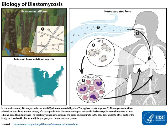
- Specifically, the fungus is native to the boreal regions of northern Ontario, southeastern Manitoba, and southern Quebec south of the St. Lawrence River in Canada; the Appalachian Mountains and other connected eastern mountain chains in the United States; the western shore of Lake Michigan and the state of Wisconsin; the entire Mississippi Valley; and the valleys of some major tributaries, including the Ohio River.
- It also appears only sporadically throughout the Arabian Peninsula and the Indian subcontinent, as well as the rest of Africa north and south of the Sahara Desert. The growth in artificial culture at 25 °C (77 °F) is assumed to be representative of its growth in nature, albeit this has never been confirmed.
- But when it grows within an infected human or animal, it changes into a large-celled budding yeast. Once recognized, blastomycosis responds well to systemic antifungal medications; nonetheless, misdiagnosis is widespread even in high-endemic regions.

Morphology of Blastomyces dermatitidis
Blastomyces dermatitidis is a dimorphic fungus that causes blastomycosis, a fungal infection in humans and animals. The fungus exists in two different morphological forms, the yeast phase, and the mold phase.
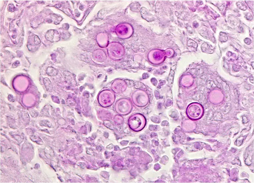
- Yeast Phase: In the yeast phase, Blastomyces dermatitidis appears as round to oval, thick-walled cells that measure 8 to 15 μm in diameter. These cells are single-celled and reproduce through budding. The cells are characterized by their double-contoured walls and their large, single, and round nuclei. The cytoplasm of the cells is granular and often contains large vacuoles. The yeast cells of Blastomyces dermatitidis are typically found in infected tissues and clinical specimens.
- Mold Phase: In the mold phase, Blastomyces dermatitidis appears as a filamentous fungus that produces branching hyphae. The hyphae are septate, meaning they have cross-walls that divide the hyphae into individual cells. The mold form of Blastomyces dermatitidis produces conidiophores, which are specialized hyphae that bear conidia, asexual spores. The conidiophores arise from the mycelium and are characterized by their long, unbranched stalks that terminate in clusters of small, round, smooth-walled conidia.
Overall, the morphology of Blastomyces dermatitidis is unique and can vary depending on its phase of growth. The yeast phase is characterized by round to oval thick-walled cells with a double-contoured wall and large, single, and round nuclei, while the mold phase produces branching hyphae and conidia on specialized hyphae. Understanding the morphology of Blastomyces dermatitidis is essential for the diagnosis and treatment of blastomycosis.
Life cycle of Blastomyces
Blastomyces is a type of mold that spreads via the air by means of spores. The spores require a microscope to observe. Tiny spores can be released into the air when soil or organic material is disturbed. Blastomycosis can occur in humans and animals if the spores are inhaled. Spores inhaled into the lungs can develop into yeast because of the host’s body temperature. Yeast can remain in the lungs, or it can travel through the blood to the skin, bones and joints, organs, and central nervous system (brain and spinal cord).

Habitat of Blastomyces dermatitidis
- Blastomyces dermatitidis is a dimorphic fungus that can exist in two forms: a mold form and a yeast form. In its mold form, it grows in the soil, particularly in areas with decomposing organic matter such as wooded areas, forests, and riverbanks. It can also be found in bird and bat guano.
- Once inhaled, the mold form of Blastomyces dermatitidis can convert to its yeast form and cause infection in the lungs. The yeast form can also be found in infected tissues of humans and animals.
- Blastomyces dermatitidis is commonly found in parts of North America, including the Mississippi River basin, the Ohio River valley, the Great Lakes region, and parts of the southeastern United States. It can also be found in parts of Africa and India.
Cultural characteristics of Blastomyces dermatitidis
- Blastomyces dermatitidis is a dimorphic fungus that may either produce molds that produce conidia or yeasts when grown at 37 degrees Celsius. Molds are grown in mycological cultures, while yeasts are grown in host tissues and certain types of culture media.
- Colonies of B. dermatitidis are white to light brown in color when grown at room temperature on Sabouraud’s agar. Hyphae with branching structures that hold spherical, ovoid, or piriform conidia on slim terminal or lateral conidiophores make up these structures. It’s possible that SDA media will result in larger chlamydospores.
- At 37 degrees Celsius, B. dermatitidis grows as a thick-walled, multinucleated, spherical yeast that produces single buds, either in host tissues or in culture. Yeast colonies have a waxy, wrinkly texture and a large attachment base from which both the bud and the parent yeast grow. Before detaching from the parent yeast, the bud will often grow to the same size.
- As a thermally dimorphic fungus, B. dermatitidis exhibits distinct phenotypes depending on the temperature at which it is grown. Common medium for cultivating B. dermatitidis in the lab include Sabouraud agar, brain-heart infusion agar, and Dixon agar.
- Colonies of B. dermatitidis grown on Sabouraud agar first appear white, cottony, or fuzzy but eventually age to tan, yellow, or brown. Colony surface texture can change, becoming either smoother or more wrinkled, and more or less granular. The colony’s underside is often a brownish color.
- Colonies of B. dermatitidis grown in brain-heart infusion agar tend to be moist and smooth, with an initial color range from white to tan that can gradually turn brown.
- Dark brown or black colonies with a velvety or fuzzy texture characterize B. dermatitidis cultures grown on Dixon agar. The colony’s underside is often a dark brown to black color.
- It’s worth noting that B. dermatitidis can take on different phenotypes in different medium and under different environmental conditions. However, using these features, B. dermatitidis can be more easily identified in the lab.
Pathogenesis of Blastomyces dermatitidis
- Blastomyces dermatitidis is a dimorphic fungus that can cause a severe lung infection known as blastomycosis. The pathogenesis of blastomycosis involves the inhalation of spores or conidia of the fungus into the lungs, where they can convert into the pathogenic yeast form and cause infection.
- When inhaled, B. dermatitidis spores or conidia reach the alveoli of the lungs, where they are engulfed by alveolar macrophages. In healthy individuals, the macrophages can usually destroy the spores or conidia. However, in immunocompromised individuals, the fungus can evade the host’s immune system and convert into the yeast form. The yeast form of the fungus can multiply within the lung tissue and form granulomas, which are masses of immune cells that surround the fungal cells to limit their spread.
- In severe cases, the infection can spread from the lungs to other organs, such as the skin, bones, and central nervous system. The spread of the infection is facilitated by the formation of pyogranulomatous lesions, which are areas of tissue damage caused by the immune response to the fungus.
- The severity of blastomycosis can vary depending on the host’s immune status, age, and other underlying health conditions. In some cases, the infection can be asymptomatic, while in others, it can cause severe respiratory distress and even death.
- In summary, the pathogenesis of Blastomyces dermatitidis involves the inhalation of spores or conidia into the lungs, the conversion of the fungus into the yeast form, the formation of granulomas and pyogranulomatous lesions, and the potential spread of the infection to other organs.
Virulence Factors of Blastomyces dermatitidis
Blastomyces dermatitidis, the causative agent of blastomycosis, possesses several virulence factors that contribute to its ability to cause disease in humans and animals. Some of the key virulence factors of B. dermatitidis include:
- Thermal dimorphism: B. dermatitidis is a thermally dimorphic fungus, meaning it can grow in two different forms depending on the temperature. At lower temperatures, it grows as a mold, producing infectious conidia that can be inhaled into the lungs. At higher temperatures, such as those found in the host’s body, it grows as a yeast, allowing it to establish an infection in the host.
- Adhesion molecules: B. dermatitidis possesses adhesion molecules on its cell surface that allow it to bind to host tissues and establish an infection. These adhesion molecules include mannoproteins, beta-glucans, and chitin.
- Melanin: B. dermatitidis produces melanin, a pigment that contributes to its virulence by protecting it from host immune responses. Melanin can also scavenge free radicals and inhibit the formation of reactive oxygen species, which are produced by host immune cells.
- Secreted enzymes: B. dermatitidis produces several secreted enzymes that can damage host tissues and help the fungus invade host cells. These include phospholipases, proteases, and elastases.
- Superoxide dismutase: B. dermatitidis produces superoxide dismutase, an enzyme that protects it from oxidative stress caused by host immune cells. This allows the fungus to survive and proliferate within the host.
Overall, these virulence factors allow B. dermatitidis to establish an infection in the host and evade host immune responses, leading to the development of blastomycosis. Understanding these virulence factors is important for developing new treatments and vaccines to combat this fungal infection.
Transmission of Blastomyces dermatitidis
- Blastomyces dermatitidis is primarily a soil fungus that is found in specific geographic locations such as the Midwestern and South Central United States, particularly in areas with moist soil rich in organic matter, such as river valleys, lake shores, and decaying woodlands. The fungus grows as a mold in the soil and can be aerosolized in the form of tiny, infectious spores that can be inhaled by humans or animals.
- Blastomyces dermatitidis is considered a primary pathogen, which means it can cause disease in healthy individuals who are exposed to it. The spores of the fungus can enter the lungs, where they are phagocytosed by alveolar macrophages. In susceptible individuals, the fungus can escape the macrophages and multiply in the lungs, leading to acute or chronic pulmonary blastomycosis.
- Blastomycosis is not contagious and cannot be transmitted from person to person, but it can be acquired by inhalation of fungal spores present in the environment. In addition to inhalation, infection can also occur through direct inoculation of the fungus into the skin or other tissues, typically through traumatic injury or occupational exposure.
- Overall, the transmission of Blastomyces dermatitidis is primarily through inhalation of spores present in the environment, particularly in areas where the fungus is endemic. It is important to take precautions when working in such environments, such as wearing masks and gloves to prevent exposure to the fungus.
Clinical Features of Blastomycosis
When people get sick with pneumonia caused by B. dermatitidis, they usually show flu-like symptoms such as coughing and chest pain. The chest x-rays may not show any specific signs, and many cases go untreated. Some people may get very sick and have breathing problems and even die, but this is rare. It’s hard to know how often people get the acute pneumonia versus other types of illness caused by B. dermatitidis, but most people who get it will recover on their own.
Blastomycosis can affect different parts of the body, and here are the common sites and symptoms:
- Chronic Pulmonary Infection:
- Symptoms: low-grade fever, weight loss, chronic cough, and sputum production
- Often mistaken for tuberculosis or primary lung malignancy
- Skin and Subcutaneous Tissue:
- Symptoms: papulosquamous, eruptive, and verrucous lesions, cutaneous ulcers, and subcutaneous nodules
- Occurs in 40-80% of cases
- Osteoarticular:
- Symptoms: pathologic fractures, localized bone pain, and destructive bony lesions
- Occurs in approximately 5-50% of patients
- Genitourinary:
- Symptoms: prostatitis or epididymitis in males, and uterus and adnexa involvement in females
- Occurs in 10-30% of patients
- Central Nervous System:
- Symptoms: mass lesion(s) or chronic meningitis
- Involvement occurs in up to 5% of non-immunocompromised patients
Laboratory Diagnosis of Blastomycosis
Blastomycosis is a fungal infection caused by the dimorphic fungus Blastomyces dermatitidis. The infection is acquired through inhalation of spores, primarily in areas where the fungus is endemic. Blastomycosis can cause a variety of symptoms ranging from flu-like symptoms to severe respiratory and systemic illness. Laboratory diagnosis of blastomycosis involves a combination of clinical, radiological, and laboratory findings. Here are the laboratory diagnostic tests used to identify and confirm Blastomycosis:
- Microscopic Examination: To do so, a KOH wet mount is performed on the clinical material to check for yeast cells or blastospores. After being cultured, yeast cells can be observed to have a diameter of between 3 and 5 µm. Blastospores are ovoid spherical shaped spores that are the result of culture.
- Culture Identification: The sample is cultured on Sabouraud Dextrose Agar (SDA). Ovoid spherical spores are produced by the Blastomyces dermatitidis colony that grows white or brown on SDA. Yeast-like cells with strong walls, several nuclei, and a spherical shape are the ideal outcome of tissue culture.
- Histological Examination: The infected area is biopsied and sent off for a histological analysis. The granuloma’s neutrophilic contacts with the host tissues can be seen, together with any polymorphonuclear leukocytes and pyogranulomatous response, using a stain called hematoxylin and eosin. The fungus is easily visible when stained with Gomori’s methenamine silver (GMS) or periodic acid-Schiff (PAS). The yeast cell wall is stained a deep black by GMS, while the inside is stained a rose-like color on a green backdrop. The yeast cells are stained red by PAS on a pink or light green background, depending on the counterstain. Histological stains reveal yeast cells ranging in size from 2-10 µm to 25-40 µm, each of which is composed of short septate hyphae and hyaline.
- Immunological Diagnosis: This can be done using a battery of immunological assays that look for the blastomycin antigen or antibodies against it. The blastomycin antigen can be detected with a complement fixation test. Blastomyces dermatitidis antigen is present when there is a high complement fixation titer. Blastomyces dermatitidis antigen A can be detected using immunodiffusion. There is also a skin test for detecting blastomycin. Antibodies against Blastomyces dermatitidis antigen A can be detected by enzyme-linked immunosorbent assay.
Treatment of Blastomycosis
- The treatment of blastomycosis depends on the severity and location of the infection. Mild pulmonary blastomycosis clears spontaneously and does not require antifungal therapy. However, for more severe or disseminated infections, antifungal therapy is necessary.
- Amphotericin B (Fungizone), itraconazole (Sporanox), or ketoconazole (Nizoral) are the drugs of choice for treatment. Amphotericin B should be preferred particularly in immunocompromised patients. Itraconazole is effective for mild to moderate pulmonary, cutaneous, and osteoarticular blastomycosis, and ketoconazole is used for mild to moderate blastomycosis.
- Surgery may be necessary for the drainage of large pulmonary abscesses along with antifungal therapies. Follow-up care should be provided to monitor the response to treatment and to detect any potential relapses.
Prevention and Control of Blastomyces dermatitidis
Prevention and control of Blastomyces dermatitidis infection involve avoiding exposure to the fungus in endemic areas. Here are some measures that can be taken to prevent and control the spread of Blastomyces dermatitidis:
- Education: Education of the public and healthcare professionals regarding the risk factors, symptoms, and prevention of blastomycosis is crucial in endemic areas. This includes measures to prevent exposure to the fungus, such as avoiding areas where the fungus is likely to be present and wearing protective clothing.
- Environmental measures: Reducing environmental exposure to the fungus is an important aspect of prevention. This includes measures such as removing organic debris, such as dead leaves and vegetation, from outdoor areas, and avoiding digging in soil in endemic areas.
- Personal protective measures: People who work in outdoor environments where the fungus is present should wear appropriate protective clothing, including gloves and masks. In addition, individuals with weakened immune systems or underlying medical conditions should take extra precautions to avoid exposure to the fungus.
- Early diagnosis and treatment: Early diagnosis and treatment of blastomycosis can help prevent the spread of the disease. It is important to seek medical attention promptly if symptoms of blastomycosis are present, especially in individuals who live in or have recently traveled to endemic areas.
- Animal control measures: Dogs are particularly susceptible to blastomycosis, and the infection can be spread to humans through infected animal tissue or respiratory secretions. Preventive measures include keeping dogs out of areas where the fungus is likely to be present, such as wooded areas and riverbanks, and prompt treatment of infected animals.
By implementing these measures, it is possible to reduce the incidence of blastomycosis and prevent its spread in endemic areas.
FAQ
What is Blastomyces dermatitidis?
Blastomyces dermatitidis is a dimorphic fungus that causes blastomycosis, a fungal infection that affects the lungs and other parts of the body.
How do people get infected with Blastomyces dermatitidis?
People get infected with Blastomyces dermatitidis by inhaling spores of the fungus present in the air. The fungus is commonly found in soil, particularly in areas near waterways and in forests.
What are the symptoms of blastomycosis?
The symptoms of blastomycosis can vary, but they often include fever, cough, chest pain, muscle aches, fatigue, and weight loss. The infection can also affect other parts of the body, including the skin and bones.
Who is at risk of developing blastomycosis?
People who live or work in areas where Blastomyces dermatitidis is endemic are at a higher risk of developing the infection. People with weakened immune systems, such as those with HIV/AIDS, are also at increased risk.
How is blastomycosis diagnosed?
Blastomycosis is diagnosed through laboratory tests, including microscopic examination of sputum, pus, or tissue biopsies; culture identification on Sabouraud Dextrose Agar; and immunological tests, such as complement fixation or ELISA.
How is blastomycosis treated?
Blastomycosis is treated with antifungal medications, such as amphotericin B, itraconazole, or ketoconazole. The choice of medication and duration of treatment depend on the severity of the infection and the patient’s overall health.
Can blastomycosis be prevented?
Blastomycosis can be prevented by avoiding exposure to contaminated soil, especially in areas where the fungus is known to be endemic. People who work in occupations that may expose them to the fungus should take appropriate precautions, such as wearing protective clothing and masks.
Is blastomycosis contagious?
Blastomycosis is not contagious and cannot be transmitted from person to person.
Can animals be infected with Blastomyces dermatitidis?
Yes, animals can be infected with Blastomyces dermatitidis, particularly dogs. Dogs may develop respiratory symptoms similar to those seen in humans, as well as skin lesions.
Is blastomycosis a rare disease?
Blastomycosis is considered a rare disease, although it is more common in certain areas of the United States, particularly in the Midwest and South. The incidence of blastomycosis has been increasing in recent years due to changes in land use and other environmental factors.
References
- Miceli A, Krishnamurthy K. Blastomycosis. [Updated 2022 Aug 21]. In: StatPearls [Internet]. Treasure Island (FL): StatPearls Publishing; 2023 Jan-. Available from: https://www.ncbi.nlm.nih.gov/books/NBK441987/
- https://en.wikipedia.org/wiki/Blastomyces_dermatitidis
- https://www.infectiousdiseaseadvisor.com/home/decision-support-in-medicine/infectious-diseases/blastomyces-dermatitidis/
- https://www.health.state.mn.us/diseases/blastomycosis/index.html
- http://www.antimicrobe.org/f02.asp
- https://microbewiki.kenyon.edu/index.php/Blastomyces_dermatitidis
- Text Highlighting: Select any text in the post content to highlight it
- Text Annotation: Select text and add comments with annotations
- Comment Management: Edit or delete your own comments
- Highlight Management: Remove your own highlights
How to use: Simply select any text in the post content above, and you'll see annotation options. Login here or create an account to get started.