What is Holobasidium?
- Holobasidium refers to a specific structure found within certain fungi, characterized by its single-celled, non-segmented nature. This club-shaped cellular structure plays a crucial role in the reproductive process of these fungi, facilitating the development and release of spores. One of the defining features of a holobasidium is the presence of four sterigmata, which are small projections that support the developing spores.
- Within the category of holobasidium, there exist two distinct types, namely chiastobasidium and stichobasidium. The primary difference between these types lies in the orientation of microtubules within the basidium. In chiastobasidium, microtubules are arranged in a perpendicular fashion relative to the basidium’s long axis, whereas in stichobasidium, the arrangement is parallel.
- The shape of a basidium is typically likened to that of a club, with a gradual widening towards the outer end and a narrower connection at the stem. The broadest part is often described as a middle hemispherical dome. However, variations in shape do exist within different genera, such as Paullicorticium, Oliveonia, and Tulasnella, where basidia can resemble an inverted egg or even adopt a barrel-like form with a broad base.
What is Basidium?
- A basidium is a microscopic structure instrumental in the reproductive cycle of Basidiomycota fungi, serving as a sporangium or spore-producing organ. Found primarily within the hymenophore of a fungus’s fruiting body, basidia play a crucial role in spore formation and dispersal, a defining characteristic of this fungal division.
- The development of basidia occurs within the tertiary mycelium, an advanced stage of fungal growth that evolves from the secondary mycelium. This tertiary mycelium is dikaryotic, meaning it contains two distinct nuclei in each cell, which is a result of the fusion of two compatible fungal hyphae.
- Basidia typically give rise to four basidiospores, although this number can range between two to eight in some species. These spores are borne on slender projections called sterigmata and are released into the environment when mature, a process that is often forceful to aid in their dispersal.
- Morphologically, basidia are commonly club-shaped, with a broader apex and a narrower base. Before reaching maturity, these structures are referred to as basidioles. Based on their cellular composition, basidia can be categorized into two types: holobasidia, which are single-celled, and phragmobasidia, which consist of multiple cells.
What is Phragmobasidium?
- Phragmobasidium refers to a type of basidium characterized by its septate, or partitioned, structure. This means that the basidium is divided into distinct cells by cross-walls, known as septa. Such a configuration is notably present in the rust fungi belonging to the order Puccinales, where the basidium comprises four cells, each separated by these cross-sectional walls. The arrangement of these cells often resembles a cross shape.
- In addition to rust fungi, certain jelly fungi within the order Tremellales also exhibit a similar septate structure in their basidia, with walls arranged in a cross-like pattern. This structural feature is significant as it distinguishes phragmobasidia from other types of basidia, such as holobasidia, which are not septate and consist of a single cell.
- The presence of phragmobasidium is a critical trait for the identification and classification of certain fungal groups. Understanding this structure is important for those studying fungal biology and ecology, as it provides insights into the diversity of reproductive strategies among fungi and helps in the identification of various fungal orders and species.
Difference Between Holobasidium and Phragmobasidium
Holobasidium and Phragmobasidium are two distinct types of basidia, which are reproductive structures found in fungi belonging to the division Basidiomycota. These structures play a crucial role in the life cycle of these fungi, particularly in spore production and dispersal. Understanding the differences between these two types of basidia is essential for mycologists and those interested in fungal biology. Here’s a comparative overview:
Holobasidium:
- Structure: Holobasidium is a non-septate basidium, meaning it lacks internal cross-walls or septa, and is comprised of a single cell.
- Shape: Typically, it has a club-like shape, wider at the top and narrower at the base.
- Spore Attachment: It usually bears four sterigmata, which are projections from the basidium that hold the developing spores.
- Types: There are variations within holobasidia, such as chiastobasidium, where microtubules are arranged perpendicular to the basidium’s long axis, and stichobasidium, with microtubules parallel to the long axis.
- Occurrence: Holobasidia are common in many Basidiomycota fungi, including mushrooms, puffballs, and bracket fungi.
Phragmobasidium:
- Structure: Phragmobasidium is a septate basidium, characterized by the presence of internal cross-walls or septa that divide it into multiple cells.
- Shape: The shape can vary, but it is often described as resembling a cross due to the arrangement of the septa.
- Spore Attachment: Like holobasidia, phragmobasidia bear sterigmata for spore development, but the number and arrangement can be influenced by the septate structure.
- Types: Phragmobasidia are particularly notable in rust fungi (order Puccinales) and some jelly fungi (order Tremellales), where the septate nature is a key identifying feature.
- Occurrence: They are less common than holobasidia and are specific to certain groups of fungi, such as the rusts and jelly fungi.
Key Differences:
- Cellularity: Holobasidium is single-celled (non-septate), while Phragmobasidium is multicellular (septate).
- Internal Structure: The presence of septa in Phragmobasidium divides it into multiple cells, in contrast to the single-cell structure of Holobasidium.
- Fungal Association: Holobasidia are found in a broader range of Basidiomycota fungi, whereas Phragmobasidia are characteristic of specific groups like rust and jelly fungi.
- Reproductive Strategy: The structural differences may reflect variations in reproductive strategies between fungi with holobasidia and those with phragmobasidia.
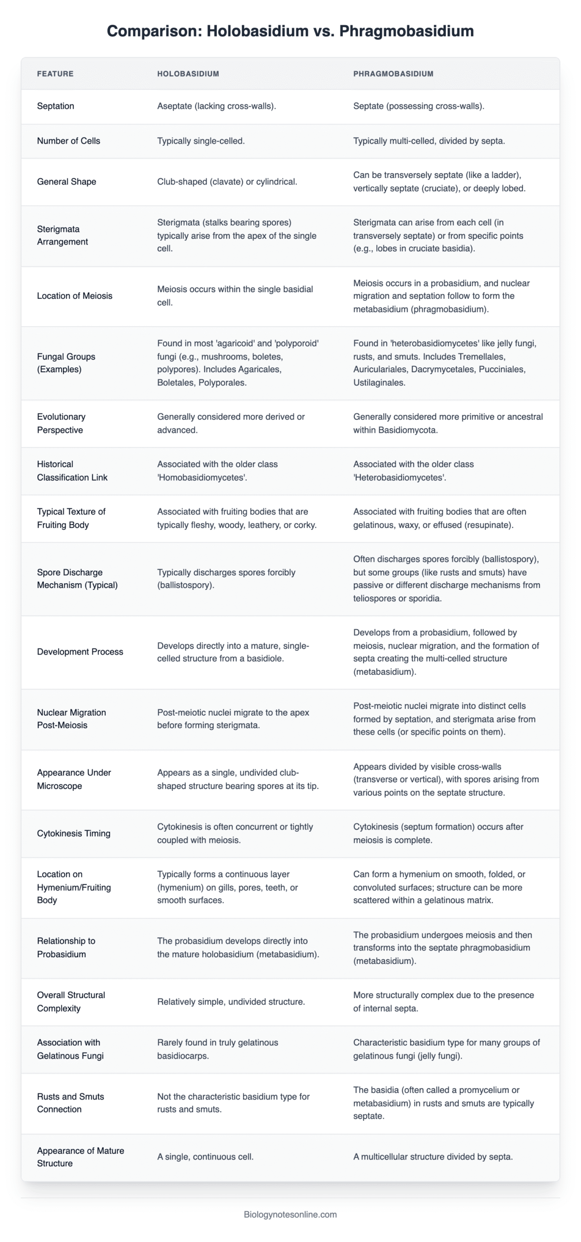
Comparison: Holobasidium vs. Phragmobasidium
| Feature | Holobasidium | Phragmobasidium |
|---|---|---|
| Septation | Aseptate (lacking cross-walls). | Septate (possessing cross-walls). |
| Number of Cells | Typically single-celled. | Typically multi-celled, divided by septa. |
| General Shape | Club-shaped (clavate) or cylindrical. | Can be transversely septate (like a ladder), vertically septate (cruciate), or deeply lobed. |
| Sterigmata Arrangement | Sterigmata (stalks bearing spores) typically arise from the apex of the single cell. | Sterigmata can arise from each cell (in transversely septate) or from specific points (e.g., lobes in cruciate basidia). |
| Location of Meiosis | Meiosis occurs within the single basidial cell. | Meiosis occurs in a probasidium, and nuclear migration and septation follow to form the metabasidium (phragmobasidium). |
| Fungal Groups (Examples) | Found in most ‘agaricoid’ and ‘polyporoid’ fungi (e.g., mushrooms, boletes, polypores). Includes Agaricales, Boletales, Polyporales. | Found in ‘heterobasidiomycetes’ like jelly fungi, rusts, and smuts. Includes Tremellales, Auriculariales, Dacrymycetales, Pucciniales, Ustilaginales. |
| Evolutionary Perspective | Generally considered more derived or advanced. | Generally considered more primitive or ancestral within Basidiomycota. |
| Historical Classification Link | Associated with the older class ‘Homobasidiomycetes’. | Associated with the older class ‘Heterobasidiomycetes’. |
| Typical Texture of Fruiting Body | Associated with fruiting bodies that are typically fleshy, woody, leathery, or corky. | Associated with fruiting bodies that are often gelatinous, waxy, or effused (resupinate). |
| Spore Discharge Mechanism (Typical) | Typically discharges spores forcibly (ballistospory). | Often discharges spores forcibly (ballistospory), but some groups (like rusts and smuts) have passive or different discharge mechanisms from teliospores or sporidia. |
| Development Process | Develops directly into a mature, single-celled structure from a basidiole. | Develops from a probasidium, followed by meiosis, nuclear migration, and the formation of septa creating the multi-celled structure (metabasidium). |
| Nuclear Migration Post-Meiosis | Post-meiotic nuclei migrate to the apex before forming sterigmata. | Post-meiotic nuclei migrate into distinct cells formed by septation, and sterigmata arise from these cells (or specific points on them). |
| Appearance Under Microscope | Appears as a single, undivided club-shaped structure bearing spores at its tip. | Appears divided by visible cross-walls (transverse or vertical), with spores arising from various points on the septate structure. |
| Cytokinesis Timing | Cytokinesis is often concurrent or tightly coupled with meiosis. | Cytokinesis (septum formation) occurs after meiosis is complete. |
| Location on Hymenium/Fruiting Body | Typically forms a continuous layer (hymenium) on gills, pores, teeth, or smooth surfaces. | Can form a hymenium on smooth, folded, or convoluted surfaces; structure can be more scattered within a gelatinous matrix. |
| Relationship to Probasidium | The probasidium develops directly into the mature holobasidium (metabasidium). | The probasidium undergoes meiosis and then transforms into the septate phragmobasidium (metabasidium). |
| Overall Structural Complexity | Relatively simple, undivided structure. | More structurally complex due to the presence of internal septa. |
| Association with Gelatinous Fungi | Rarely found in truly gelatinous basidiocarps. | Characteristic basidium type for many groups of gelatinous fungi (jelly fungi). |
| Rusts and Smuts Connection | Not the characteristic basidium type for rusts and smuts. | The basidia (often called a promycelium or metabasidium) in rusts and smuts are typically septate. |
| Appearance of Mature Structure | A single, continuous cell. | A multicellular structure divided by septa. |

- Text Highlighting: Select any text in the post content to highlight it
- Text Annotation: Select text and add comments with annotations
- Comment Management: Edit or delete your own comments
- Highlight Management: Remove your own highlights
How to use: Simply select any text in the post content above, and you'll see annotation options. Login here or create an account to get started.