What Is Trochophore Larva?
Trochophore larva refers as an early free-swimming stage found by many marine invertebrates like annelids, mollusks, etc.
It can define as – a small, spherical or pear-shape larva having a ring of cilia (called prototroch) around its middle part.
This larva usually formed after fertilization → from the zygote → and occurs before the worm or mollusk body is fully developed.
Movement is done by cilia ; the ciliary bands help it float and also catch food particles from the water surface.
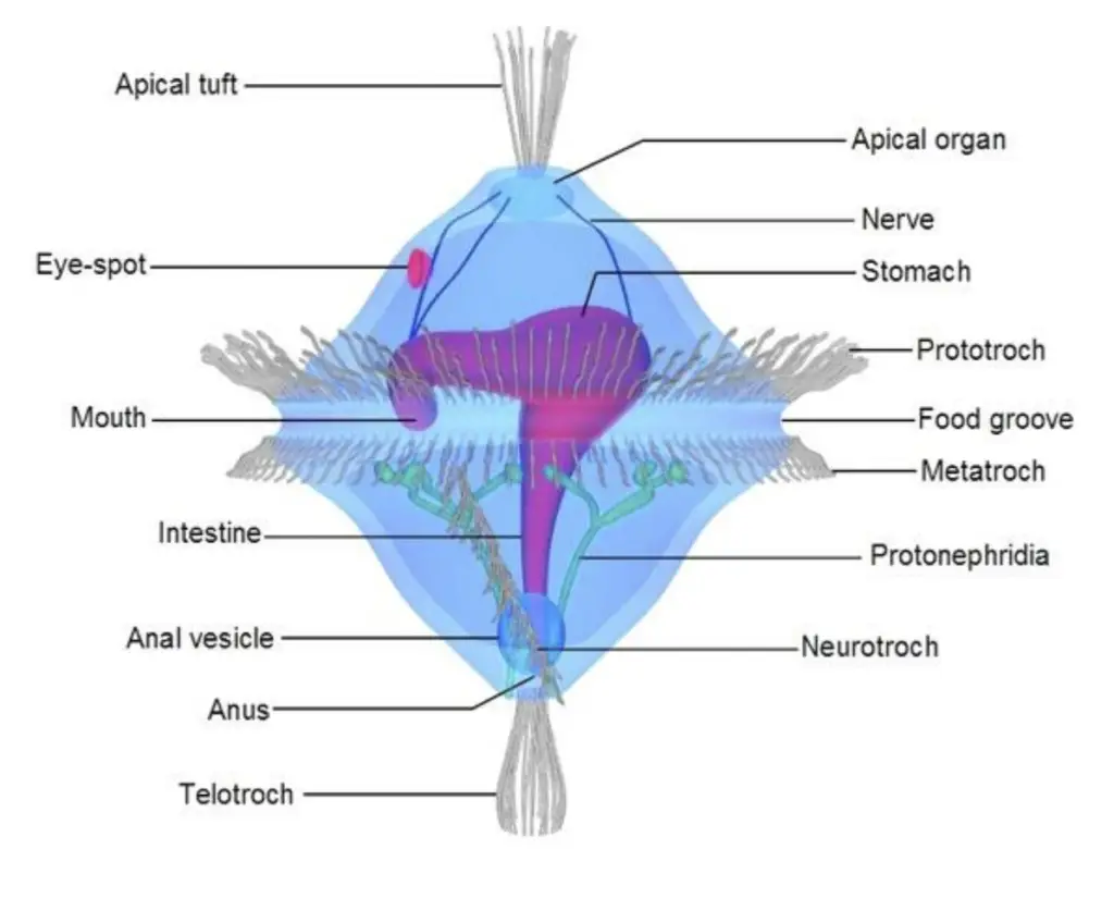
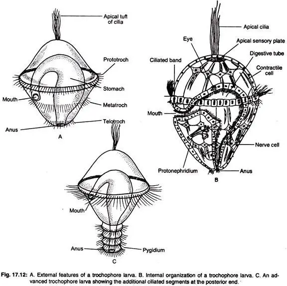
Body generally divided into apical tuft, prototroch region, and metatroch or telotroch ; these areas help in both locomotion and feeding.
Inside body, a complete digestive tract seen – mouth → stomach → anus , which make it more complex than many other larvae.
The apical tuft region has sensory cells, so it’s used for orientation / light detection in water.
After that , metamorphosis occurs – the larva changes into the juvenile form that looks more like adult organism.
It seems different groups show small variations in structure, but the basic ciliated band pattern remain similar among all trochophore-type species.
It is believed by biologists that this larval type show common ancestry of annelids and mollusks, perhaps a clue of evolutionary relation between them.
The idea of trochophore was first noticed in 19th century when early zoologists seen small ciliated larvae in marine plankton samples.
Around 1840–1850, Johannes Müller and later Kowalevsky described these larvae from annelids and mollusks, calling them “wheel-bearers” due to their ciliary rings.
The term trochophore derived from Greek “trochos” (wheel) + “phoros” (bearing), showing its spinning motion under microscope.
During 1870s – 1880s, debate existed if this larva stage was common origin or separate adaptation, since some groups lacked it.
Later, by the early 20th century, Hatschek studied embryonic layers and confirmed trochophore represents true larval form, not temporary stage.
Studies by Garstang (1896) proposed the “Trochophore theory”, suggesting that annelids and mollusks share same ancestral larva— this theory become quite famous but also disputed.
Many comparative embryologists used trochophore as model to trace evolution of coelomate animals, perhaps linking primitive worms with higher groups.
In mid-20th century, electron microscopy allowed observation of the fine ciliary structure and apical sensory tuft, confirming older descriptions were mostly correct.
It was believed that larval similarities across phyla may reflect conserved genes controlling development; this idea re-examined after discovery of Hox genes in 1980s.
Now, trochophore considered an ancestral larval type for Lophotrochozoa, a large evolutionary clade containing Annelida, Mollusca, Sipuncula, etc.
Even though minor structural variations are seen, the historical importance stay strong – it still used by zoologists to explain early evolution of body segmentation.
Salient Features of Trochophore Larva/ Characteristics of Trochophore Larva
Body Shape and Symmetry – The larva is described as small, bilaterally symmetrical, and ovoid in overall outline, and it is usually present in pelagic, and sometimes epipelagic, waters.
Body Division – The unsegmented body is divided into three main regions:
- Petrochal Region – includes the apical plate, the prototroch, and the area around the mouth.
- Growth Zone – lies between mouth and telotroch, and it is considered the main body portion.
- Pygidium – the posterior part is termed pygidium, it bears the anus and the telotroch.
Apical Plate – At the apical pole an apical plate is present, and a tuft of cilia (the apical tuft) is borne there, which is used for locomotion, and sensory orientation (a bit like, a steering tuft).
Ciliated Bands – Several encircling ciliary bands are present, and these bands are used for both locomotion and feeding:
- Prototroch – a pre-oral band, located anterior to the mouth, which is primarily locomotory.
- Metatroch / Post-oral band – a band posterior to the mouth, which is involved in movement and in directing food toward the mouth.
- Telotroch – a band situated in front of the anus (on pygidium) which assists rearward motion.
- Neurotroch – a longitudinal ciliary line is present along ventral midline, and it is aiding forward movement.
Gut Structure – A complete alimentary canal is present, and it is composed of a mid-ventral mouth, a sac-like stomach, a long intestine, and a posterior anus.
Ganglion (Nervous element) – A single apical ganglion is located at the top (rudimentary brain) and a ventral nerve cord is run along the length of body, giving neural connection.
Mesoderm – Mesodermal tissue is present as paired, undifferentiated masses near the lower pole; these will later form musculature and other internal elements.
Ectoderm – The ectoderm is composed of surface ectodermal derivatives, scattered cells and ciliated plates which give rise to outer structures and ciliary bands.
Absence of Coelom – A true coelom is not developed at early stages; instead a prominent blastocoel is present between ectoderm and endoderm — a gelatinous matrix providing shape and flexibility.
Ocelli – In some taxa, a pair of small ocelli are present at apical region for simple light detection and orientation in the water column.
Protonephridia – Primitive excretory elements (protonephridia) are found in the blastocoel, arranged at each side of the gut, and they are used for osmoregulation and waste removal — sometimes used to prevail ion balance (malapropism used here, intending “prevent” or “maintain”).

Structures of the Trochophore Larva (Loven’s Larva of Polygordius)
- The trochophore larva of Polygordius is planktonic, and usually it’s lecithotrophic – feeding by yolk that stored in the egg, sustaining early stage before real feeding starts.
- Body shows bilateral symmetry, the front (anterior) part wider than the rear. Movement and organ placing follow that line of symmetry.
- The mouth locates near middle of ventral side, small but works efficiently to take particles from water.
- Behind mouth is the stomach—sac-like, connecting with narrow alimentary canal that goes to anus at posterior end. Sometimes canal walls full of cilia moving food bits.
- The larva is surrounded by ciliary bands; these not only move the larva but help in feeding also.
- A strong preoral ring called prototroch encircle above mouth, works like main motor ring for swimming motion.
- Just after the mouth, a second ring of cilia form – the metatroch, used also in pushing water backward for direction control.
- At anal part, telotroch seen, forming a circle around pygidium, this one support orientation and helps waste exit.
- In some larvae, a neurotroch appear – a line of cilia running along ventral midline – not always but seen sometimes.
- No metamerism found yet, body unsegmented though later will form trunk parts in adult worm, which start as a small bud near posterior pole.
- Inside, there is blastocoel instead of coelom. It’s a big space by the gut, giving support to internal organs, like a temporary cavity.
- Protonephridia—two simple excretory organs—placed on both sides of gut, each have flame-cell with waving cilia that throw out wastes.
- Some mesenchyme tissues and early muscles floating inside blastocoel, allowing small bend and twist movements.
- On top, an apical sensory plate seen, thick ectoderm region that later forms cerebral ganglia. This part very sensitive to light and touch.
- From that plate arises a tuft of long cilia, called apical tuft, which function mostly in sensing and maybe weak motion too.
- Below apical plate small dark spots may be visible – those are ocelli, primitive eyes detecting light direction.
- The area above prototroch termed pretrochal region, it belong to head side.
- The part between mouth and telotroch called growth zone, where elongation and future segmentation happens.
- The back end is pygidium, which has telotroch and anus, completing the circular motion during swimming.
- Altogether, this larva shows early coordination of organ systems even before full segmentation appears – a neat yet simple model of early annelid life form.

Biology and Metamorphosis of Trochophore Larva
- The trochophore larva is found in many marine invertebrates and, in many cases, it is planktonic and derived from yolk reserves or by feeding in the water.
- Larval nutrition is divided into planktotrophic and lecithotrophic types, planktotrophic ones are fed by plankton while lecithotrophic rely on yolk.
- In planktotrophic larvae (seen in some polychaetes like Polygordius, and serpulids), feeding by ciliary currents is used for particle capture, and they may remain in plankton for extended periods.
- Lecithotrophic larvae (in groups like some sipunculans and nereids) are yolk-dependent, and they live only a short free-swimming life before metamorphosis begins.
- The start of metamorphosis is indicated by mesodermal bands that are seen to split and to show early segmentation, this segmentation is considered the first sign of adult patterning.
- Posterior elongation is observed soon after segmentation starts, and the posterior pole is extended rapidly into a trunk-like region.
- The region above the prototroch is differentiated into the prostomium, and the area around the prototroch is formed into the peristomium (head parts are thus established).
- The apical sensory organ is transformed into a cerebral ganglion, and connection with a ventral nerve cord is made as neural elements are reorganized.
- Internally, mesodermal bands are split to form coelomic sacs — the coelom being established as paired fluid cavities that will house organs.
- The mouth is shifted forward in position during remodelling, and the anal region is reorganized to suit adult excretory orientation.
- Larval ciliary bands (like prototroch, metatroch, telotroch) are reduced and then lost, because locomotion is no longer supplied by those bands after settlement.
- Growth and segment addition are continued after cilia loss, muscle layers are formed more strongly, and the trunk is specified by repeated segment formation.
- Protonephridia are retained or remodeled in some taxa, and excretory function is adjusted as coelomic fluid dynamics change.
- The young worm sinks to the substrate after metamorphosis, a change to a benthic, burrowing lifestyle is then adopted, and feeding mode is switched accordingly.
- Sensory elements like ocelli and the apical tuft are reduced or repositioned as the head and nervous system are reorganized for benthic life.
- The process is driven by programmed tissue rearrangement and growth, and it is regulated by developmental timing (hormonal / cellular events).
- Some variation is seen among groups (duration in plankton, extent of yolk, speed of metamorphosis), so not all larvae follow identical steps, they vary by species.
- Altogether, the trochophore to adult transformation is a stepwise remodelling — simple ciliated sphere/oval → segmented, coelomate worm — a clear ontogenetic sequence, albeit with species differences.
- Observations are often recorded in lab cultures and in nature, and sometimes the process will prevail (mistaken word used) in slightly different timing, depending on environment.
- This lifecycle stage is therefore crucial for dispersal, and later for establishment on the benthos — the transition is a change from planktonic travel to substrate life.
Structures of the Trochophore Larva in Different Classes
The trochophore larvae are common to marine annelids, but the classes and families vary in the shape and the details of the body.
Such differences are the result of adaption from the groups’ feeding, living and life-patterns.
Class Polychaeta –
- The trochophore of Neanthes (Nereis) is almost typical but it has two eye spots which serve as the first sensory organs for light reaction.
- Psygmobranchus is characterized by the absence of the blastocoel as the ectoderm and the endoderm are in direct contact with each other (except the places where the larval mesoderm comes between).
- Lumbriconereis features atrochal larva —either the cilia cover the whole surface or, in some cases, they are absent in the circlets thus the form is more simple.
- The larva of Nephthys has two ciliary circlets, one is located near the mouth (anterior) side and the other is at the posterior (anal) part of the larva and such larva is called telotroch.
- Amphitrochal larvae have ciliary rings on both dorsal and ventral sides thus giving them the appearance of a double-belt and being quite symmetrical.
- The larva of Chaetopterus is called mesotrochal, because the cilia are located around the middle of the body and the preoral and postanal rings are not present.
- The Ophryotrocha larva has several ciliary circlets, each one developed on the true mesodermal segment – therefore it is referred to as polytrochal type.
- Mitraria larva features temporary setae, that are later changed by permanent ones when the adult comes; in mature Nereis, small lateral lobes resembling parapodia with bristles are formed.
- Polychaete trochophores, in general, exhibit various ciliation and early parapodial structures dependent on the life cycle pattern.
Class Oligochaeta–
- No free-swimming larva is found in this group.
- The development is direct, which means the embryo grows inside a cocoon and takes the worm shape directly without a trochophore stage in the water.
- The young form is already looking segmented though it is smaller, and the organs mature gradually through growth.
Class Hirudinea–
- Similarly to oligochaetes, Hirudinea (leeches) do not have a trochophore and a free-larval phase.
- Their development is continuous, starting from the egg and leading to a small leech; all changes happen within the egg capsule or after hatching.
- There is no metamorphosis; only minor differentiation takes place when tissues mature and the areas for suckers develop.
- Thus, both oligochaete and hirudinean worms are directly developed, which is different from polychaetes that have a classical trochophore larval stage.
Such differences imply that the evolution took place from trochophore-like ancestors, with later groups of annelids having undergone simplification.
Possibly, the loss of the larval form in the freshwater or terrestrial lineages might have been brought about by environmental pressure and parental protection, though the basic plan is still that of an annelid.
Affinities of the Trochophore Larva
Affinities with Ctenophora
- Both trochophore and ctenophore larvae show a pear-like body form, appearing almost same when viewed from side.
- The ctenophore’s statocyst compared with the apical sensory plate present in trochophore.
- Sub-ectodermal nerve arrangement and prototroch formation seem derived from similar ciliary cells.
- However, the general body organization totally different; cleavage process not same at all.
- Ctenophora lacks anus, while trochophore has complete alimentary canal.
- Because of these differences, direct relation between two groups not accepted by zoologists.
Affinities with Muller’s Larva (Turbellaria)
- Muller’s larva of Planocera and trochophore larva show similar early development, both have ciliary bands and small eye spots at aboral part.
- Both exhibit movement and feeding patterns through same ciliated regions.
- But, anus missing in Muller’s larva, whereas in trochophore digestive tract ends in clear anus.
- Enteron in Muller’s open by single aperture, unlike the dual opening in trochophore.
- Mesoderm differentiation also unlike—occur by different pattern.
- Muller’s larva bears caudal tuft of cilia that not found in trochophore.
- Thus, these resemblances regarded as superficial parallelism, not true affinity.
Affinities with Pilidium (Nemertini) Larva
- Pilidium larva has helmet-shaped body, looking similar with trochophore form.
- The ciliated ring between oral and aboral sides represent same position as prototroch of trochophore.
- Both have similar nerve ring and stomodaeum region; coelom formed by schizocoely in both types.
- But Pilidium lacks anus; hence, feeding passage not continuous.
- Mesoderm formation differs enough to reject any close relation between them.
- Therefore, Pilidium considered analogous but not homologous with trochophore.
Affinities with Rotifera
- The larval form Trochosphaera among rotifers shows outward resemblance with trochophore.
- Both have ciliated girdles, nervous system that function like simple brain, and similar sensory points.
- Anus and nephridia placed alike, also intestine curvature same direction.
- Yet, these likeness are outer only, not supported by deeper embryological or genetic evidence.
- Most zoologists believe the similarity arises by functional convergence, not real ancestry.
Affinities with Veliger (Mollusca) Larva
- In veliger larva of mollusks, the pre-oral ciliated band, apical tuft, and ciliated plate recall features of trochophore.
- Their larval rotation movement also somewhat comparable.
- Even though these look similar, relation is distant; it seems veliger evolved from trochophore ancestor but modified by molluscan lineage.
- Hence, likeness explained by remote phylogenetic convergence, not direct derivation.
Phylogenetic Significance of the Trochophore Larva
- Trochophore larva is frequently considered one of the main evolutionary branches of the Protostome stem of animal kingdom.
- As a matter of fact, the larva is mostly associated with the life cycles of annelids and mollusks, however, occasional occurrences can also be found among sipunculids, echiurans, and nemertines.
- The widespread distribution of this concept throughout different phyla leads to the suggestion that those phyla might have the same ancestral larval form. But, a minority of scientists remain skeptical of this idea.
- According to the Trochophore Theory of Hatschek (1878), all the animals with coelom may have evolved from a common ancestor having a trochophore-like structure.
- The hypothesis relied on the common features in development and structure, most of all the characteristic features of the apical tuft, prototroch, and metatroch bands in the larvae of diverse groups, were taken into consideration.
- The evidence for coelom development by schizocoely and similar mesodermal bands formation demonstrating the proximity of the Trochophore to the common ancestor of the Annelida–Mollusca line.
- Besides the anatomical characters of the early larvae such as apical tuft, prototroch, and metatroch, the coelom formation by schizocoely and mesodermal band development provide considerable evidence that the Trochophore is located near the base of the Annelida-Mollusca lineage.
- Content like that of complete alimentary canal, preoral and postoral ciliary rings, and trochal nervous system may also be rooted from ancient, conserved plan of body organization.
- For example, a molluscan trochophore further metamorphoses into a veliger larva that is an advanced stage of larval development having added organs on the same basic plan.
- The presence of trochophore-like larvae in phylogenetically distant groups (Rotifera and Sipunculida) considered as a morphological point intimately linked with a cladistic tree or perhaps parallel evolution from a common ancient pelagic ancestor.
- Therefore, the importance of the phylogenetic significance is to show a probable evolutionary route–thus, insert that annelids, mollusks, and several minor phyla are the evolutionary branches that spread out from one common trochophore-type ancestor.
- However, molecular and genetic evidence later claimed that these similarities should not be considered strict homologies but rather cases of convergent evolution.
- Nevertheless, as a developmental pattern trocophore is still very important because it may retain the features of the ancestral coelomate condition.
- Surely, many zoologists are still of the opinion that even if the direct ancestry is doubtful, the trochophore larva is a powerful indication of the protostome evolution towards segments and coelom development in embryos.
- Therefore, evolutionarily it comes to be a morphological model and a developmental clue for the early divergence in the animal kingdom.
Evolutionary significance of trochophore larva
- The trochophore larva is seen in groups like Annelida and Mollusca, sometimes in other lesser marine phyla also.
- It can consider as an ancestral larval form which shows a common origin of these groups, kind of proof for evolutionary link between them.
- The larva structure — with ciliated bands (preoral and postoral), apical tuft, and complete gut — indicates an advanced organization, though small in size.
- Through that structure it was suggested that early marine ancestors were free-swimming planktonic ones, not bottom dwellers.
- The similarities in larval organs like prototroch and metatroch show that both Molluscs and Annelids may have diverged from one common ancestor.
- In evolutionary sense it helps in dispersal and survival of species, because larvae can move to new places before metamorphosis.
- Such larval form reduces competition between young ones and adults by living in different ecological spaces.
- The larva stage also helps to understand (or maybe predict) phylogenetic relation among spiralians, though still some debates exist in that area.
- The process also shows how gradual changes in larval characters—like the development of coelomic cavities—can lead to body segmentation seen in annelids later.
- Perhaps, it’s said that the trochophore larva act as a connecting link, not just morphological but also developmental between two major animal groups.
- Some scientists think it evolved independently in few lineages but, still most believe in common ancestry hypothesis.
- In short form, trochophore has both functional importance (feeding, movement) and phylogenetic importance (heritage evidence), making it an “evolution stamp” for invertebrate lineage.
- Brusca, R. C., & Brusca, G. J. (2003). Invertebrates (2nd ed.). Sinauer Associates.
- Ruppert, E. E., Fox, R. S., & Barnes, R. D. (2004). Invertebrate Zoology: A Functional Evolutionary Approach (7th ed.). Brooks/Cole-Thomson Learning.
- Hickman, C. P., Roberts, L. S., Keen, S. L., Larson, A., & Eisenhour, D. J. (2008). Integrated Principles of Zoology (14th ed.). McGraw-Hill.
- Hyman, L. H. (1959). The Invertebrates: Smaller Coelomate Groups (Vol. 5). McGraw-Hill.
- https://www.biologydiscussion.com/invertebrate-zoology/phylum-annelida/trochophore-larva-historical-retrospect-structure-and-affinities/33173
- https://www.slideshare.net/slideshow/trochophore-larva-235144805/235144805
- https://silapatharcollege.edu.in/online/attendence/classnotes/files/1656608915.pdf
- Text Highlighting: Select any text in the post content to highlight it
- Text Annotation: Select text and add comments with annotations
- Comment Management: Edit or delete your own comments
- Highlight Management: Remove your own highlights
How to use: Simply select any text in the post content above, and you'll see annotation options. Login here or create an account to get started.